Research in Pharmacy
www.jrespharm.comHow to cite this article: Atay Balkan İ, Taşkın T, Acar Şah E, Akaydın G, Yeşilada E. Comparative study of the anti-inflammatory, antioxidant and
Comparative study of the anti-inflammatory, antioxidant
and urease inhibitory activities of Eryngium kotschyi Boiss.
and E. campestre L. var. virens (Link) Weins extracts
İrem ATAY BALKAN 1 * , Turgut TAŞKIN 2 , Esra ACAR ŞAH 3 , Galip AKAYDIN 4 ,
Erdem YEŞİLADA 5
1 Department of Pharmacognosy, Hamidiye Faculty of Pharmacy, University of Health Sciences Turkey, Üsküdar 34668 İstanbul, Turkey.
2 Department of Pharmacognosy, Faculty of Pharmacy, Marmara University, Haydarpasa 34668 İstanbul, Turkey. 3 Department of Pharmacognosy, School of Pharmacy, İstanbul Medipol University, Beykoz 34810 İstanbul, Turkey. 4 Department of Biology Education, Hacettepe University, Beytepe 06800 Ankara, Turkey
5 Department of Pharmacognosy, Faculty of Pharmacy, Yeditepe University, Ataşehir 34755 İstanbul, Turkey. * Corresponding Author. E-mail: [email protected] (İ.A.); Tel. +90-216-418 96 16/28 04.
Received: 16 December 2019 / Revised: 07 April 2020/ Accepted: 13 April 2020
ABSTRACT: Despite widespread traditional usage of Eryngium species in Anatolia (Turkey), only a limited number of scientific studies existson E. kotschyi, an endemic species. Previously, extracts from E. campestre and E. kotschyi were reported to have significant inflammatory effects in vivo. This study aimed to investigate the in vitro anti-inflammatory, antioxidant, urease inhibitory activities of ethanol extracts of E. kotschyi and E. campestre var. virens roots as well as distilled water and ethanol extracts of E. kotschyi aerial parts. The NO and cytokine inhibitory effects were evaluated by Griess and ELISA assays. The antioxidant activities were tested on DPPH•, ABTS•+ and CUPRAC assays. The EtOH extract of E. kotschyi roots (EKr EtOH) and aerial parts (EKae EtOH) inhibited 50.08% and 41.52% of NO production at 100 µg/ml, respectively. The EtOH extract of the roots of E. campestre var. virens (ECr EtOH) and EKr EtOH provided 36.22% and 65.23% IL-6 inhibition and 44.24% and 56.84% IL-1α inhibition at 100 µg/ml. EKae EtOH exerted highest antioxidant activity on ABTS•+ (2.4±0.0005 µM trolox/mg extract) and CUPRAC (0.97±0.07 mM trolox/mg extract). This extract was also found the richest among all in terms of phenolics content (6.1±0.001 mg/GAE/g extract). EKr EtOH and ECr EtOH exhibited strongest DPPH• (IC50 = 1.132±0.057 mg/ml) radical scavenging and ferric reducing/antioxidant power (0.36±0.005 mM Fe2+/mg extract) activity respectively. The extracts exerted low urease inhibitory activity. Consequently, the results of this study might contribute to the elucidation of the mechanism of in vivo anti-inflammatory activity of the extracts.
KEYWORDS: Eryngium kotschyi; Eryngium campestre var. virens; anti-inflammatory; antioxidant; urease inhibition, phenolics.
1. INTRODUCTION
Eryngium (Apiaceae) species are known as ‘boğadikeni’ in Anatolia and represented by 25 species (1). Eryngium kotschyi Boiss., which is one of the endemic Eryngium species of Turkey, is a perennial plant which
has amethyst colored, dense paniculate flowers [2, 3] . Eryngium species are traditionally used for a broad spectrum of diseases in Anatolia; notably roots are used for inflammatory diseases such as sinusitis, urinary infections, oedema or inflammations [4, 5].
Results of a literature survey showed that there are only a limited number of studies on E. kotschyi except of the studies that report cytotoxic, in vivo antinociceptive and anti-inflammatory activities of the plant [3, 6, 7]. On the other hand, Eryngium campestre L. var. virens (Link) Weins is a perennial plant which is usually about 30-60 cm in length. Cytotoxic, antibacterial, antitumor, anti-inflammatory and antinociceptive activities were previously reported for E. campestre [3, 8, 9, 10]. Triterpen saponins, flavonoids and coumarins were previously isolated from E. campestre while triterpene saponins were isolated from E. kotschyi [6, 11, 12, 13, 14, 15, 16]. In a previous work, the ethanol (EtOH) extract of the roots of E. kotschyi and E. campestre as well as dH2O and EtOH extracts of the aerial parts of E. kotschyi exerted significant anti-inflammatory effects on carrageenan induced oedema model [3].
İD İD İD İD
Inflammation occurs as a defense mechanism of the body against various destructive stimuli (chemical, biological or physical), however when it becomes persistent as chronical inflammation, it contributes to the progression of many diseases. These diseases include inflammatory bowel diseases, neurodegenerative disorders, rheumatoid arthritis, heart attacks, Alzheimer’s disease, and cancers [17, 18, 19]. Although inflammatory responses manifest itself in distinct ways among various inflammatory diseases, they can all be characterised by common mediators including prostaglandins, chemokines, inflammatory cytokines, and toxic molecules such as nitric oxide and free radicals [17, 18]. Among them nitric oxide (NO) is produced by many cell types in the body as a crucial mediator involving in critical physiological functions such as the regulation of vascular tone, neurotransmission and immune system, while in high concentrations NO and its reactive derivatives such as peroxynitrite involves in the pathogenesis of inflammation, sepsis and carcinogenesis [20]. Therefore, inhibition of NO production is endorsed to be an important measure to asses the activity of possible drug candidates against inflammation [21]. Likewise, cytokines are also of important value which are divided into two subgroups depending on their ability to induce or reduce inflammation; proinflammatory and anti-inflammatory cytokines, succesfully. Interleukin (IL)-1 and IL-6 which are classified as proinflammatory cytokines, involve in processes such as inflammation, tissue destruction and shock, and induce the transcription of proinflammatory genes [22, 23]. It was shown that IL-6 levels are raised in many inflammatory diseases such as systemic lupus erythematosus, psoriasis, rheumatoid arthritis, Crohn's disease, systemic juvenile idiopathic arthritis and ankylosing spondylitis [24].
Oxidative stress occurs when the antioxidant defence mechanisms of the body failes to balance the effects of reactive oxygen species (ROS). The progression of various diseases including inflammatory diseases, cancer and diabetes is found to be associated with oxidative stress [25]. Researches indicate that oxydative stress is induced by inflammatory process or vice versa and reduces cellular antioxidant capacity [26].
Helicobacter pylori is a major cause of chronic inflammation of stomach. Peptic ulcer and neoplastic
processes on gastric mucosa have been shown to be related with the activity of the pathogens in stomach. Urease activity of H. pylori allows it to survive in the acidic condition of stomach. The pathogen's urease pathway have been widely targeted by drug discovery studies [27].
In order to further elucidate the previously reported mechanism on the in vivo anti-inflammatory activity of the extracts from the roots and aerial parts of E. kotschyi, the present study was designed to investigate the in vitro inflammatory and antioxidant potentials of these extracts. The in vitro anti-inflammatory and antioxidant activities of the root extract of E. campestre var. virens was also studied. Moreover, to obtain an opinion on the potential benefits of these extracts against Helicobacter pylori infection their urease inhibitory activities were studied. The antioxidant activities and the phenolic contents of the plant extracts were repeatedly shown to be correlated. The phenolic compounds are known to be strong antioxidants [28]. For that reason, the chemical compositions of the extracts were also compared based on their total phenolic contents.
2. RESULTS
2.1. Total phenolic contents
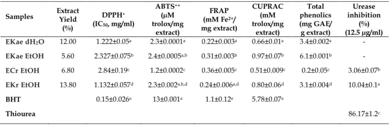
The highest total phenolic content was determined in EKae EtOH (6.1±0.001 mgGAE/g extract). The total phenolic contents of EKae dH2O (3.4±0.002 mg GAE/g extract) and EKr EtOH (3.1±0.004 mgGAE/g extract) were found to be close. On the other hand, ECr EtOH (0.2±0.05 mg GAE/g extract) was found to contain the lowest levels of phenolics.
2.2. Effects of the extracts on cell viability
The non-toxic concentrations of the extracts on Raw 264.7 macrophages were determined by WST-1 assay (Figure 1). The indicated concentrations (12.5, 25, 50 and 100 µg/ml) of extracts with lipopolysachharides (LPS, 1 µg/ml) were applied to cells, and incubated at 37 ºC for 24 h. The viability of the cells were not significantly decreased by the treatment with the EtOH extract of the aerial parts of E. kotschyi [EKae EtOH] (Figure 1A). On the other hand, the dH2O extract of the aerial parts of the same plant [EKae dH2O] applied at the same concentration slightly reduced the cellular viability of the cells at the highest two concentrations (Figure 1C). The cell viabilities were 85.70±4.21% and 80.65±7.42% at 50 and 100 µg/ml concentrations, respectively. The EtOH extract of the roots of E. kotschyi [EKr EtOH] only slightly decreased the cell viability (80.96±3.60%) at 100 µg/ml concentration (Figure 1B). The EtOH extract of the roots of E. campestre var. virens [ECr EtOH] did not significantly decreased cellular viability (Figure 1D). L-N6-(1-iminoethyl) lysine (L-NIL) at 10 µM did not significantly reduce the viability of Raw 264.7 cells.
(A) (B)
(C) (D)
Figure 1. Effects of E. kotschyi extracts [aerial EtOH (A), root EtOH (B), aerial dH2O (C)] and E. campestre var.
virensroot EtOH extract (D) on the viability of Raw 264.7 macrophages as determined by WST-1 assay. Indicated concentrations of extracts were applied to cells with LPS (1 µg/ml) for 24 h. C: L-NIL applied at 10 µM concentration. (-): Only media treated control group. Error bars represents the mean ± SEM value for three experiments. Values of * p ≤ 0.05, ** p ≤ 0.01 and *** p ≤ 0.001 were considered statistically significant compared to only media treated control group.
2.3. Effects of the extracts on NO production
Raw 264.7 cells were co-incubated with the indicated concentrations (12.5, 25, 50 and 100 µg/ml) of the extracts and LPS (1 µg/ml) for 24 h to determine the NO inhibitory activities. As shown in Figure 2, the treatment of cells with LPS resulted in a significant increasement of NO levels compared to only media treated control group. Although EKae dH2O (Figure 1C) and ECr EtOH (Figure 1D) did not exert remarkable inhibition at these concentrations, the other two extracts exerted significant inhibitions. EKr EtOH inhibited 4.31%, 12.47%, 19.62 %and 50.08% of NO production at 12.5, 25, 50 and 100 µg/ml concentrations respectively. On the other hand, EKae EtOH inhibited 13.85%, 24.61%, 33.44%, 41.52% of NO production at the same concentrations. It is noteworthy that despite the fact that EKr EtOH was found to be more active at high concentration (100 µg/ml), the activities of EKae EtOH was found to be higher at lower concentrations. L-NIL which is a known inhibitor of NO, exerted 86.94% inihibition at 10 µM concentration.
2.4. Effects of the extracts on the production of IL-6 and IL-1α on LPS-induced Raw 264.7 macrophages Raw 264.7 macrophages were co-incubated with both LPS (1 µg/ml) and the indicated concentrations (12.5, 50 and 100 µg/ml) of the extracts and for 24 h. After that the media was collected and IL-6 levels were measured. As shown in Figure 3 the treatment of cells with LPS significantly increased IL-6 levels compared to only media treated control group. ECr EtOH inhibited 11.05%, 18.10%, 36.22% of IL-6 production at these concentrations (Figure 3D). EKr EtOH exerted remarkable inhibition (65.23%) only at the highest concentration (Figure 3B). On the other hand, the activities of EKae dH2O and EKae EtOH were found weak. Dexamethasone which is a known inhibitor of IL-6 production, exerted 80.14% inihibition at 5 µM concentration.
All extracts were tested for their IL-1α inhibitory potentials at 100 µg/ml concentrations. EKae dH2O and EKae EtOH did not significantly inhibit IL-1α productions. EKr EtOH significantly inhibited IL-1α in a concentration dependent manner. This extract provided 38.94% and 56.84% decrease in IL-1α levels at 50 and 100 µg/ml concentraions compared to only LPS treated control group (Figure 4 B). ECr EtOH inhibited 50.92%, 44.24% of IL-1α production at these concentrations (Figure 4C).
0 20 40 60 80 100 - 12.5 25 50 100 C C e ll vi ab il it y% EKae EtOH (µg/ml) 0 20 40 60 80 100 - 12.5 25 50 100 C e ll vi ab il it y% EKr EtOH (µg/ml) ** 0 20 40 60 80 100 - 12.5 25 50 100 C e ll vi ab il it y% EKae dH2O (µg/ml) ** ** 0 20 40 60 80 100 - 12.5 25 50 100 C e ll vi ab il it y% ECr EtOH (µg/ml)
(A) (B)
(C) (D)
Figure 2. Effect of E. kotschyi extracts [aerial EtOH (A), root EtOH (B), aerial dH2O (C)] and E. campestre var.
virens root EtOH extract (D) on the NO productions of LPS induced Raw 264.7 macrophages as determined by Griess Assay. The indicated concentrations of extracts was co-incubated with LPS (1 µg/ml) for 24 h. C: L-NIL (10 µM) was applied. (-): Only media treated control group. Error bars represents the mean ±SEM values for three experiments. Values of * p ≤ 0.05, ** p ≤ 0.01 and *** p ≤ 0.001 vs. LPS treated control group and # p ≤ 0.05, ## p ≤ 0.01 and ### p ≤ 0.001 vs. only media treated group were considered statistically significant.
(A) (B)
(C) (D)
Figure 3. Effect of E. kotschyi extracts [aerial EtOH (A), root EtOH (B), aerial dH2O (C)] and E. campestre var.
virens root EtOH extract (D) on the IL-6 productions of LPS induced Raw 264.7 macrophages. IL-6 concentrations were determined by ELISA. Cells were co-incubated with the indicated concentrations of extracts and LPS (1 µg/ml) for 24 h. Dexamethasone (Dex) was applied at 10 µM concentration. (-): Only media treated control group. Error bars represent the mean ± SEM. Values of * p ≤ 0.05, ** p ≤ 0.01 and *** p ≤ 0.001 vs. LPS treated control group and # p ≤ 0.05, ## p ≤ 0.01 and ### p ≤ 0.001 vs. only media treated group were considered statistically significant.
0 20 40 60 80 100 120 - - 12.5 25 50 100 C (L P S t re at e d %) NO LPS (1 µg/ml) EKae EtOH (μg/ml) ## * * * * 0 20 40 60 80 100 120 - - 12.5 25 50 100 (L P S t re at e d %) NO LPS (1 µg/ml) EKr EtOH (μg/ml) ## ** ** 0 20 40 60 80 100 120 140 - - 12.5 25 50 100 (L P S t re at e d %) NO LPS (1 µg/ml) EKae dH2O (μg/ml) ## 0 20 40 60 80 100 120 - - 12.5 25 50 100 (L P S t re at e d %) NO LPS (1 µg/ml) ECr EtOH (μg/ml) ## 0 50000 100000 150000 - - 12.5 50 100
IL
-6
(p
g/
m
l)
LPS (1 µg/ml) EKae EtOH (μg/ml) # 0 50000 100000 150000 200000 - - 12.5 50 100IL
-6
(p
g/
m
l)
LPS (1 µg/ml) EKr EtOH (μg/ml) # * 0 50000 100000 150000 200000 - - 12.5 50 100IL
-6
(p
g/
m
l)
LPS (1 µg/ml) EKae dH2O (μg/ml) # * * * 0 50000 100000 150000 - - 12.5 50 100 Dex IL -6 (p g/ m l) LPS (1 µg/ml) ECr EtOH (μg/ml) # * * * *(A) (B)
(C)
Figure 4. Effect of E. kotschyi extracts [aerial EtOH (A), root EtOH (B), aerial dH2O (A)] and E. campestre var.
virens root EtOH extract (C) on the IL-1α productions of LPS induced Raw 264.7 macrophages. IL-1α levels were determined by ELISA. Cells were treated with the indicated concentrations of extracts and LPS (1 µg/ml) for 24 h. (-): Only media treated control group. Error bars represents the mean ±SEM values. Values of * p ≤ 0.05, ** p ≤ 0.01 and *** p ≤ 0.001 vs. LPS treated control group and # p ≤ 0.05, ## p ≤ 0.01 and ### p ≤ 0.001 vs. only media treated group were considered statistically significant.
2.5. DPPH• radical scavenging activity
The highest DPPH• radical scavenging activity is detected for EKr EtOH, followed by EKae dH2O, EKae EtOH and ECr EtOH (Table 1). The IC50 values are 1.132±0.057, 1.222±0.05, 2.327±0.075, 2.84±0.19 mg/ml respectively. The free radical scavenging activities of the extracts were found to be lower than BHT (0.15±0.026 mg/ml). A linear relationship was not observed between DPPH• radical scavenging activity and the amount of total phenolic contents.
Table 1. The antioxidant and urease inhibitory activities of plant extracts. Samples Extract Yield
(%) DPPH• (IC50, mg/ml) ABTS•+ (µM trolox/mg extract) FRAP (mM Fe2+/ mg extract) CUPRAC (mM trolox/mg extract) Total phenolics (mg GAE/ g extract) Urease inhibition (%) (12.5 µg/ml) EKae dH2O 12.00 1.222±0.05a 2.3±0.0001a 0.22±0.003a 0.66±0.01a 3.4±0.002a -
EKae EtOH 5.60 2.327±0.075b 2.4±0.0005a,b 0.31±0.003b 0.97±0.07b 6.1±0.001b - ECr EtOH 6.80 2.84±0.19c 1.2±0.0002c 0.36±0.005c 0.51±0.009c 0.2±0.05c 3.06±0.07b EKr EtOH 13.80 1.132±0.057d 2.3±0.002a,b,d 0.24±0.006a,d 0.80±0.06d 3.1±0.004d 10.04±0.1a
BHT 0.15±0.026e 13±0.001e 1.1±0.12e 5.78±0.07e
Thiourea 86.17±1.2c
Values are mean of triplicate determination (n = 3) ± standard deviation; means with different superscripts (a-e) are
significantly different, p<0.05, GAE–Gallic acid equivalents. 2.6. ABTS•+ radical cation scavenging activity
As shown in Table 1, ABTS•+ radical scavenging activities of the extracts were as follows the EKae EtOH, EKae dH2O, EKr EtOH, ECr EtOH. The values were 2.4±0.0005, 2.3±0.0001, 2.3±0.002 and 1.2±0.0002 µM trolox/mg extract, respectively. The free radical scavenging activities of both extracts was found to be less
0 50 100 150 - - EtOH dH2O IL -1 α % LPS (1 µg/ml) EKae (μg/ml) # 0 5 10 15 20 - - 12.5 50 100 IL -1 α (p g/ m l) LPS (1 µg/ml) EKr EtOH (μg/ml) # * * 0 5 10 15 20 25 - - 12.5 50 100 IL -1 α (p g/ m l) LPS (1 µg/ml) ECr EtOH (μg/ml) # * *
than BHT (13±0.0001 µM trolox/mg). A linear relationship was observed between ABTS•+ radical scavenging activity and the amount of total phenolic contents.
2.7. Ferric reducing antioxidant/power capacity
As shown in Table 1, ECr EtOH (0.36±0.005 mM Fe2+/mg extract) exhibited the highest ferric reducing antioxidant capacity followed by EKae EtOH (0.31±0.003 mM Fe2+/mg extract), EKr EtOH (0.24±0.006 mM Fe2+/mg extract) and EKae dH2O (0.22±0.003 mM Fe2+/mg extract). The activities of the extracts were less than BHT. A linear relationship was not observed between ferric reducing antioxidant capacity and the amount of total phenolic contents.
2.8. Cupric ion reducing antioxidant capacity
The cupric reducing antioxidant capacity of the extracts were as follows EKae EtOH (0.97±0.07 mM trolox/mg extract), EKr EtOH (0.80±0.06 mM trolox/mg extract), EKae dH2O (0.66±0.01 mM trolox/mg extract) and ECr EtOH (0.51±0.009 mM trolox/mg extract). A linear relationship was observed between cupric reducing antioxidant capacity and the amount of total phenolic contents.
2.9. Urease inhibitory activity
The urease inhibitory activities of the extracts were tested at 12.5 µg/ml concentration. EKr EtOH exerted 10.04±0.1% inhibition of urease activity. On the other hand, ECr EtOH showed 3.06±0.07% inhibition. Both extracts exerted lower activity than thiourea, a reference compound, which provided 86.17±1.2% inhibition at the same concentration. While the dH2O and EtOH extracts of the aerial parts of E. kotschyi did not exert significant urease inhibition (Table 1).
3. DISCUSSION
Despite the common traditional usage of Eryngium species for a wide spectrum of diseases in Anatolia, a literature survey revealed that, only a limited number of scientific studies have been carried out so far particularly on E. kotschyi, which is one of the endemic species to Turkish flora [2, 4, 5]. The in vivo anti-inflammatory and anti-nociceptive activities of E. kotschyi and E. campestre were previosly reported [2; 29]. From the MeOH extract of the roots of E. kotschyi, bioactivity guided fractionation procedures led to the identification several active fractions and a moderate effective triterpen saponin [29]. In a previous study Kupeli et al. evaluated the in vivo anti-inflammatory activities of the aerial and root parts of various Eryngium species and reported that dH2O and EtOH extracts of the aerial parts of E. kotschyi as well as the EtOH extract of the root parts of E. kotschyi exerted significant in vivo anti-inflammatory activities [3]. In order to cast light onto the anti-inflammatory activity mechanism of these extracts, the present study investigated the in vitro effects of the extracts on NO, IL-6 and IL-1α production on LPS induced Raw 264.7 macrophages. EKae EtOH which was determined to be the richest extracts of phenolics, significantly inhibited NO production of LPS induced Raw 264.7 cells, but did not exert significant inhibition on IL-6 and IL-1α levels. On the other hand, EKae dH2O which is the second richest extract in phenolics, failed to inhibit NO production, but slightly reduced IL-6 levels. It’s noteworthy that the aerial parts of of E. kotschyi did not exert high anti-inflammatory activity on the tested parameters except that the EtOH extract of the aerial parts exerted signifacant NO inhibitory activity. In the present study the roots of E. kotschyi were also investigated along with the roots of
E. campestre var. virens. EtOH extracts of both plants exerted significant inhibitory activity on IL-1α
productions. Although the ECr EtOH did not effect NO production, EKr EtOH significantly inhibited NO productions. ECr EtOH which contains the lowest levels of phenolics, significantly inhibited IL-6 production. The EtOH extract of the roots of E. campestre var. virens only exerted inhibitory activities on cytokine secrection. The EtOH extract of the roots of E. kotschyi exerted inhibitory activity on all parameters and it was the most active one against NO. Compared to aerial parts the roots parts of both plants exerted higher activities on all parameters except the EtOH extract of the aerial parts of E. kotschyi which exerted significant NO inhibitory activity. Literature survey revealed there is only a limited number of studies on the in vitro anti-inflammatory effects of the plants particularly against the activated macrophages. In a previous study, the EtOH extract from the aerial parts of E. campestre significantly inhibited NO secretion of cytokine stimulated murine brain endothelial cells and LPS induced murine monocyte/macrophage-like cell line P388D1 [10].
The extracts prepared from the aerial and root parts of E. kotschyi and the roots of E. campestre var. virens were also compared by means of their antioxidant activities and total phenolic contents. EKae EtOH having the richest phenolics content exerted the highest antioxidant activity by ABTS•+ and CUPRAC assays. On the
other hand, EKr EtOH and EKae dH2O exerted the highest antioxidant potential on DPPH• assay. ECr EtOH showed weak antioxidant potential on all assays except FRAP assay, although possessing the least phenolic content among all. In a recent study, the MeOH extract and subextracts obtained from the MeOH extract (ethyl acetate, n-butanol and water) of the flowering aerial parts of E. kotschyi was investigated for their total phenolic and total flavonoid compounds as well as their antioxidant capacity (DPPH●, ABTS+● and FRAP methods). In this study, the highest total phenolic and total flavonoid content was found in ethyl acetate subextract which also showed the highest antioxidant capacity [30]. In a previous study, the boiling water extracts of the aerial parts and roots of E. kotschyi were investigated for their antioxidant potentials by DPPH• radical scavenging assay, phosphomolybdenum assay and reducing power assays. The boiling water extract of E. kotschyi aerial and root parts exerted high antioxidant capacity with IC50 values 39.38 µg/ml and 39.42 µg/ml by DPPH• radical scavenging assay, and 50.90% and 49.62% as equivalent to α-tocopherol (mg/g) by phosphomolybdenum assay. Reducing power analysis verified the antioxidant capacity [31]. In the present study, the dH2O extract prepared at room temperature from the aerial parts of E. kotschyi was also found to exert DPPH• activity (IC50 = 1.222±0.05 mg/ml). Difference with the results might be due to the differences between the extraction techniques. The DPPH• method better responds to polar compounds in the extract. The increasing temperature for the extraction procedure seems to increase the DPPH• activity of the extract. Our results suggest that E. kotschyi aerial parts is a potential candidate for further investigations in anti-inflammatory activity as well as its promising antioxidant activity.
The antioxidant activity of the aerial parts of E. campestre was reported in a number of studies [32, 33, 34, 35, 36, 37]. However only a few studies reported the antioxidant activity of its roots. Different solvent extracts (ethyl acetate, n-butanol and aqueous extracts) prepared from roots of E. campstre was reported to exert good antioxidant activities due to its polyphenolic compounds [33]. The MeOH extract of the roots was also evaluated by an antioxidant activity assay based on the Briggs–Rauscher (BR) reaction as well as TEAC and DPPH• assays and IC50 value for DPPH• assay was estimated as 216.0 ± 4.0 μg/ml [38].
The urease inhibitory activites of the extracts were also evaluated as a measure to investigate
anti-Helicobacter pylori activities of the extracts. Only EKr EtOH exerted 10.04% urease inhibition. The remaining
extracts were not active at 12.5 µg/ml concentration. All extracts exerted lower activity than Thiourea. 4. CONCLUSION
It’s revealed that the EtOH extracts of the root parts of E. kotschyi and E. campestre var. virens exerted the highest inhibitory activities on NO, IL-6 and IL-1α productions. Containing relatively lower levels of phenolic compounds, these extracts also exerted the highest antioxidant activities on DPPH• and FRAP assays, respectively. Further investigations are needed to explain the chemical composition of these extracts to explain the observed activity. The extracts prepared from the aerial parts of E. kotschyi (EtOH and dH2O) exerted high antioxidant activities on ABTS•+, FRAP and CUPRAC assays, and on DPPH• and ABTS•+ assays, respectively. These extracts were also determined to contain the highest levels of total phenolics that may contribute to explain the antioxidant activity of these extracts. On the other hand, the EtOH extracts of the aerial part of E.
kotschyi exerted significant inhibitory activity on NO production.
Consequently, the results of this paper might shed light on the effects of the extracts on inflammatory mediators which might account for the previously reported in vivo anti-inflammatory activity of the E. kotschyi extracts. Besides the inhibitory activities of the extracts on NO and cytokines, the antioxidant activity of the extracts might contribute to the anti-inflammatory activity of the extracts. The total phenolic contents of the extracts were also reported. As far as we know this is the first report on the in vitro anti-inflammatory and urease inhibitory activities of E. kotschyi.
5. MATERIALS AND METHODS 5.1. Reagents and Chemicals
LPS, dexamethasone, dimethylsulfoxide (DMSO), Griess reagent, Folin Ciocalteu’s phenol reagent, 2,4,6-tripyridyl-s-triazine, gallic acid, and 2,2-diphenyl-1-picryl-hydrazyl (DPPH•) were obtained from SigmaAldrich (St. Louis, MO). Dulbecco’s modified Eagle’s medium (DMEM), penicillin-streptomycine solution and Fetal bovine serum (FBS) was purchased from Invitrogen/Gibco (Grans Island, NY, USA). Enzyme linked immunosorbent assay (ELISA) kits for TNFα and IL-1α were from R&D Systems (MN, USA) and eBiosciences respectively. L-NIL was sourced from Cayman. Butylated hydroxytoluene and 2,2’-azino-bis(3-ethylbenzothiazoline-6-sulfonate) were obtained from Fluka. WST-1 reagent was purchased from Roche
Applied Science (Mannheim, Germany).Urease (lyophilized 5 U/MG EC 3.5.1.5) were sourced from Merck KGaA. All other reagents were of analytical grade.
5.2. Plant material
The aerial parts and roots of E. kotschyi Boiss., Akaydın 13365 were collected from Sarıveliler and the roots of E. campestre L. var. virens (Link) Weins, Akaydın 13338 were collected from Kazancı (Karaman-Turkey) in July, 2010. The material was identified by G.A. (one of the authors). Voucher specimen are deposited at the Herbarium of the Faculty of Education, Hacettepe University, Ankara, Turkey.
5.3. Extraction
Dried and powdered roots of E. kotschyi and roots of E. campestre var. virens (10 g, each) as well as aerial parts of E. kotschyi were separately extracted two times with 90% EtOH (200 ml) at room temperature. The extracts were filtered through a filter paper. Evaporation of the solvents to dryness under reduced pressure yielded crude E. kotschyi root EtOH extract [EKr EtOH], E. campestre var. virens root EtOH extract [ECr EtOH] and E. kotschyi aerial parts EtOH extract [EKae EtOH] (Table 1). The extracts were filtered and the organic solvent was evaporated to dryness under reduced pressure (40°C). On the other hand, the dried and powdered aerial parts of E. kotschyi (10 g) was extracted two times with dH2O (200 ml). The extract was filtered and lyophilized [EKae dH2O] (Table 1) [3].
5.4. Determination of total phenolic contents
FCR method was used to measure the total phenolic contents. Extract (5 µL) and water (225 µL) was mixed in the plate. Afterwards, Folin-Ciocalteu reagent (diluted 1/3 with distilled water, 5 µL) and 2% sodium carbonate solution (15 µL) were added to the mixture. It was then lefted at room temperature (RT) for 2 hours. Finally the absorbance was read at 760 nm against the reference using a micro plate reader. The results were calculated and expressed as mg gallic acid equivalents/g extract [39].
5.5. Cells and cell culture
Dulbecco's modified Eagle's medium (DMEM) was supplemented with 10% Fetal Bovine Serum (FBS), 4 mM L-glutamin, 100 IU/ml penicillin and 100 µg/ml streptomycin to prepare cell culture media. The Raw 264.7 macrophages (ATCC TIB-71) were grown in the cell culture media at 37°C in a humidified atmosphere containing 5% CO2.
5.6. WST-1 assay for cell viability
The viability of Raw 264.7 macrophages were tested by using a WST-1 assay kit (Roche Applied Sciences). This assay depends on the reducement of a tetrazolium-based salt dye to a purple colored formazan salt by metabolically active cells. Raw 264.7 cells (22.500 cells/well) were seeded into 96-well plates in 10% FBS-DMEM. The cells were treated with the indicated concentrations of the extracts ranging from 12.5 µg/ml to 100 µg/ml with 1 µg/ml LPS, and incubated for 24 hrs (37°C). The supernatant was then discarded and WST-1 reagent was added directly to the wells [final concentration of 5% (v/v)]. The cells were incubated with the reagent at 37°C for an additional 60 mins. Finally, absorbance was measured between 420-480 nm (λmax 450 nm). All extracts and standards were dissolved in DMSO. They were then diluted with DMEM. The concentration of DMSO in the cell culture medium was not more than 0.1% (v/v).
5.7. Griess assay
The NO inhibitory activities were evaluated by Griess assay on LPS induced Raw 264.7 macrophages. The 96-well plates were used to seed Raw 264.7 cells at a density of 22.500 cells/well, in 10% FBS-DMEM. The indicated (12.5 µg/ml-100 µg/ml) concentrations of the extracts with 1 μg/ml LPS was applied to cells for 24 hrs at 37°C. Accumulated nitrite in the culture medium was determined by Griess Assay. For this purpose, 50 μl of cell culture medium and 50 μl of 1% (w/v) sulfanilamide in 5% (v/v) phosphoric acid was mixed and incubated for 10 mins. After that, 50 μl of 0.1% (w/v) naphtylethylenediamine-HCl (Promega) was added. The mixture was then incubated at room temperature for an additional 10 mins. The absorbance was measured at 550 nm by a microplate reader. The amount of nitrite was calculated from the serial dilution standard curve prepared from NaNO2.
5.8. Enzyme immunoassay for quantification of cytokines (IL-1α, IL-6)
The cytokine inhibitory activities were evaluated by ELISA on LPS induced Raw 264.7 macrophages. The 96-well plates were used to seed Raw 264.7 cells at a density of 22.500 cells/well, in 10% FBS-DMEM. The extracts and 1 μg/ml LPS were together applied to cells for 24 hrs (37°C). The inhibitory effects of extracts on the production of cytokines were determined by Sandwich ELISA method. The supernatants were harvested and tested in agreement with the manufacturer's instructions for the relevant ELISA kit.
5.9. DPPH• radical scavenging activity
The free radical scavenging capacity of extracts were determined by DPPH• test. The DPPH• solution (0.1 mM, 240 µL) was added to extracts (10 µL) prepared at varying concentrations ranging from 5 mg/ml to 0.5 mg/ml. The mixture was then lefted at RT for 30 min. The absorbance was measured against the reference at 517 nm. The results obtained from three experiments were given as IC50 = mg/ml [40].
5.10. Trolox equivalent antioxidant activity
Briefly, 50 µL of each extract was taken into the plate and 50 µL of water was added. Afterwards, 150 µL of ABTS•+ working solution were added to the mixture. The absorbance of the mixture was measured against the reference using a microplate reader at 734 nm for 6 min. The standard curve was prepared using trolox. The results were expressed as µM trolox/mg extract [41].
5.11. Ferric reducing/antioxidant power (FRAP) assay
The ferric reducing ability of extracts were evaluated using the FRAP method. The FRAP reagent (10 µL) and extracts (190 µL) were mixed. The absorbance of the mixture was measured after 4 mins against the reference at 593 nm. The FeSO4.7H2O standard cureve was prepared and FRAP values were expressed as a mM Fe2+/mg extract [42].
5.12. Cupric reducing antioxidant capacity (CUPRAC) assay
CUPRAC method was used to asses the antioxidant capacity. The neocuproine ethanolic solution (7.3.10-3 M, 60 µL), Cu (II) (1.10-2 M, 60 µL), and 1 M NH4Ac buffer solution (60 µL) were mixed in plate. Extracts (each in 60 µL) and EtOH (pure, 10 µL) were added to the initial mixture to make the total volume of 250 µL and it was vortexed for 10 s. The absorbance was measured exactly after 60 mins against a reagent blank at 450 nm. The results were reported as trolox equivalents (mM trolox/mg extract) [43].
5.13. Urease inhibitory activity
Urease enzyme (500 µL) was mixed with the working solution (100 µL). The mixture was incubated in the incubator at 37°C for 30 min. After that, 1100 µL of urea was added and the mixture was incubated for an additional 30 min. in the incubator (37°C). The reagents; R1 (1% phenol, 0.005% sodium nitroprusside) and R2 (0.5% NaOH, 0.1% sodium hypochlorite) were added to the mixture respectively. It was then incubated for 2 hrs in the incubator at 37°C. The absorbance was read against the reference at 635 nm [44].
5.14. Statistical analysis
The data obtained from triplicate experiments were reported as mean ± standard deviation (SD) or mean ± standard error ofthe mean (SEM). Graphpad Prism 5 Demo or PASW Statistics were used to analyse data. The determination of the significance of the differences between means were achieved by Tukey’s Multiple Comparison Test or Mann Whitney U test. The p-values less than 0.05 were considered statistically significant. Author contributions: Concept – İ.A.B., E.Y.; Design – İ.A.B, T.T.; Supervision – E.Y.,İ.A.B., T.T., G.A.; Resource – İ.A.B., T.T.; Materials – İ.A.B., T.T., G.A., E.Y.; Data Collection and/or Processing - İ.A.B., T.T., E.A.Ş.; Analysis and/or Interpretation - İ.A.B., T.T., E.Y.; Literature Search – İ.A.B., T.T., E.A.Ş.; Writing – İ.A.B., T.T., E.Y.; Critical Reviews – İ.A.B., T.T., E.A.Ş., G.A., E.Y.
Conflict of interest statement: The authors declared no conflict of interest in the manuscript.
REFERENCES
[1] Ecevit Genç G, Akalın Uruşak E, Wörz A. A New Species of Eryngium (Apiaceae) from Turkey: Eryngium
[2] Davis PH. The Flora of Turkey and the East Aegean Islands Vol. 4, Edinburgh University Press, Edinburg, 1972. [3] Küpeli E, Kartal M, Aslan S, Yesilada, E. Comparative Evaluation Of The Anti-İnflammatory And Antinociceptive
Activity of Turkish Eryngium species. J Ethnopharmacol. 2006; 107: 32-37. [CrossRef]
[4] Sezik E, Yeşi̇lada E, Tabata M, Honda G, Takaishi Y, Fujita T, Tanaka T, Takeda Y. Traditional Medicine in Turkey VIII. Folk Medicine in East Anatolia; Erzurum, Erzi̇ncan, Aǧri, Kars, Igdir Provinces. Econ Bot. 1997; 51 (3): 195-211. [CrossRef]
[5] Yesilada E, Sezik E. Part 28. A Survey On The Traditional Medicine in Turkey: Semi-Quantitative Evaluation Of The Results. In: Singh K, Govil JN, Hashmi S, Singh G. (Eds). Recent Progress in Medicinal Plants. Vol. VII. Ethnomedicine and Pharmacognosy-II. Studium Press, LLC, Houston, Texas, 2003, pp. 389-412.
[6] Aslan Erdem S, Mitaine Offer AC, Miyamoto T, Kartal M, Lacaille Dubois MA. Triterpene Saponins from Eryngium
kotschyi. Phytochemistry. 2015; 110: 160-165. [CrossRef]
[7] Yurdakök B, Baydan E. Cytotoxic Effects of Eryngium kotschyi and Eryngium maritimum on Hep2, HepG2, Vero and U138 MG Cell Lines. Pharm Biol. 2013; 51 (12): 1579-1585. [CrossRef]
[8] Usta C, Yildirim AB, Turker AU. Antibacterial And Antitumour Activities Of Some Plants Grown in Turkey. Biotechnol Biotec Eq. 2014; 28 (2): 306–315. [CrossRef]
[9] Çelik A, Aydınlık N, Arslan I. Phytochemical Constituents and Inhibitory Activity towards Methicillin‐Resistant
Staphylococcus aureus Strains of Eryngium Species (Apiaceae). Chem Biodivers. 2011; 8: 454-459. [CrossRef]
[10] Strzelecka M, Bzowska M, Kozieł J, Szuba B, Dubiel O, Riviera Núńez D, Heinrich M, Bereta J. Anti-Inflammatory Effects of Extracts from Some Traditional Mediterranean Diet Plants. J Physiol Pharmacol. 2005; 56 (1):139-56. [11] Kartal M, Mitaine Offer AC, Abu Asaker M, Miyamoto T, Calis I, Wagner H, Lacaille Dubois MA. Two New
Triterpene Saponins from Eryngium campestre. Chem Pharm Bull. 2005; 53(10): 1318-1320. [CrossRef]
[12] Kartal M, Mitaine Offer AC, Paululat T, Abu Asaker M, Wagner H, Mirjolet JF, Guilbaud N, Lacaille-Dubois MA. Triterpene Saponins from Eryngium campestre. J Nat Prod. 2006; 69 (7):1105-8. [CrossRef]
[13] Hohmann J, Pall Z, Gunther G, Mathe I. Flavonolacyl Glycosides of the Aerial Parts of Eryngium campestre. Planta Med. 1997; 63(1):96. [CrossRef]
[14] Kartnig T, Wolf J. Flavonoids from the Aboveground Parts of Eryngium campestre. Planta Med. 1993; 59(3): 285. [CrossRef]
[15] Erdelmeier CA, Sticher O. Coumarin Derivatives from Eryngium campestre. Planta Med. 1985; 51(5):407-409. [CrossRef]
[16] Sticher O, Erdelmeier C. Cumarinderivate aus den Wurzeln von Eryngium campestre. Planta Med. 1982; 45(7): 160-161. [CrossRef]
[17] Feldmann M, Maini RN. TNF Defined as A Therapeutic Target for Rheumatoid Arthritis and Other Autoimmune Diseases. Nat Med. 2003; 9: 1245-1250. [CrossRef]
[18] Heller RA, Schena M, Chai A, Shalon D, Bedilion T, Gilmore J, Woolley DE, Davis RW. Discovery and Analysis of Inflammatory Disease-Related Genes Using cDNA Microarrays. P Natl A Sci USA. 1997; 94: 2150-2155. [CrossRef] [19] Coussens LM, Werb Z. Inflammation and Cancer. Nature. 2002; 420(6917): 860-867. [CrossRef]
[20] Tsai SH, Lin-Shiau SY, Lin JK. Suppression of Nitric Oxide Synthase And The Down-Regulation of The Activation of NF-kappaB in Macrophages by Resveratrol. Br J Pharmacol. 1999; 126(3): 673–680. [CrossRef]
[21] Chen YC, Shen SC, Lee WR, Hou WC, Yang LL, Lee TJF. Inhibition of Nitric Oxide Synthase Inhibitors and Lipopolysaccharide Induced Inducible NOS And Cyclooxygenase-2 Gene Expressions By Rutin, Quercetin, And Quercetin Pentaacetate in Raw 264.7 Macrophages. J Cell Biochem. 2001; 82: 537-548.[CrossRef]
[22] Dinarello CA. Proinflammatory Cytokines. Chest. 2000; 118: 503-508.[CrossRef]
[23] Zang JM, An J. Cytokines, Inflammation and Pain. Int Anesthesiol Clin. 2009; 45 2): 27-37.[CrossRef] [24] Gabay C. Interleukin-6 and Chronic Inflammation. Arthritis Res Ther. 2006; 6: 1-6.[CrossRef]
[25] Geronikaki AA, Gavalas AM. Antioxidants and Inflammatory Disease: Synthetic and Natural Antioxidants with Anti-inflammatory activity. Comb Chem High Throughput Screen. 2006; 9(6): 425-442.[CrossRef]
[26] Khansari N, Shakiba Y, Mahmoudi M. Chronic Inflammation and Oxidative Stress as a Major Cause Of Age-Related Diseases and Cancer. Recent Pat Inflamm Allergy Drug Discov. 2009; 3(1):73-80.[CrossRef]
[27] McGowan CC, Cover TL, Blaser MJ. Helicobacter pylori and Gastric Acid: Biological and Therapeutic Implications. Gastroenterology. 1996; 110: 926-938. [CrossRef]
[28] Leopoldini M, Russo N, Toscano M. The Molecular Basis of Working Mechanism of Natural Polyphenolic Antioxidants. Food Chem. 2011; 125(2): 288-306.
[29] Aslan Erdem S, Arıhan O, Mitaine Offer AC, İskit AB, Kartal M, Lacaille-Dubois MA. Antinociceptive Effects of Turkish Endemic Eryngium kotschyi Boiss. Roots by Bioactivity Guided Fractionation. Rec. Nat. Prod. 2016; 10(2): 168-175.
[30] Paşayeva L, Köngül Şafak E, Arıgün T, Fatullayev T, Tugay O. In vitro Antioxidant Capacity and Phytochemical Characterization of Eryngium kotschyi Boiss. J Pharm Pharmacogn Res. 2020; 8(1): 18-31.
[31] Yurdakök B, Gencay YE, Baydan E, Erdem SA, Kartal M. Antibacterial and Antioxidant Activity of Eryngium kotschyi and Eryngium maritimum. J Food Agric Environ. 2014; 12(2): 35-39. [CrossRef]
[32] Matejić JS, Stojanović-Radić ZZ, Ristić MS, Veselinović JB, Zlatković BK, Marin PD, Džamić AM. Chemical Characterization, in vitro Biological Activity Of Essential Oils And Extracts of Three Eryngium L. Species And Molecular Docking of Selected Major Compounds. J Food Sci Technol. 2018; 55(8): 2910-2925. [CrossRef]
[33] Bouzidi S, Benkiki N, Hachemi M, Haba H. Investigation of in vitro Antioxidant Activity and in vivo Antipyretic and Anti-Inflammatory Activities of Algerian Eryngium campestre L. Curr Bioact Compd. 2017; 13(4): 340-346. [CrossRef]
[34] Gunes MG, Isgor BS, Isgor YG, Shomalı Moghaddam N, Geven F, Yıldırım O. The Effects of Eryngium campestre Extracts on Glutathione-S-Transferase, Glutathione Peroxidase and Catalase Enzyme Activities. Turk J Pharm Sci. 2014; 11: 339-346.
[35] Hawas UW, Abou El-Kassem LT, Awad HM, Taie HAA. Anti-Alzheimer, Antioxidant Activities and Flavonol Glycosides of Eryngium campestre L. Curr Chem Biol. 2013; 7: 188-195.[CrossRef]
[36] Sucio S, Parvu AE. Comparative Study On The Effects Of Eryngıum Sp. Extracts in An Acute Inflammatıon Model in Rat. Annals of the RSCB. 2012; 17(2): 86-91.
[37] The Local Food-Nutraceuticals Consortium. Understanding Local Mediterranean Diets: A Multidisciplinary Pharmacological and Ethnobotanical Approach. Pharm Res. 2005; 52: 353-366. [CrossRef]
[38] Dall'Acqua S, Cervellati R, Loi MC, Innocenti G. Evaluation of in vitro Antioxidant Properties Of Some Traditional Sardinian Medicinal Plants: Investigation of The High Antioxidant Capacity of Rubus ulmifolius. Food Chem. 2008; 106: 745-749.[CrossRef]
[39] Ozsoy N, Can A, Yanardağ R, Akev N. Antioxidant Activity of Smilax excelsa L. Leaf Extracts. Food Chem. 2008; 110(3): 571-583.[CrossRef]
[40] Fu W, Chen J, Cai Y, Lei Y, Chen L, Pei L, Zhou D, Liang X, Ruan J. Antioxidant, Free Radical Scavenging, Anti-Inflammatory and Hepatoprotective Potential of the Extract From Parathelypteris Nipponica (Franch. Et Sav.) Ching. J Ethnopharmacol. 2010; 130(3): 521-528.[CrossRef]
[41] Re R, Pellegrini N, Proteggente A, Pannala A, Yang M, Rice-Evans C. Antioxidant Activity Applying An Improved ABTS Radical Cation Decolorization Assay. Free Radic Biol Med. 1999; 26(9-10): 1231–1237. [CrossRef]
[42] Benzie IF, Strain JJ. The Ferric Reducing Ability of Plasma (FRAP) As a Measure Of “Antioxidant Power”: The FRAP Assay. Anal Biochem. 1996; 239(1): 70-76. [CrossRef]
[43] Apak R, Güclü K, Ozyurek M, Karademir SE. Novel Total Antioxidant Capacity Index for Dietary Polyphenols And Vitamins C and E, Using Their Cupric Ion Reducing Capability in The Presence of Neocuproine: CUPRAC Method. J Agric Food Chem. 2004; 52: 7970-7981. [CrossRef]
[44] Ghous T, Akhtar K, Nasim FUH, Choudhry MA. Screening Of Selected Medicinal Plants for Urease Inhibitory Activity. BLM. 2010; 2(4): 64-69. [CrossRef]
![Figure 1. Effects of E. kotschyi extracts [aerial EtOH (A), root EtOH (B), aerial dH 2 O (C)] and E](https://thumb-eu.123doks.com/thumbv2/9libnet/5457639.105242/3.892.154.741.111.512/figure-effects-kotschyi-extracts-aerial-etoh-etoh-aerial.webp)
![Figure 3. Effect of E. kotschyi extracts [aerial EtOH (A), root EtOH (B), aerial dH 2 O (C)] and E](https://thumb-eu.123doks.com/thumbv2/9libnet/5457639.105242/4.892.150.745.641.1023/figure-effect-kotschyi-extracts-aerial-etoh-etoh-aerial.webp)