O R I G I N A L A R T I C L E – P A N C R E A T I C T U M O R S
Substaging of Lymph Node Status in Resected Pancreatic Ductal
Adenocarcinoma Has Strong Prognostic Correlations: Proposal
for a Revised N Classification for TNM Staging
Olca Basturk, MD1,8, Burcu Saka, MD2, Serdar Balci, MD3, Lauren M. Postlewait, MD4, Jessica Knight, MPH5, Michael Goodman, MD, PhD5, David Kooby, MD4, Juan M. Sarmiento, MD6, Bassel El-Rayes, MD7,
Hyejeong Choi, MD3, Pelin Bagci, MD3, Alyssa Krasinskas, MD3, Brian Quigley, MD3, Michelle D. Reid, MD3, Gizem Akkas, MD3, Shishir K. Maithel, MD4, and Volkan Adsay, MD3
1
Department of Pathology, New York University, New York, NY;2Department of Pathology, Istanbul Medipol University, Istanbul, Turkey;3Department of Pathology, Emory University School of Medicine and Winship Cancer Institute, Atlanta, GA;4Department of Surgical Oncology, Emory University School of Medicine and Winship Cancer Institute, Atlanta, GA; 5
Department of Epidemiology, Emory University School of Medicine and Winship Cancer Institute, Atlanta, GA; 6Department of General Surgery, Emory University School of Medicine and Winship Cancer Institute, Atlanta, GA; 7Department of Medical Oncology, Emory University School of Medicine and Winship Cancer Institute, Atlanta, GA; 8Present Address: Memorial Sloan Kettering Cancer Center, New York, NY
ABSTRACT
Background. The current tumor-node-metastasis staging system for the pancreas does not incorporate the number of lymph nodes (LNs) with metastasis.
Methods. Among 1649 pancreaticoduodenectomies, 227 stringently defined pancreatic ductal adenocarcinomas (PDACs) that had undergone a specific approach of LN harvesting were analyzed for the prognostic value of LN substaging protocols used for other gastrointestinal (GI) organs.
Results. The median number of LNs harvested was 18, and the median number of LNs with metastasis was 3.
Lymph node metastasis was detected in 175 cases (77 %). The number of LNs involved correlated significantly with clinical outcome. When cases were substaged with the protocol already in use for the upper GI organs (N0: no metastasis, N1: metastasis to 1–2 LNs; N2: metastasis to C3 LNs), the median overall survival times were 35, 21, and 18 months, and the respective 3-year survival rates were 46, 34, and 20 % (p = 0.004). Analysis of the Surveillance, Epidemiology and End Results (SEER) database also confirmed the survival differences between these substages (median overall survival times of 23, 15, and 14 months and respective 3-year survival rates of 37, 22, and 18 %; p \ 0.0001). The substaging protocol for the lower GI organs (N0: no metastasis; N1: metastasis to 1–3 LNs; N2: metastasis to C4 LNs) also was significant, with median overall survival times of 35, 21, 18 months and respective 3-year survival rates of 46, 26, and 23 %; p = 0.009). The association between higher N stage and shorter survival persisted with multivariate modeling for both protocols, although the prognostic value of the upper GI protocol appeared to be slightly stronger according to the Akaike Information Criterion method.
Conclusion. In conclusion, with proper LN harvesting, the LN metastasis rate in PDACs is very high (77 %). Sub-staging of LN metastasis has significant prognostic value and needs to be considered in the N staging of PDACs. The protocol already in use for other upper GI tract organs, which currently also is proven significant for ampulla,
This study was presented in part at the annual meeting of the United States and Canadian Academy of Pathology in San Diego, March 2014.
Electronic supplementary material The online version of this article (doi:10.1245/s10434-015-4861-0) contains supplementary material, which is available to authorized users.
Ó Society of Surgical Oncology 2015 First Received: 9 March 2015; Published Online: 11 September 2015 V. Adsay, MD
e-mail: [email protected] DOI 10.1245/s10434-015-4861-0
would be preferable, although the lower GI tract protocol also is applicable.
Current nodal N staging of pancreatic ductal adenocar-cinoma (PDAC) by the tumor-node-metastasis (TNM) guidelines of the American Joint Committee on Cancer (AJCC) and the Union for International Cancer Control (UICC) distinguishes only between negative and node-positive disease1and does not capture the prognostic impact of the number of lymph nodes (LNs) with metastasis. For many organs, including the gastrointestinal (GI) tract, the number of LNs with metastasis has been incorporated into the staging protocols, with significant prognostic value.1 Cases involving the upper GI tract with LN metastasis are substaged as N1 (metastasis to 1–2 LNs) and N2 (metastasis to C3 LNs), and cases involving the lower GI tract with LN metastasis are substaged as N1 (metastasis to 1–3 LNs) and N2 (metastasis to C4 LNs). Recently, Kang et al. and Balci et al. tested the upper GI protocol for ampullary carcinomas in their institutional cohorts and in the SEER database. Both studies found their results correlating independently and strongly with clinical outcome.2–5
The prognostic value of the LN status in PDAC has been controversial, with some studies failing to disclose an association and others showing significant correlation.6–18 These different results are attributable to methods defining what constitutes PDAC as well as to how the LNs are sampled, evaluated, and documented. This is reflected in the wide range of reported frequencies of LN metastasis, from 57 to 81 % of cases.6,10,12–14,16,17,19–33 In many studies, no pathologic verification was performed to exclude non-PDAC carcinomas (e.g., acinar, neuroen-docrine) or secondary cancers [ampullary, duodenal, common bile duct (CBD)].
The LN count also is markedly affected by the method of LN harvesting in the macroscopy rooms of pathology departments. In the recent analysis of the SEER database by Tomlinson et al.,34 the median number of LNs exam-ined was only 7 although the number of LNs required to attain the optimal separation of survival curves was 15.35 On the other hand, more exhaustive grossing approaches that involve indiscriminate total sampling of pancreatic surfaces for purposes of examining the margins6,36–38 inevitably lead to overcounting of LNs because individual LNs are not grossly separated out.
In this study, the number of LNs with metastasis and its prognostic significance as well as the potential value of N substaging protocols of both the upper and lower GI tract were analyzed in well-characterized PDACs, all of which were processed using a uniform LN harvesting protocol.39
METHODS
This study was approved by the appropriate institutional review committees. Among 1649 pancreatoduodenectomy specimens at Wayne State University (1998–2006) and Emory (2006–2013) University, 227 specimens processed according to a uniform grossing protocol39,40with a tissue diagnosis of conventional PDAC established by the authors were included in this study. Accordingly, unusual variants of carcinomas of ductal differentiation such as medullary, colloid, or intraductal papillary mucinous neoplasms with invasive adenocarcinoma were excluded from the study, together with nonductal tumors (neuroendocrine, solid-pseudopapillary, acinar, pancreatoblastoma tumors), sec-ondary carcinomas, cases with questionable tumor origin, and patients who had received neoadjuvant therapy. Cases with a follow-up period shorter than 3 months also were excluded, as has become customary.
All the patients underwent diagnostic laparoscopy before surgery. None of the patients in this study under-went ‘‘extended pancreatectomy.’’ Standard Whipple procedure was performed for 70 % and pylorus-preserving pancreaticoduodenectomy for 30 % of the patients. More-over, the LN dissection technique has been standardized to ensure that all the hepatic artery and the retroportal LNs are dissected. The tissue to the right of the superior mesenteric artery starting on the adventitia and for 180° on that side also was removed, but no circumferential dissection of the superior mesenteric artery was performed because findings have shown that this does not improve the survival of patients but could significantly jeopardize their quality of life.
All cases were processed according to a practical pro-tocol devised for a more accurate LN harvesting, the details of which have been published previously.39,40 With this standardized approach, currently routine at many institu-tions, in the gross laboratory, the LN areas are removed before dissection of the pancreatic head (Fig.1) to ensure complete harvesting. In other words, the potential LN regions are dissected and put aside before further cutting of the pancreatic head to avoid the under-evaluation of tissues that contain the LNs. In essence, all this simple protocol advocates is the removal of the LN regions before they can be lost. After this protocol was put in place, the LN yield jumped from 6 to 14 in the one institution and 7 to 14 in the other institution hosting this study.39,40 This simple approach has been adopted and used with ease by ordinary gross room personnel in conventional gross rooms using routine procedures.
In addition, all cases were subjected to detailed patho-logic reevaluation by the authors, including reexamination of all LNs for metastatic carcinoma. Tumor (T) and node
(N) stages were reassigned according to the current AJCC guidelines.1
The study analyzed the prognostic significance of the protocols used in both the upper GI tract (N0: no LN metastasis; N1: metastasis to 1–2 LNs; N2: metastasis to C3 LNs) and the lower GI tract (N0: no LN metastasis; N1: metastasis to 1–3 LNs; N2: metastasis to C4 LNs). The quality of each protocol relative to each of the other pro-tocols was estimated using Akaike Information Criteria (AIC). The data also were analyzed separately for those cases that had 12 or more LNs examined (the minimum number required by CAP’ guidelines).1,41
External validation of the upper GI protocol, recently also found to be correlated strongly with survival in ampullary carcinomas,2–5was performed using the SEER database by two of the authors who had specific expertise with this database. The analysis included resected PDACs reported between 2004 and 2010. These years were selec-ted because the AJCC 6th-edition criteria1were integrated into SEER in 2004. Only cases that had complete infor-mation relevant to survival were included.
Kaplan–Meier survival curves were estimated and compared between substages using the log-rank test. Multivariate analysis was conducted using Cox propor-tional hazard models adjusted for age, race, sex, primary tumor site, and AJCC T stages. The proportional hazard assumption was evaluated using log–log survival curves. All analyses were conducted using SAS software version 9.3 and SPSS version 22.
RESULTS Study Population
The study enrolled 227 patients with a confirmed diag-nosis of ordinary PDAC, all processed according to a
uniform grossing protocol. The mean age of the patients was 65 years (range 33–87 years), and 57 % were females (n = 129). The median PDAC size was 3.1 cm (range 0.6– 7 cm). The majority of the PDACs (95 %) showed carci-noma foci in the orange-peeled soft tissue (pT3).
Follow-up information during a period of 3 to 154 months (median, 15 months; patients with less than 3 months of follow-up data were eliminated from the study) was avail-able for all the patients. At the last follow-up assessment, 143 patients (63 %) had died of disease during a median follow-up period of 14 months. A median follow-follow-up assessment at 21.5 months found that 84 patients (37 %) were alive with disease. The clinicopathologic characteristics of the study cohort/SEER data are summarized in Table1.
Lymph Node Analysis
The median number of regional LNs examined from a given patient, including LNs sent as separate specimens, was 18 (range 3–45). The median number of LNs harvested from the pancreaticoduodenectomy specimen itself was 17 (range 2–43), and 81 % of these specimens had 12 or more LNs. In analysis of the SEER database, the median number of regional LNs examined was 11 (range 1–89), and only 47 % of the specimens had 12 or more LNs (Table1).
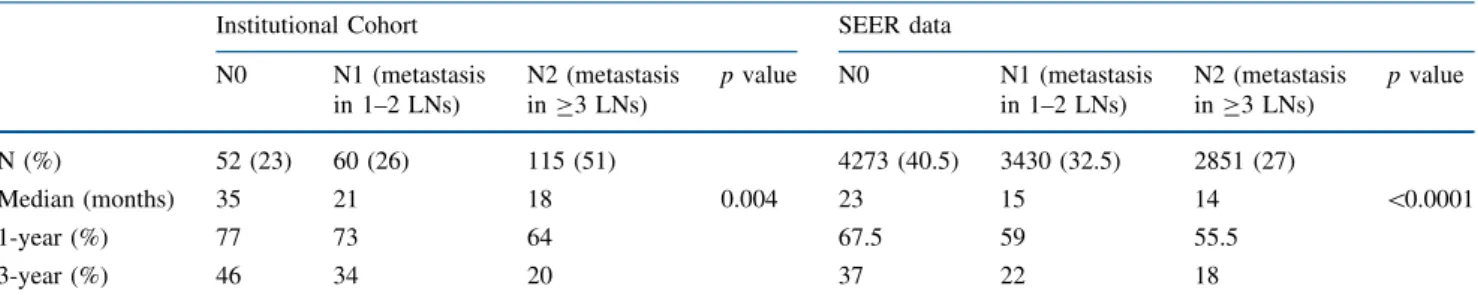
During the pathologic reevaluation of the cases, 6 cases originally classified as N-negative were reclassified as N-positive. After this thorough reexamination, 175 cases (77 %) were classified as N-positive. The SEER database had 6281 patients (59.5 %) with LN metastases. The median number of LNs with metastasis was 3 (range 1–20) in our cohort and 1 (range 1–47) in the SEER database. When N-positive cases were reclassified according to the upper GI protocol (N1: metastasis to 1–2 LNs; N2: metastasis to C 3 LNs), 60 cases (26 %) were N1 and 115
FIG. 1 Orange-peeling method of lymph node examination. Peri-pancreatic soft tissue is removed facilitating complete removal of the areas that potentially contain lymph nodes. a Posterior surface of a pancreaticoduodenectomy before the orange peeling. b View after the
posterior pancreaticoduodenal lymph node (LN) area (groove between the pancreatic head and the duodenal wall) has been shaved off. c View after the posterior pancreatic LN area also has been shaved off
(51 %) were N2, and when these cases were reclassified according to the lower GI protocol (N1: metastasis to 1–3; N2: metastasis to C4 LNs), 90 cases (40 %) were N1 and 85 cases (37 %) were N2.
Notably, the frequency of LN metastasis in the seven arbitrarily defined regions of peripancreatic tissue was 41 % in the posterior pancreatoduodenal region, 37 % in the posterior pancreatic region, 34 % in the anterior pan-creatic region, 24 % in the anterior pancreatoduodenal region, 24 % in the common bile duct region, 31 % in the superior head region, and 25 % in the inferior head region. The superior mesenteric artery (uncinate) margin sections, submitted entirely as a perpendicular margin, often har-bored LNs, with 78 % of all cases harboring LNs in this region and 38 % harboring LN metastases.
Survival Analyses
The median overall survival with N-negative cases was significantly longer than with N-positive cases both in our cohort (35 vs 19 months; p = 0.004) and in the SEER database (23 vs 15 months; p \ 0.0001) (Table2). When tested with the upper GI protocol, the median overall sur-vival times for N0 vs N1 vs N2 were respectively 35, 21, and 18 months, and the respective 3-year survival rates
were 46, 34, and 20 % in our cohort (p = 0.004) (Table3). These values were respectively 23, 15, and 14 months and 37, 22, and 18 % in the SEER database (p \ 0.0001) (Table3). The lower GI protocol also had statistically significant correlation, with median overall survival times for N0 vs N1 vs N2 of 35, 21, and 18 months and respective 3-year survival rates of 46, 26, and 23 %; (p = 0.009) (Table4).
These results were even more evident when separate analyses were performed for cases with 12 or more LNs harvested. In our cohort, the median overall survival time was 47.5 months for the N-negative cases and 20 months for the N-positive cases (p = 0.004). In the SEER data-base, the corresponding survival times were 30 and 17 months (p \ 0.0001) (Supplementary Table 1). Also, the median overall survival times for the upper GI protocol N0, N1, and N2 cases were respectively 47.5, 30, and 20 months, and the respective 3-year survival rates were 51, 37, and 21 % (p = 0.003) in our cohort (Supplemen-tary Table 2), whereas the corresponding median overall survival times were respectively 30, 20, and 15 months, and 3-year survival rates were respectively 44, 29, and 19 % (p \ 0.0001) in the SEER database (Supplementary Table 2). When the lower GI protocol was tested in cases with 12 or more LN harvested, it also showed significant correlation, with median overall survival times of for N0 vs N1 vs N2 of 47.5, 25, 19 months and respective 3-year survival rates of 51 %, 27 %, 27 % (p = 0.01) (Supple-mentary Table 3).
Kaplan–Meier survival curves comparing these sub-stages are illustrated in Fig. 2 and Supplementary Fig. 1. Based on log-rank tests of each protocol, the main driver of the p value appeared to be N0 versus N2 (p = 0.001 for the upper GI protocol and p = 0.004 for the lower GI proto-col). However, for the lower GI protocol, a significant difference between N0 and N1 was evident (p = 0.02) but not between N1 and N2 (p = 0.3). Although the upper GI protocol showed a trend between N0 and N1 and between N1 and N2, neither reached statistical significance (p = 0.1 for both). Notably, the p value of the test for trend was 0.001 for the upper GI protocol and 0.003 for the lower GI protocol.
In the multivariable Cox regression model adjusted for age, race, sex, tumor site, tumor size, T stage (AJCC 7th ed), and distant metastasis, the association between higher proposed N stage and shorter survival persisted. Using the N0 group as a reference, for the upper GI protocol, the adjusted hazard ratios (HRs) and 95 % confidence intervals (CIs) for N1 and N2 were respectively 1.2 (0.7–2.1; p = 0.4) and 1.6 (1.0–2.6; p = 0.06) for all the cases and 1.4 (0.7–2.5; p = 0.3) and 2.0 (1.1–3.6; p = 0.01) for the cases with 12 or more LNs harvested. For the lower GI protocol, the adjusted HRs (95 % CIs) for N1 and N2 were
TABLE 1 Clinicopathologic features of Institutional Cohort and SEER data Institutional Cohort N = 227 (%) SEER data N = 10,554 (%) Gender Female 129 (57) 5327 (50.5) Male 98 (43) 5227 (49.5) Age Year 65 (33–87) 66 (4–85)
Tumor size (median, mm) 31 (6–70) 33 (1–99) T stage (AJCC 7th ed)
T1 4 (2) 723 (7)
T2 4 (2) 1913 (18)
T3 213 (95.5) 7031 (67)
T4 2 (1) 887 (8)
LN Stage (AJCC 7th ed)
N-negative 52 (23) 4273 (40.5)
N-positive 175 (77) 6281 (59.5)
Median number of LNs examined (range)
18 (3–45) 11 (1–89)
Median number of LNs with metastasis (range)
3 (1–20) 1 (1–47)
Outcome
Dead 143 (63) 6524 (62)
Alive 84 (37) 4030 (38)
respectively 1.4 (0.9–2.3; p = 0.18) and 1.6 (1.0–2.8; p = 0.07) for all the cases and 1.6 (0.9–2.8; p = 0.1) and 2.0 (1.1–3.7; p = 0.03) for the cases with 12 or more LNs harvested.
In these models, age, and tumor size also were signifi-cantly associated with survival. For the upper GI protocol, the HR (95 % CI) was 1.2 (1.0–1.4; p = 0.02) for a 10-year increase in age and 1.2 (1.0–1.4; p = 0.03) for a 10-mm increase in tumor size. For the lower GI protocol, these HRs (95 % CIs) were respectively 1.2 (1.0–1.4; p = 0.02) and 1.2 (1.0–1.4; p = 0.02).
Quality of the Protocols
Using the Akaike Information Criterion (AIC), which estimates the quality of each protocol relative to each of the other protocols, the upper GI protocol appeared to be the preferred model because it had the minimum AIC value:
Current AJCC N staging protocol AIC = 1272.6 Upper GI N staging protocol AIC = 1270.4
Lower GI N staging protocol AIC = 1274.1 DISCUSSION
In this study, the prognostic implication of the LN substaging protocols used by the AJCC/UICC for upper and lower GI organs were tested in a well-characterized cohort of resected pancreatic head PDACs that had been subjected to a uniform and simple LN harvesting protocol. To have a more uniform cohort for analysis, only ordinary PDACs were included, whereas other carcinoma types as well as secondary tumors were carefully excluded, which may account for the slightly lower survival rates in this cohort than those reported in the literature.6,42,43 In fact, when studies published with stringently defined cohorts are evaluated separately, our results are in accordance with those results.12,32,35,44,45
In this stringently defined and processed cohort, 77 % of the resected PDACs were found to have metastatic carci-noma in the LNs. In the literature, this number ranges from
TABLE 2 Current AJCC pancreas n stages and median survivals of Institutional Cohort and SEER data (all cases)
Survival Institutional cohort cases SEER data
N-negative N-positive p value N-negative N-positive p value
N (%) 52 (23) 175 (77) 4273 (40.5) 6281 (59.5)
Median (months) 35 19 0.004 23 15 \0.0001
1 year (%) 77 67 67.5 57
3 year (%) 46 24 37 20.5
TABLE 3 UPPER GI PROTOCOL N substages and median survivals of Institutional Cohort and SEER data (all cases)
Institutional Cohort SEER data
N0 N1 (metastasis in 1–2 LNs) N2 (metastasis in C3 LNs) p value N0 N1 (metastasis in 1–2 LNs) N2 (metastasis in C3 LNs) p value N (%) 52 (23) 60 (26) 115 (51) 4273 (40.5) 3430 (32.5) 2851 (27) Median (months) 35 21 18 0.004 23 15 14 \0.0001 1-year (%) 77 73 64 67.5 59 55.5 3-year (%) 46 34 20 37 22 18
TABLE 4 LOWER GI PROTOCOL N substages and median survivals of Institutional Cohort (all cases) Institutional Cohort
N0 N1 (metastasis in 1–3 LNs) N2 (metastasis in C4 LNs) p value
N (%) 52 (23) 90 (40) 85 (37)
Median (months) 35 21 18 0.009
1-year (%) 77 73 61
57 to 81 %,10,12–14,16,17,20–28 although in studies from experienced institutions this typically is more than 70 % of patients.12,14,20 Along the same lines, the median number of regional LNs examined per case was 18, and the median number of LNs involved by metastatic PDAC was 3. These median values are higher than most values reported in the literature,21,23,26–28,30,45–49 with some exceptions.6,12,22,24,50,51
In the SEER database, which serves as a reflection of general practice rather than the practice of tertiary care centers alone, the median number of LNs examined in all recorded PDACs is reported to be only seven in recent studies.34,35When we sorted out the SEER cases that had more thorough pathologic reporting, we found this number to be slightly higher (n = 11), and the median number of LNs with metastasis still was 1. These figures still are significantly lower than those in our cohort and in other major studies.
In our institutional cohort, the LN sampling was per-formed using the orange-peeling approach, which gives a more thorough LN harvest and thus higher rates of LNs identified and fewer overlooked LN metastases. This is a very straightforward protocol involving separation of LN regions before cutting of the pancreatic head so LNs are not lost, and numerous institutions currently have adopted this protocol.
Before establishment of this approach, the median number of regional LNs examined at our own institutions also was very low, 6 and7,40similar to the median number in the SEER database.34,35The application of a better LN harvesting approach also may account for the higher per-centage of N2 cases in our institutional cohort (N1: 26 %; N2: 51 %) than in the SEER database, which had more N1 cases (N1: 32.5 %; N2: 27 %).
It should be noted that it should not matter which grossing approach is used as long as complete harvesting of LNs is achieved and the requirement of College of American Surgeons (minimum of 12 LNs) is fulfilled. Our median harvested LN number of 17 was similar to those reported from most experienced institutions.
This study confirms the significant prognostic implica-tions of LNs with metastasis in PDACs. In this uniform cohort of PDACs, the median survival of the patients with N-negative disease was 35 months compared with 19 months for patients with N-positive disease. In the SEER database, these figures were respectively 23 and 15 months. Moreover, when the cases with the ACS-re-quired number of 12 or more LNs were analyzed separately, these results became even more striking, both in the institutional cohort and the SEER database, empha-sizing the importance of proper grossing and documentation. 0 0.0 0.2 0.4 0.6 0.8 1.0 N0 N1 N2 52 60 115 51.5 59.5 111 36.5 44.5 64 25.5 21.5 33.5 15.5 10 19.5 9.5 8 7 6.5 4.5 4.5 10 20 30
Time to event (months) Upper GI Protocol ALL CASES p-value = 0.004 N0 N1 N2 Cumulati v e Sur vi v a l
N at risk during each 20-month interval
40 50 60 0 0.0 0.2 0.4 0.6 0.8 1.0 N0 N1 N2 52 90 85 51.5 88.5 82 36.5 64 44.5 25.5 33 22 15.5 19 10.5 9.5 9 6 6.5 5.5 3.5 10 20 30
Time to event (months) Lower GI Protocol p-value = 0.009 N0 N1 N2 Cumulati v e Sur vi v a l
N at risk during each 20-month interval
40 50 60
FIG. 2 Comparison of survival between proposed substages in an institutional cohort, which includes all cases including left upper gastrointestinal (GI) protocol (N0 no LN metastasis, N1 metastasis in 1–2 LNs, N2 metastasis in C3 LNs) and right lower GI protocol (N0
no LN metastasis, N1 metastasis in 1–3 LNs; N2 metastasis in C4 LNs). The number of patients taken into consideration at each interval is shown in the table below the Kaplan–Meier curves
The primary aim of this study was to determine the prognostic value of LN substaging systems currently part of the AJCC guidelines for some organs including the GI tract1 and recently documented also for ampullary carci-noma.2–5Indeed, subclassification of node-positive PDAC cases into N1 (metastasis to 1–2 LNs) versus N2 (metas-tasis to C3 LNs), as already used by AJCC/UICC for the upper GI tract, was found to have significant differences in survival, both in the institutional cohort and the SEER database. The protocol used for the lower GI tract also was found to have significant correlation with outcome. How-ever, the upper GI protocol appears to be the preferred model because it has the minimum AIC value. Also, con-sidering the pancreas to be part of the upper GI tract and because the same protocol was recently tested and proven very valuable also in the ampulla (another upper GI organ), the upper GI protocol ought to be the choice for this purpose.
Remarkable negative clinical implications of a higher number of LNs with metastasis are not surprising because it after all reflects the tumor burden. These findings support the lymph node ratio (LNR) concept, shown by findings to have substantial biologic significance in pancreatic cancer as well as in other malignancies.27,53–57This study did not test the LNR because this factor is not currently trans-ferrable to routine clinical practice. However, LNR may become a consideration in the future because it may prove to be a better assessment of LN status.
In summary, LN status is one of the most important prognosticators in ordinary PDAC, and thus its proper documentation is crucial. When careful LN harvesting protocols such as the orange-peeling approach is used, nearly 80 % of ordinary PDACs of the pancreatic head that are resected prove to have LN metastasis. Separating the node-positive cases into N1 and N2 subsets has significant prognostic value and should be incorporated into the next AJCC/UICC LN staging protocol. For this, the protocol already in use for the other upper GI organs, whose find-ings also show to be an independent prognostic factor for ampullary cancers, would be preferable.
ACKNOWLEDGMENTS This study was supported in part by a grant from National Institutes of Health (#5P50 CA62924) awarded to Volkan Adsay. Lauren M. Postlewait has received funding from the Katz Foundation. For the remaining authors, no support was declared. CONFLICT OF INTEREST There are no conflicts of interest.
REFERENCES
1. Edge S, Byrd DR, Campton CC, Fritz AG, Greene FL, Trotti A. AJCC Cancer Staging Manual. 7 ed. Springer, New York, 2010. 2. Kang HJ, Eo SH, Kim SC, et al. Increased number of metastatic lymph nodes in adenocarcinoma of the ampulla of Vater as a
prognostic factor: a proposal of new nodal classification. Surgery. 2014;155:74–84.
3. Falconi M, Crippa S, Dominguez I, et al. Prognostic relevance of lymph node ratio and number of resected nodes after curative resection of ampulla of Vater carcinoma. Ann Surg Oncol. 2008;15:3178–86.
4. Roland CL, Katz MH, Gonzalez GM, et al. A high positive lymph node ratio is associated with distant recurrence after surgical resection of ampullary carcinoma. J Gastrointest Surg. 2012;16:2056–63.
5. Balci S, Basturk O, Saka B, et al. Substaging nodal status in ampullary carcinomas has significant prognostic value: proposed revised staging based on an analysis of 313 well-characterized cases. Ann Surg Oncol. 2015.
6. Strobel O, Hinz U, Gluth A, et al. Pancreatic adenocarcinoma: number of positive nodes allows to distinguish several N cate-gories. Ann Surg. 2015;261:961–9.
7. Fouquet T, Germain A, Brunaud L, Bresler L, Ayav A. Is per-ineural invasion more accurate than other factors to predict early recurrence after pancreatoduodenectomy for pancreatic head adenocarcinoma? World J Surg. 2014;38:2132–7.
8. Jamieson NB, Denley SM, Logue J, et al. A prospective com-parison of the prognostic value of tumor- and patient-related factors in patients undergoing potentially curative surgery for pancreatic ductal adenocarcinoma. Ann Surg Oncol. 2011; 18:2318–28.
9. Moghanaki D, Mick R, Furth EE, et al. Resection status, age, and nodal involvement determine survival among patients receiving adjuvant chemoradiotherapy in pancreatic adenocarcinoma. Jop. 2011;12:438–44.
10. Kanda M, Fujii T, Nagai S, et al. Pattern of lymph node metas-tasis spread in pancreatic cancer. Pancreas. 2011;40:951–5. 11. Nakagohri T, Kinoshita T, Konishi M, Takahashi S, Gotohda N.
Nodal involvement is strongest predictor of poor survival in patients with invasive adenocarcinoma of the head of the pan-creas. Hepatogastroenterology. 2006;53:447–51.
12. House MG, Gonen M, Jarnagin WR, et al. Prognostic significance of pathologic nodal status in patients with resected pancreatic cancer. J Gastrointest Surg. 2007;11:1549–55.
13. Pai RK, Beck AH, Mitchem J, et al. Pattern of lymph node involvement and prognosis in pancreatic adenocarcinoma: direct lymph node invasion has similar survival to node-negative dis-ease. Am J Surg Pathol. 2011;35:228–34.
14. Winter JM, Cameron JL, Campbell KA, et al. 1423 pancreati-coduodenectomies for pancreatic cancer: a single-institution experience. J Gastrointest Surg. 2006;10:1199–210; discussion 1210–191.
15. Richter A, Niedergethmann M, Sturm JW, Lorenz D, Post S, Trede M. Long-term results of partial pancreaticoduodenectomy for ductal adenocarcinoma of the pancreatic head: 25-year experience. World J Surg. 2003;27:324–9.
16. Kang MJ, Jang JY, Chang YR, Kwon W, Jung W, Kim SW. Revisiting the concept of lymph node metastases of pancreatic head cancer: number of metastatic lymph nodes and lymph node ratio according to N stage. Ann Surg Oncol. 2014;21:1545–51. 17. Konstantinidis IT, Deshpande V, Zheng H, et al. Does the
mechanism of lymph node invasion affect survival in patients with pancreatic ductal adenocarcinoma? J Gastrointest Surg. 2010;14:261–7.
18. Gleisner AL, Spolverato G, Ejaz A, Pawlik TM. Time-related changes in the prognostic significance of the total number of examined lymph nodes in node-negative pancreatic head cancer. J Surg Oncol. 2014;110(7):858–63.
19. Asiyanbola B, Gleisner A, Herman JM, et al. Determining pattern of recurrence following pancreaticoduodenectomy and adjuvant 5-flurouracil-based chemoradiation therapy: effect of number of
metastatic lymph nodes and lymph node ratio. J Gastrointest Surg. 2009;13:752–759.
20. Sohn TA, Yeo CJ, Cameron JL, et al. Resected adenocarcinoma of the pancreas-616 patients: results, outcomes and prognostic indicators. J Gastrointest Surg.. 2000;4:567–79.
21. Clark EJ, Taylor MA, Connor S, et al. Validation of a prognostic nomogram in patients undergoing resection for pancreatic ductal adenocarcinoma in a UK tertiary referral centre. HPB Oxford. 2008;10:501–5.
22. Buc E, Couvelard A, Kwiatkowski F, et al. Adenocarcinoma of the pancreas: does prognosis depend on mode of lymph node invasion? Eur J Surg Oncol. 2014;40:1578–85.
23. Showalter TN, Winter KA, Berger AC, et al. The influence of total nodes examined, number of positive nodes, and lymph node ratio on survival after surgical resection and adjuvant chemoradiation for pancreatic cancer: a secondary analysis of RTOG 9704. Int J Radiat Oncol Biol Phys. 2011;81:1328–35.
24. Murakami Y, Uemura K, Sudo T, et al. Number of metastatic lymph nodes, but not lymph node ratio, is an independent prog-nostic factor after resection of pancreatic carcinoma. J Am Coll Surg. 2010;211:196–204.
25. Sergeant G, Ectors N, Fieuws S, Aerts R, Topal B. Prognostic relevance of extracapsular lymph node involvement in pancreatic ductal adenocarcinoma. Ann Surg Oncol. 2009;16:3070–9. 26. Riediger H, Keck T, Wellner U, et al. The lymph node ratio is the
strongest prognostic factor after resection of pancreatic cancer. J Gastrointest Surg. 2009;13:1337–44.
27. Sierzega M, Popiela T, Kulig J, Nowak K. The ratio of meta-static/resected lymph nodes is an independent prognostic factor in patients with node-positive pancreatic head cancer. Pancreas. 2006;33:240–5.
28. Berger AC, Watson JC, Ross EA, Hoffman JP. The metastatic/ examined lymph node ratio is an important prognostic factor after pancreaticoduodenectomy for pancreatic adenocarcinoma. Am Surg. 2004;70:235–40; discussion 240.
29. Dusch N, Weiss C, Strobel P, Kienle P, Post S, Niedergethmann M. Factors predicting long-term survival following pancreatic resection for ductal adenocarcinoma of the pancreas: 40 years of experience. J Gastrointest Surg. 2014;18:674–81.
30. Slidell MB, Chang DC, Cameron JL, et al. Impact of total lymph node count and lymph node ratio on staging and survival after pancreatectomy for pancreatic adenocarcinoma: a large, popula-tion-based analysis. Ann Surg Oncol. 2008;15:165–74.
31. Opfermann KJ, Wahlquist AE, Garrett-Mayer E, Shridhar R, Cannick L, Marshall DT. Adjuvant radiotherapy and lymph node status for pancreatic cancer: results of a study from the Surveil-lance, Epidemiology, and End Results (SEER) Registry data. Am J Clin Oncol. 2012;37(2):112-6.
32. He J, Ahuja N, Makary MA, et al. 2564 resected periampullary adenocarcinomas at a single institution: trends over three dec-ades. HPB Oxford. 2014;16:83–90.
33. Winter JM, Brennan MF, Tang LH, et al. Survival after resection of pancreatic adenocarcinoma: results from a single institution over three decades. Ann Surg Oncol. 2012;19:169–75.
34. Tomlinson JS, Jain S, Bentrem DJ, et al. Accuracy of staging node-negative pancreas cancer: a potential quality measure. Arch Surg. 2007;142:767–723; discussion 773–764.
35. Schwarz RE, Smith DD. Extent of lymph node retrieval and pancreatic cancer survival: information from a large U.S. popu-lation database. Ann Surg Oncol. 2006;13:1189–200.
36. Verbeke CS, Leitch D, Menon KV, McMahon MJ, Guillou PJ, Anthoney A. Redefining the R1 resection in pancreatic cancer. Br J Surg. 2006;93:1232–7.
37. Verbeke CS, Menon KV. Redefining resection margin status in pancreatic cancer. HPB Oxford. 2009;11:282–9.
38. Maksymov V, Hogan M, Khalifa MA. An anatomical-based mapping analysis of the pancreaticoduodenectomy retroperi-toneal margin highlights the urgent need for standardized assessment. HPB Oxford. 2013;15:218–23.
39. Adsay NV, Basturk O, Saka B, et al. Whipple made simple for surgical pathologists: orientation, dissection, and sampling of pancreaticoduodenectomy specimens for a more practical and accurate evaluation of pancreatic, distal common bile duct, and ampullary tumors. Am J Surg Pathol. 2014;38:480–93. 40. Adsay NV, Basturk O, Altinel D, et al. The number of lymph
nodes identified in a simple pancreatoduodenectomy specimen: comparison of conventional vs orange-peeling approach in pathologic assessment. Mod Pathol. 2009;22:107–12.
41. Bilimoria KY, Bentrem DJ, Lillemoe KD, Talamonti MS, Ko CY, Pancreatic Cancer Quality Indicator Development Expert Panel ACoS: assessment of pancreatic cancer care in the United States based on formally developed quality indicators. J Natl Cancer Inst. 2009;101:848–59.
42. Gonzalez RS, Bagci P, Kong KT, et al. Distal common bile duct adenocarcinoma: analysis of 47 cases and comparison with pancreatic and ampullary ductal carcinomas (abstract). Mod Pathol. 2012;25:109A.
43. Saka B, Tajiri T, Ohike N, et al. Clinicopathologic comparison of ampullary versus pancreatic carcinoma: preinvasive component, size of invasion, stage, resectability and histologic phenotype are the factors for the significantly favorable outcome of ampullary carcinoma. Mod Pathol. 2013;26:429A–429A.
44. Gutierrez JC, Franceschi D, Koniaris LG. How many lymph nodes properly stage a periampullary malignancy? J Gastrointest Surg. 2008;12:77–85.
45. Valsangkar NP, Bush DM, Michaelson JS, et al. N0/N1, PNL, or LNR? The effect of lymph node number on accurate survival prediction in pancreatic ductal adenocarcinoma. J Gastrointest Surg. 2013;17:257–66.
46. Brennan MF, Kattan MW, Klimstra D, Conlon K. Prognostic nomogram for patients undergoing resection for adenocarcinoma of the pancreas. Ann Surg. 2004;240:293–8.
47. Pawlik TM, Gleisner AL, Cameron JL, et al. Prognostic relevance of lymph node ratio following pancreaticoduodenectomy for pancreatic cancer. Surgery. 2007;141:610–18.
48. Opfermann KJ, Wahlquist AE, Garrett-Mayer E, Shridhar R, Cannick L, Marshall DT. Adjuvant radiotherapy and lymph node status for pancreatic cancer: results of a study from the Surveil-lance, Epidemiology, and End Results (SEER) Registry data. Am J Clin Oncol. 2014;37:112–6.
49. Huebner M, Kendrick M, Reid-Lombardo KM, et al. Number of lymph nodes evaluated: prognostic value in pancreatic adeno-carcinoma. J Gastrointest Surg. 2012;16:920–6.
50. Esposito I, Kleeff J, Bergmann F, et al. Most pancreatic cancer resections are R1 resections. Ann Surg Oncol. 2008;15:1651–60. 51. Hartwig W, Hackert T, Hinz U, et al. Pancreatic cancer surgery in the new millennium: better prediction of outcome. Ann Surg. 2011;254:311–9.
52. Lash TL, Cole SR. Immortal person-time in studies of cancer outcomes. J Clin Oncol. 2009;27:e55–6.
53. Ashfaq A, Pockaj BA, Gray RJ, Halfdanarson TR, Wasif N. Nodal counts and lymph node ratio impact survival after distal pancreatectomy for pancreatic adenocarcinoma. J Gastrointest Surg. 2014;18:1929–35.
54. Yamamoto Y, Ikoma H, Morimura R, et al. The clinical impact of the lymph node ratio as a prognostic factor after resection of pancreatic cancer. Anticancer Res. 2014;34:2389–94.
55. Wentz SC, Zhao ZG, Shyr Y, et al. Lymph node ratio and pre-operative CA 19-9 levels predict overall survival and recurrence-free survival in patients with resected pancreatic adenocarcinoma. World J Gastrointest Oncol. 2012;4:207–15.
56. Robinson SM, Rahman A, Haugk B, et al. Metastatic lymph node ratio as an important prognostic factor in pancreatic ductal ade-nocarcinoma. Eur J Surg Oncol. 2012;38:333–9.
57. La Torre M, Cavallini M, Ramacciato G, et al. Role of the lymph node ratio in pancreatic ductal adenocarcinoma: impact on patient stratification and prognosis. J Surg Oncol. 2011;104:629–33.