IDENTIFICATION OF THE ROLE OF THE NUCLEAR MATRIX PROTEIN C1D IN DNA REPAIR AND RECOMBINATION
A THESIS SUBMITTED TO
THE DEPARTMENT OF MOLECULAR BIOLOGY AND GENETICS AND THE INSTITUTE OF ENGINEERING AND SCIENCE OF
BILKENT UNIVERSITY
IN PARTIAL FULFILLMENT OF THE REQUIREMENTS FOR THE DEGREE OF DOCTOR OF PHILOSOPHY
BY
TUBA ERDEMİR APRIL, 2002
I certify that I have read this thesis and that in my opinion it is fully adequate, in scope and in quality, as a thesis for the degree of Doctor of Philosophy.
__________________________
Prof. Dr. Kuyaş Buğra
I certify that I have read this thesis and that in my opinion it is fully adequate, in scope and in quality, as a thesis for the degree of Doctor of Philosophy.
__________________________
Prof. Dr. Mehmet Öztürk
I certify that I have read this thesis and that in my opinion it is fully adequate, in scope and in quality, as a thesis for the degree of Doctor of Philosophy.
__________________________
Prof. Dr. Beyazıt Çırakoğlu
I certify that I have read this thesis and that in my opinion it is fully adequate, in scope and in quality, as a thesis for the degree of Doctor of Philosophy.
__________________________
Assist. Prof. Dr. Uğur Yavuzer
I certify that I have read this thesis and that in my opinion it is fully adequate, in scope and in quality, as a thesis for the degree of Doctor of Philosophy.
__________________________
ABSTRACT
IDENTIFICATION OF THE ROLE OF THE NUCLEAR MATRIX PROTEIN C1D IN DNA REPAIR AND RECOMBINATION
TUBA ERDEMIR
P.hD in Molecular Biology and Genetics Supervisor: Dr. Uğur Yavuzer
April 2002, 120 pages
The nuclear matrix protein C1D is an activator of the DNA dependent protein kinase (DNA-PK), which is essential for the repair of DNA double-strand breaks (DNA-DSBs). C1D is phosphorylated very efficiently by DNA-PK, and its mRNA and protein levels are induced upon γ-irradiation, suggesting that C1D may play a role in repair of DNA-DSBs in vivo. In an attempt to identify the possible biological functions of C1D and the nuclear matrix, two approaches were employed. One of the strategies was to apply yeast-two hybrid system to screen for polypeptides that interact with C1D. This screening revealed a number of cDNA clones that encode mainly proteins involved in recombination, DNA repair and transcription. Among the identified proteins, TRAX (Translin Associated protein X) was chosen for further analysis. Although, the biological function of TRAX remains unknown, its bipartite nuclear targeting sequences suggest a role for TRAX in the movement of associated proteins including Translin, into nucleus. After cloning the full-length TRAX ORF into bacterial and mammalian expression vectors, the specificity of the interaction between C1D and TRAX was confirmed both in vivo and in vitro conditions. Notably, it was shown that C1D and TRAX interact in mammalian cells only after γ-irradiation. In addition, it was demonstrated that the induced interaction of TRAX and C1D is not due to the alterations in their subcellular localizations but possibly through post-translational modifications in response to γ-irradiation.
The second strategy was identification of the S. cerevisiae C1D homolog. Because of the high homology between yeast and mammalian C1D proteins, S.
cerevisiae was used as a model organism to reveal the function of Yc1d. YC1D gene
was knocked-out in yeast and the phenotypic and functional consequences of disruption of the YC1D gene was analyzed. It was found that yc1d mutant strain was slightly sensitive to γ-irradiation and was defective in DNA-DSB repair, thus, raising the possibility that yeast C1D and human C1D might be functional homologs.
ÖZET
NÜKLEER MATRİKS PROTEİNİ C1D’NİN DNA TAMİR VE REKOMBİNASYONDAKİ ROLÜNÜN BELİRLENMESİ
TUBA ERDEMIR
Doktora Tezi, Moleküler Biyoloji ve Genetik Bölümü Tez Yöneticisi: Dr. Uğur Yavuzer
Nisan 2002, 120 sayfa
Nükleer matriks proteini C1D, DNA çift sarmal kırıklarının (DNA-DSBs) tamiri için gerekli olan, DNA-PK’nın aktivatörüdür. C1D’nin DNA-PK tarafından fosforlanması ve mRNA ile protein miktarının γ-radyasyonu sonucunda artması, in
vivo koşullarda C1D’nin DNA-DSB’in tamirinde rol alabileceğini göstermektedir.
C1D’nin ve nükleer matriksin olası biyolojik fonksiyonlarını tanımlamak amacıyla iki yaklaşımda bulunuldu. Birinci strateji, C1D ile etkileşen polipeptidleri maya-ikili hibrid sistemini uygulayarak taramaktı. Bu tarama sonucunda rekombinasyon, DNA tamiri ve transkripsiyonda rol alan proteinleri kodlayan bir grup cDNA klonu açığa çıkarıldı. Bulunan proteinler arasında TRAX (Translinle etkileşen X proteini), daha detaylı olarak incelenmek üzere seçildi. TRAX’ın biyojik fonksiyonu bilinmemesine rağmen, içerdiği ikili çekirdek hedefleme dizini sayesinde, aralarında Translin’in de bulunduğu proteinleri, hücre çekirdeğine taşıdığı düşünülmektedir. TRAX cDNAsının proteine kodlanan kısmının tümü, bakteri ve memeli ekspresyon vektörlerine klonlandıktan sonra, TRAX ve C1D arasındaki etkileşimin özgün olup olmadığı hem in vivo hem de in vitro koşullarda doğrulandı. Özellikle, memeli hücrelerinde C1D ve TRAX’ın ancak γ-radyasyonu sonrasında etkileşimde bulundukları gösterildi. Buna ek olarak, γ-radyasyonuna bağlı olarak oluşan TRAX ve C1D arasındaki bu etkileşimin, bu proteinlerin hücre içi lokasyonlarını değiştirmelerine değil, fakat oluşan olası bir post-translasyonel degişimden kaynaklandığı gösterildi.
İkinci strateji C1D’nin S.cerevisiae homoloğunu tanımlamaktı. Maya ve memeli C1D’si arasındaki yüksek benzerlikten ötürü, Yc1d’nin fonksiyonunu tanımlamak amacıyla, S.cerevisiae model organizma olarak kullanıldı. YC1D geni maya genomundan yok edilerek, bunun fenotipik ve fonksiyonel sonuçları araştırıldı.
ACKNOWLEDGEMENTS
First of all, I would like to gratefully acknowledge enthusiastic supervision of Dr. Uğur Yavuzer during this work. It was a great chance for me to work with her. She was always there when I needed. As an advisor, she was full of inspiration, insight, intelligence and enthusiasm. I hope a small part of her depth of understanding breath of knowledge, her patience and humour rubbed off on me. I am eternally thankful for her.
I would like to thank Prof. Dr. Mehmet Öztürk and Bilkent University for providing us the environment to study molecular biology in Turkey.
I would like to thank to Mehmet Öztürk for His-tagged Hepatitis C core protein expressing construct, Stan Hollenberg for providing the yeast strains, Steve Elledge for cDNA library and yeast plasmids, Marie Ricciardone and Birsen Cevher for the sequencing data, Emine Erçıkan Abalı for Pfu enzyme, Allison Rattray for HO-endonuclease expression plasmids, Bleda Bilican for flourescnce microscopy experiments and Colin R. Goding for restriction enzymes, consumables, and valuable discussions.
Special thanks to Sevim Baran, Füsün Elvan, Abdullah Ünlü for their helps. Most of all, I thank Tolga Çağatay who stand by me through some very rough times as well as the best days of my life and special thanks for being the member of the Tuba’s computer SOS team (Tolga, Hani, Nuri)
I am indebted to my friends Arzu Atalay, Cemaliye Akyerli, Bleda Bilican, Hilal Özdağ, Hani-Al-Otaibi, Esra Yıldız, Kenny Fountain, Buket, Nuri Öztürk, Reşat, Aslı Müftüoğlu, who have been good friends over the years and who have made the life more enjoyable.
I thank to my family; Kudret, Hakan, Özden Erdemir and Emire, Fethi and Emre Dinçer, and Çevikels for challenging me to go as far as I can and for their unfailing support and endless optimism.
Finally, I am eternally thankful to my husband, Alphan. Alphan has made my life a million times better than it used to be. He is source of encouragement, support and happiness. Thanks for waiting for me for hours at lab, understanding me why I was so upset when I didn’t see blue yeast colonies on plate, providing me an enjoyable and lovely home atmosphere and loving me as I am.
TABLE OF CONTENTS
Page SIGNATURE PAGE……….………...……ii ABSTRACT………... ...iii ÖZET……….………..…iv ACKNOWLEDGEMENTS..…….………. vTABLE OF CONTENTS……….……… vii
LIST OF TABLES……….………xii
LIST OF FIGURES……….... xiii
ABBREVATIONS………xv
INTRODUCTION …….………..………1
CHAPTER 1.………..………..2
1.1. DNA DAMAGE……….………..2
1.2. DNA DOUBLE STRAND BREAKS (DNA-DSBS)…….…………..4
1.3. PATHWAYS OF DSB REPAİR………...…...5
1.3.1. Homologous Recombination..………..5
1.3.2. Single-Strand Annealing………..………….………7
1.3.3. Non-Homologous End Joining………...…….….8
1.4. ROLE OF DNA-PK AND ITS COMPONENTS IN NHEJ……..…...8
1.5. NUCLEAR MATRIX INVOLVEMENT IN REGULATION OF DNA-PK AND DNA-DSB REPAIR………...………..….12
MATERIALS AND METHODS……….…...18
CHAPTER 2………...19
2.1. BACTERIAL STRAINS………...……….19
2.1.1. Growth and Maintenance of Bacteria……….…...…….20
2.2. YEAST STRAINS………...………...……20
2.2.1 Growth and Maintenance of Yeast……….…22
2.3. MAMMALIAN CELLS…………..……….……..23
2.4. OLIGONUCLEOTIDES………....24
2.4.1. Synthesis of Oligonucleotides……….…………24
2.4.2. List of Oligonucleotides Used in This Study……….……….24
2.5. RECOMBINANT DNA TECHNIQUES….……….…….26
2.5.1. Polymerase Chain Reaction………....26
2.5.2. DNA Purification From Agarose Gel……….27
2.5.3. Quantification of DNA……….……..27
2.5.4. DNA Sequencing……….…...27
2.5.5. Enzymatic Manipulation of DNA………..28
2.5.5.1.Restriction Enzyme Digestion of DNA………..28
2.5.5.2.Dephosphorylation of DNA Ends………...28
2.5.5.3.DNA Ligation……….28
2.5.5.4.Repairing of 5’-Overhanging Ends to Generate Blunt Ends………...………..28
2.5.6. Preparation of Plasmid DNA………..29
2.5.6.1.Introduction of Plasmid DNA into E.Coli Cells Using CaCl2………..………...……29
2.5.6.2. High Efficiency Transformation with quick and simple method……….………..….…..29
2.6. YEAST TECHNIQUES………..………....34
2.6.1. Yeast Two Hybrid Screen………...……34
2.6.2. Two-Hybrid Assay………..………....35
2.6.3. Small Scale Yeast Transformation……….….……36
2.6.4. Colony –lift β-Galactosidase Assay Using X-gal as a substrate………..……...…….36
2.6.5. Liquid Culture Assay Using ONPG as a Substrate……….…37
2.6.6. Extraction of Yeast Proteins……….…..37
2.6.7. Colony- PCR……….…….…….38
2.6.8. In vivo Plasmid-based Repair Assay………..38
2.6.9. In vivo plasmid-based Homologous Recombination Assay……….……..39
2.6.10. HO Endonuclease Sensitivity Assay……….………..…39
2.6.11. Phenotype analyses……….…………40
2.7. CELL CULTURE TECHNIQUES………...……..40
2.7.1. Maintenance and Subculturing of Cells……….….40
2.7.1.1.Cryopreservation of Cells………….……….….……….41
2.7.1.2.Thawing of Frozen Cells……….……….……....…41
2.7.1.3.Estimation of Cell Number by haemocytometer Counting……….………..………41
2.7.2. Transient Transfections………..…….…...42
2.7.2.1.Transfection using FuGENETM 6………..……..42
2.8. BIOCHEMICAL TECHNIQUES………...42
2.8.1. Immunoprecipitation………..……….42
2.8.1.1.Cell Lysis and Sample Preparation…………....……...42
2.8.1.2.Antibody Incubation and Purification of Cellular Proteins………..………..………….43
2.8.2. Immunoblotting……….…………..43
2.8.3. In Vitro Transcription and Translation………...45
2.8.4. Ni-NTA Pull Down Assays………45
2.9. GEL ELECTROPHORESIS………...……….…46
2.9.2. Denaturing Discontinuous Polyacrylamide Gel Electrophoresis
(SDS-PAGE)………...46
2.10. MICROSCOPY AND MICROPHOTOGRAPHY………...47
2.11. RADIOCHEMICALS………..…...48
2.12. EQUIPMENT………...…48
2.13. STANDARD SOLUTIONS AND BUFFERS………...48
RESULTS………...51
CHAPTER 3: IDENTIFICATION OF C1D INTERACTING PROTEINS………..52
3.1. IDENTIFICATION OF C1D INTERACTING PROTEINS USING YEAST TWO HYBRID SCREEN………...….…….……52
3.2. FEATURES OF TRAX………...54
3.3. C1D AND FULL LENGTH TRAX INTERACT IN YEAST SPECIFICALLY……….55
3.4. ANALYSIS OF TRAX AND C1D INTERACTION IN VITRO.…..57
3.5. MAPPING THE INTERACTION DOMAINS OF TRAX………....58
3.5.1. Constructing the LZ Mutant Form of TRAX And In Vivo/In Vitro Expression………...………...58
3.5.2. TRAX Interacts with C1D Through Its Putative Leucine Zipper Region………..……...60
3.5.3. LZ Region Of TRAX Is Also Important For Interaction With Translin……….…..61
3.5.4. C1D and Translin Compete With Each Other in Binding to TRAX………..62
3.6. TRAX-C1D INTERACT IN MAMMALIAN CELLS….…….…….64
3.6.1. TRAX and C1D Interaction is γ-Irradition dependent in Mammalian Cells………...65 3.6.2. C1D and TRAX Interact Only in Response to DNA Damaging
4.2. PHENOTYPIC ANALYSIS OF YC1D MUTANT STRAIN...…….76 4.2.1. yc1d Mutant Strain is Temperature Sensitive…..…..……….76 4.2.2. yc1d Mutant Strain is slightly sensitive to γ-irradiation….…78 4.3. IDENTIFICATION OF THE FUNCTION OF YC1D GENE……....80 4.3.1. Yc1d is Involved in DSB Repair………..……….80 4.3.2. In The Absence of Yc1d, Accurate End-joining is
Impaired……….…….……82 4.3.3. Role of Yc1d in Homologous Recombination…….………...83 4.4. YKU70 AND YC1D ARE IN THE SAME EPISTASIS GROUP…..88
4.4.1. Preparation of Yeast Strains Deleted for both YC1D and
YKU70………88
4.4.2. Phenotypic Analysis of yc1d/yku70 Mutant Strain………….90
4.4.2.1.yc1d/yku70 mutant strain is also temperature sensitive………..……..………90 4.4.2.2. yc1d-yku70 double mutant strain displayed no obvious sensitivity to DNA damaging agents………….……….……….91
4.4.3 Analysis of yc1d-yku70 double mutant for NHEJ…………..94
4.4.4 Analysis of yc1d-yku70 double mutant for HR…….……….95
DISCUSSION………....……….……..…..96 CHAPTER 5: IDENTIFICATION OF C1D INTERACTING PROTEINS………..97 CHAPTER 6: IDENTIFICATION OF S.CEREVISIAE HOMOLOG OF C1D…..104
6.1.FUTURE PERSPECTIVES………109
REFERENCES……….111 APPENDICES……….………….120 APPENDIX 1: Plasmid Maps
LIST OF TABLES
…….Page Table 1 List of E.Coli strains used during the course of this study…………..……19
Table 2 List of S.cerevisiae strains used during the course of this study…….……20 Table 3 The way the constructs that were prepared during the course of this study ………32
LIST OF FIGURES
…….Page
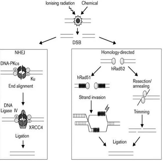
Figure 1: Pathways of DSB repair………6
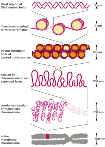
Figure 2. The levels of DNA organization in eukaryotic nucleus……….….14
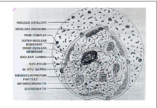
Figure 3: Schemetic view of Nucleus……….………15
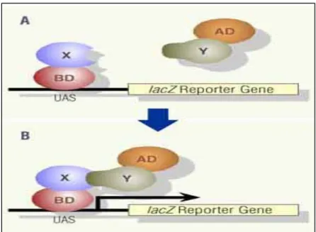
Figure 4: Yeast Two Hybrid Assay………53
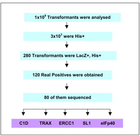
Figure 5: Summary of Yeast Two Hybrid Screen using C1D as bait……….54
Figure 6: Alignment of Clone41 Against full length TRAX………..55
Figure 7: C1D and TRAX interact specifically in yeast……….56
Figure 8: C1D and TRAX interact in vitro……….57
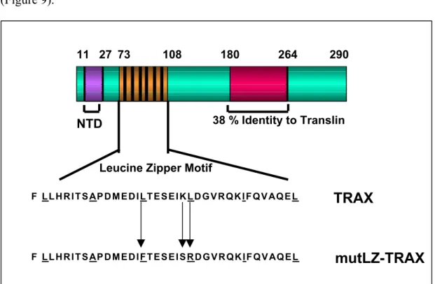
Figure 9. Domains of TRAX………..58
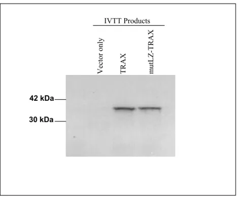
Figure 10: The IVTT of mutLZ-TRAX gives a similar size protein product to the wild type protein……….59
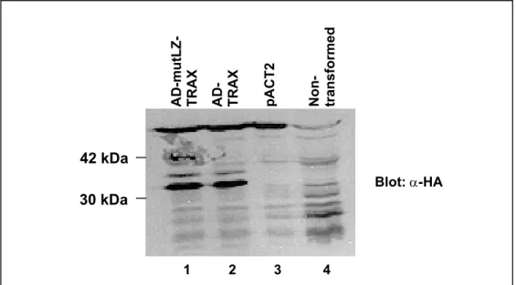
Figure 11: Verification of mutLZ-TRAX and TRAX expression in yeast strain L40 by immunobloting……….……….…….60
Figure 12: The putative Leucine zipper region of TRAX is essential for TRAX/C1D Interaction……….…………..61
Figure 13: Translin requires the full-length TRAX with intact LZ for interaction…62 Figure 14: In vitro competition assay……….63
Figure 15: In mammalian cells TRAX and C1D don’t interact under normal Conditions………..………….65
Figure 16: C1D and TRAX interact in mammalian cells in response to γ-irradiation……….66
Figure 17: TRAX and C1D don’t interact in mammalian cells in response to u.v. Irradiation……….…………..68
Figure 18: Subcellular localization of Translin, TRAX and C1D when they were expressed alone………...……70 Figure 19: Effect of TRAX-C1D, TRAX-Translin and C1D-Translin coexpression on expression pattern of TRAX, C1D and Translin………72 Figure 20: Subcellular Localizations of TRAX, Translin and C1D in triple
transfected cells………...74 Figure 21: yc1d mutant strain is temperature sensitive………..………77 Figure 22: yc1d mutant strain is not sensitive to either MMS or UV radiation,
however is slightly sensitive to γ-irradiation………..79 Figure 23: yc1d mutant strain is defective in NHEJ as demonstrated by a plasmid-
based in vivo DNA end joining assay……….81 Figure 24: Accurate DNA-end joining is impaired in yc1d and yku70 mutant
strains………..………83 Figure 25: Schematic view of in vivo homologous recombination assay……….…. 85 Figure 26: Analysis of yc1d role in HR recombination using our in vivo HR
assay………86 Figure 27: Survival of strains containing a plasmid with a galactose inducible HO-
endonuclease expressed relative to survival time at zero……….…. 87 Figure 28: Steps of PCR mediated gene knockout strategy……… 89 Figure 29: Confirmation of the integration of the disruption cassette into the YC1D
gene loci in yku70 mutant background………..……… 90 Figure 30: The yc1d-yku70 double mutant strain is temperature sensitive………....91 Figure 31: As yku70 and yc1d, yc1d-yku70 double mutant strain is not sensitive to
MMS, u.v. and γ irradiation………...……93 Figure 32: Yku70 and Yc1d are involved in the same pathway to rejoin 3’-
overhanging ends………94 Figure 33: yc1d/yku70 double mutant strain, similar to yku70 mutant strain,
ABBREVIATIONS
AD Activation domain
APS Ammonium persulfate
BME beta mercaptoethanol
bp Base pair
BS Bloom Syndrome
BUR Base unpairing region
cDNA Complementary DNA
CFP Cyan florescence protein
CPD Cyclobutane pyrimidine dimers
C-Terminus Carboxyl terminus
DBD DNA binding domain
ddH2O Deionised distilled water
DMEM Dulbecco’s modified Eagle medium
DNA Deoxynucleic acid
DNA-PK DNA dependent protein kinase
DNA-PKcs DNA dependent protein kinase catalytic subunit
dNTP deoxynucleotide triphosphate
ds DNA double stranded DNA
DSB Double strand break
DTT Dithiothretiol
EDTA Diaminoethane tetra-acetic acid
EtBr Ethidium Bromide
FCS Fetal calf serum
g gram
GFP Green fluorescence protein
GST Glutathione S-Transferase
hC1D human C1D
hnRNA Heterogenous nuclear RNA
HR Homologous recombination
HRP Horse Radish Peroxidase
Ig Immunoglobulin
IR Ionizing radiation
IVTT In vitro transcription and translation
kDa kilo Dalton
LacZ β-galactosidase
LB Luria-Bertoni media
Log Logarithmic
LZ Leucine zipper
MARs Matrix attachment regions
MBR Major breakpoint region
ml milliliter
mM millimolar
µg microgram
µl microliter
µM micromolar
MMS Methyl methane sulfate
mRNA messenger RNA
MutLZ-TRAX Leucine zipper motif mutant TRAX
NER Nucleotide excision repair
ng nanogram
NHEJ Non-homologous end joining
NM Nuclear matrix
N-terminus Amino terminus
PI3K Phosphotidyl inositol-3 kinase
pmole picomole
RFP Red fluorescence protein
RMX RAD50, MRE11, XRS2 complex
RNA Ribonucleic acid
RNAse Ribonuclease
RPA Replication protein A
rpm Revolutions per minute
Scid Severe combined immunodeficiency
SDS Sodium Dodecyl Sulfate
ss DNA Single stranded DNA
SSA Single-strand annealing
TBE Tris-Borate-EDTA solution
TBS Tris buffered saline
TBST Tris buffered saline with Tween 20
TE Tris-EDTA solution
TEMED N,N,N,N-tetramethyl-1,2 diaminoethane
TRAX Translin associated factor X
v volt
WS Werner syndrome
wt wild type
X-gal 5-bromo-4-chloro-3-indolyl β-D-galactopyranoside
yc1d Mutant yeast C1D gene
YC1D Yeast C1D gene
Yc1d Yeast C1D protein
CHAPTER 1
1.1. DNA DAMAGE
DNA damage can be defined as any modification of DNA that alters its coding properties and leads to blockage of essential cellular processes such as transcripton and replication. Cellular responses to unrepaired DNA damage may include acute cytotoxicity, programmed cell death, mutation fixation, and increased recombination leading to genomic instability. Damage to genomic DNA occurs spontaneously and can be further enhanced by environmental mutagens such as ultraviolet (u.v.) light from the sun, inhaled cigarette smoke, or incompletely defined dietary factors. Large proportions of DNA alterations are caused by spontaneous damage, which occur with an estimated rate of 1-3x10⁴ per cell per day. DNA damaging agents of different types produce different kinds of lesions in DNA. Types of DNA damage can be divided into two main groups;
• Single base modifications: These are some minor base modifications or base replacements that might simply alter the coding sequence. This simple base substitution includes deamination, depurination, reaction of reactive oxygen species (ROSs), nucleotide analog incorporation, alkylation, insertion and deletions.
• Damage that involve more than one base or, result in damage to the DNA
backbone: These are types of DNA damage that may distort DNA structure and
interfere with replication or transcription. Ultraviolet light-induced thymine dimers, intrastrand and interstrand cross-linking between two bases, single strand or double strand breaks are also examples for this type of DNA damage.
If left unrepaired, DNA damage can have serious consequences for cells, causing mutations, which may lead to cancer formation or improper functioning of the encoded proteins. Therefore, for the faithful reproduction and preservation of the genomic integrity, damaged DNA must be repaired. Since there are many different types of DNA damage, it is not surprising that the repair system employs many different protein complexes. Approximately more than 100 genes participate in various aspects of DNA repair, even in organisms with very small genomes. As a result of analysis of the human genome sequence, considering evolutionary relationships and common sequence motifs in DNA repair genes, 130 human DNA repair genes have been identified (Wood et al., 2001) .
According to the mechanisms they employ in repairing different types of damage, they can be grouped into three.
• Direct reversal of DNA damage: It is the simplest way to correct the damage. There are two well-studied proteins in this type of repair. One of them is cyclobutane pyrimidine dimers (CPD) photolyase, which reverts the covalent linkage of pyrimidine dimers in a light dependent reaction. CPD photolyases are found in bacteria, fungi, plants, and many vertebrates, but not in placental mammals. The other one is alkyl transferase that removes the methyl group from O6-methylguanine. Alkyl transferase is present in all living organisms. • Excision repair: In cases where damage is present in just one strand, the damage
can be accurately repaired by cutting the damaged DNA and replacing it with newly synthesized DNA using the complementary strand as a template. There are three types of excision repair. First one is mismatch repair, which repairs mismatched nucleotides and small loops. Second one is base excision repair that removes incorrect bases such as uracil or damaged bases like 3-methyladenine
• Double strand break repair: Among the other forms of DNA damage, double strand breaks (DSBs) are potentially the most dangerous, since they can lead to broken rearranged chromosomes, cell death, or cancer. Eukaryotes have evolved specific mechanisms to repair DSBs by either homologous recombination (HR), which requires the presence of homolog sequences or non-homologous end joining (NHEJ) by which the two ends of a DSB are joined by a process that is independent of DNA sequence homology (Nickoloff and Hoekstra, 1998).
1.2. DNA DOUBLE-STRAND BREAKS (DNA-DSBs)
DSBs are generated by exogenous agents such as ionizing radiation (IR) and certain chemotherapeutic drugs, or by spontaneously generated ROSs and mechanical stress on the chromosomes. They can also be produced when DNA replication forks encounter DNA single-stranded breaks or other types of lesions. They can occur at the termini of chromosomes due to defective metabolism of telomers. Additionally, they are generated to initiate recombination between homologous chromosomes during meiosis and also during V(D)J recombination, which is a process necessary for lymphocyte development (Critchlow and Jackson, 1998; Jackson and Jeggo, 1995; Khanna et al., 2001).
The presence of an unrepaired DSB will activate the DNA-damage response systems of a cell to arrest its progression through the cell cycle thereby, preventing possible chromosome loss or transmission of damaged DNA to the next cellular generation. The cell cycle checkpoints are believed to prevent the replication of damaged DNA (G1/S and intra S checkpoints) or segregation of damaged chromosomes (G2/M checkpoint). Once the damage is repaired, the cell cycle resumes. However if the level of DNA damage is beyond repair, DSBs can lead to programmed cell death (apoptosis). If a cell with an unrepaired DSB persists to divide, the broken chromosome ends can lead to chromosomal fragmentations, translocations and deletions, all of which can lead to carcinogenesis through activation of oncogenes, inactivation of tumour suppressor genes or loss of heterozygosity (Critchlow and Jackson, 1998; Kanaar et al., 1998). Notably, a large
proportion of cancers of lymphoid origin exhibit chromosomal rearrangements involving the immunoglobulin or T-cell receptor loci, indicating that they have arisen through inappropriate resolution of DNA DSBs corresponding to V(D)J recombination intermediates (Khanna and Jackson, 2001) .
1.3. PATHWAYS OF DSB REPAIR
To overcome deleterious effects of DSBs eukaryotes have evolved two main repair pathways, nonhomologous end joining (also called illegitimate recombination) (NHEJ) and homologous recombination (HR). Both mechanisms operate in all eukaryotic cells but the relative contribution of each mechanism varies. HR pathway requires extensive regions of DNA homology and repairs DNA accurately using information on the undamaged sister chromatid or homologous chromosome. On the contrary, DNA end-joining pathway uses no, or extremely limited sequence homology to rejoin juxtaposed ends in a manner that may not be error free. It seems that mammalian cells prefer NHEJ whereas HR is more common in the budding yeast Saccharomyces cerevisiae. One possible explanation for this preference in mammalian cells might be misalignment of repetitive DNA sequences that could lead to deletions and translocations. In addition to these two main mechanisms, DSBs can also be repaired by single strand annealing (SSA), which, can be considered as a sub-pathway of HR. In SSA; resection and annealing of short regions of complementary sequence initiates repair by SSA pathway in which ligation is preceded by the trimming of non-complementary single strand DNA tails (Karran, 2000).
functional homologues of them have been isolated from mammalian species. According to the proposed model for HR (Figure 1) (Kanaar et al., 1998; Khanna and Jackson, 2001); DSBs generated by DNA damaging agents is processed to a single stranded region with 3’-overhang by an exonuclease and then Rad51 protein associates with 3’ ssDNA-end and searches for the homologous duplex DNA to catalyse strand exchange in a reaction, which generates a joint molecule between homologous damaged and undamaged duplex DNA. In addition to Rad51 this step requires the coordinated action of Rad52, Rad54, Rad55/57 and the single strand binding protein RPA. DNA synthesis, requiring a DNA polymerase, its accessory factors and a ligase, then restores the missing information (Karran, 2000) .
Similar to yeast Rad51, mammalian Rad51 protein polymerizes on DNA to form nucleoprotein filaments that searches the homologous DNA and like yeast homologs, human Rad52 and Rad54 facilitate Rad51 strand invasion activity (Kanaar et al., 1998). Mammalian Rad51 also interacts with Brca1 and Brca2 proteins (products of breast cancer genes) (Haber, 1999), which are supposed to act as protein scaffolds to coordinate the actions of the HR machinery (Khanna and Jackson, 2001). Another protein that has been linked to HR in mammals is the protein kinase ATM, which is deficient in the cancer prone, radiosensitive, and the neurodegenerative syndrome ataxia telangiectasia (A-T). Upon IR, ATM phosphorylates proteins involved in HR such as Brca1 and c-Abl besides its other targets. The c-Abl tyrosine kinase modulates Rad51 strand exchange activity through phosphorylation (Khanna and Jackson, 2001; Rotman and Shiloh, 1999). The helicases defective in the human cancer predisposition and developmental disorders, Werner syndrome (WS) and Bloom syndrome (BS) have also been implicated in regulating HR through their ability to suppress NHEJ (Karow et al., 2000)
1.3.2. Single-Strand Annealing (SSA)
This pathway can be considered as a subpathway of HR and it doesn’t involve formation and resolution of Holliday junctions. However, SSA relies on regions of homology with which to align the strands of DNA to be joined.
S.cerevisiae genes, which define SSA, belong to the Rad52 epistasis group of HR.
The hRad50, hMre11 and Nbs1 define the human SSA pathway. Mre11 has several activities, the most important of which may be 3-5’ exonuclease activity which may remove damaged DNA to expose short lengths of single-stranded DNA. If a sister chromatid is available, then single stranded overhangs may initiate rejoining by homologous recombination. Alternatively, sites of limited homology within the
1.3.3. Non-Homologous End Joining
In contrast to HR, NHEJ is achieved without the need for extensive homology between DNA ends to be joined (Figure1). Many of the proteins involved in NHEJ have been identified in several ways such as biochemical fractionation of extracts capable of end joining reactions, genetic and biochemical analysis of the proteins required for DSB repair during V(D)J recombination and genetic analysis in yeast, rodent, and human cell lines. According to these analyses, DNA dependent protein kinase (DNA-PK) is found to be a key component of the NHEJ (Jeggo, 1997; Lees-Miller, 1996). DNA-PK is an abundant nuclear serine/threonine kinase that is activated in vitro by DNA double strand breaks and certain other discontinuities in the DNA double helix (Dvir et al., 1993; Gottlieb and Jackson, 1993).
Recently, several other yeast NHEJ components have been identified including the nuclease complex containing Rad50p, Mre11p, and Xrs2p (Boulton and Jackson, 1998; Milne et al., 1996). Most DNA DSBs are not blunt ended but have single-strand overhangs, which must be trimmed by exonucleases and/or endoucleases before rejoining. In yeast, Mre11p, Rad50p and Xrs2p complex seems to be involved in this trimming process. Mammalian cells have homologues of Rad50p and Mre11p but a different protein, Nbs1, seems to replace Xrs2p (Maser et al., 1997). Nbs1 (also called Nibrin) is a protein whose deficiency leads to a rare human genetic disorder called Nijmegan breakage syndrome (NBS) (Baumann and West, 1998; Carney et al., 1998). It is supposed that Nbs1 regulates function of Rad50p and Xrs2p through signaling the existence of DNA damage to the cell cycle checkpoint machinery. Repair by NHEJ is completed by the DNA ligase IV/ XRCC4 complex, of which yeast homologous are Lig4p and Lif1, respectively (Baumann and West, 1998; Featherstone and Jackson, 1999).
1.4. ROLE OF DNA-PK AND ITS COMPONENTS IN NHEJ
DNA-PK consists of the heterodimeric Ku proteins, of 70- and 80- kDa and a large catalytic domain of 470-kDa, termed DNA-PKCS (catalytic subunit).
According to biochemical studies, the ser/thr kinase catalytic activity of DNA-PK was shown to be triggered upon association with DNA ends through its DNA targeting component, Ku 70/80 (Dvir et al., 1993; Gottlieb and Jackson, 1993). Although weak, DNA-PKCS possesses an intrinsic DNA-end binding activity that is greatly stimulated and stabilized by Ku complex (Hammarsten and Chu, 1998).
DNA-PKCS doesn’t appear to have significant homology to other characterized proteins. However, the C-terminal region of DNA-PKCS falls into phosphatidylinositol-3 kinase (PI3K) family and more specifically, DNA-PKCS is most related to a subgroup of proteins in this family, which includes ATM, ATR, and TRRAP. These proteins have been shown to be involved in controlling transcription, cell cycle, and/or genome stability in organisms ranging from yeast to man (Hartley et al., 1995; Poltoratsky et al., 1995). Interestingly, they all have serine/threonine kinase activity rather than lipid kinase activity (Smith et al., 1999). DNA-PK also has an intrinsic phosphorylation activity. Autophosphorylation has an important role in regulation of DNA-PK activity by affecting dissociation of DNA-PKCS from the Ku complex and results in inhibition of DNA-PK catalytic function (Chan and Lees-Miller, 1996). Upon activation, DNA-PKCS phosphorylates a variety of proteins in
vitro including itself, Ku complex, Sp1, c-Jun, c-Myc, p53, Hsp90, the RNA
polymerase II subunit, RPA, XRCC4 and a recently identified nuclear matrix protein C1D (Anderson and Miller, 1992; Jackson, 1997; Leber et al., 1998; Lees-Miller, 1996; Smith et al., 1999; Yavuzer et al., 1998).
Ku was initially characterized as an antigen present in the sera of patients with autoimmune diseases (Mimori et al., 1981). Ku70 and Ku80 heterodimer binds, without sequence specificity, to a variety of DNA structures such as double stranded DNA ends, nicks, gaps, and hairpins, however, it doesn’t bind to closed circular DNA (Dynan and Yoo, 1998). Another characteristic of Ku protein is its ability to translocate along the DNA molecules in an ATP independent manner (Paillard and Strauss, 1991). This ability of Ku is supposed to function transporting DNA-PKCS to
The role of DNA-PK complex in NHEJ became evident through genetic complementation studies of rodent mutants that were shown to be sensitive to ionizing radiation and defective in repair of DNA DSBs and site-specific V(D)J recombination (Jeggo, 1997). Among the 11-complementation groups of IR-sensitive cells that were initially defined, 4 were defective in NHEJ (IR4, IR5, IR6, and IR7). It has been established that IR7 cells are deficient in DNA-PKCS, whereas IR5 and IR6 cells are deficient in the Ku80 and Ku70 respectively. Subsequent work showed that IR4 cells are defective in Xrcc4, a small protein that forms a tight and specific complex with DNA ligase IV (Smith et al., 1999). In light of the above reports, proposed model is that Ku/DNA-PKCS complex binds to DSBs and facilitates the recruitment and activation of other NHEJ components such as Xrcc4 and DNA ligase IV (Figure 1).
Mice deficient in various components of NHEJ have been generated to study NHEJ in mammals. According to these experiments, deficiency of any components of DNA-PK leads to multiple defects, including growth retardation, hypersensitivity to IR and severe combined immunodeficiency (Scid) due to severely impaired V(D)J recombination, which is an essential process during mammalian lymphoid cell development (Smith et al., 1999).
V(D)J recombination is a well characterized site-specific recombination in higher eukaryotes which occurs during the development of B and T cells. In this process, an enormous repertoire of immnunoglobulin and T-cell receptor genes is produced by bringing together variable (V), diversity (D), and joining (J) sub-exonic gene segments in various combinations (Oettinger, 1999). V(D)J recombination is initiated by the generation of blunt DNA-DSBs between recombination signal sequences and coding gene segments. Subsequently, these intermediates are resolved by the ligation of pairs of coding ends to form coding joints, and pairs of recombination signal sequence ends to yield recombination signal sequence joints (Jeggo et al., 1995). Since site specific DNA-DSBs are introduced during the process of V(D)J recombination, there is a mechanistic link between the repair of IR induced DNA damage and DNA DSBs formed during V(D)J recombination (Smith and Jackson 1999). Indeed, DNA DSB repair mutants of IR4, IR5, IR6 and IR7 can not perform V(D)J recombination effectively (Jackson and Jeggo, 1995).
So far, no mutations for DNA-PKcs have been identified in any congenital human diseases, suggesting that defects in NHEJ are rare or cause embryonic lethality in humans. However, DNA-PK deficiency leads to increased rates of neoplastic transformation in mice. Mice mutant for DNA-PKCS and/or Ku70 have a high incidence of T-cell lymphomas and especially Ku70 knockout mice have increased rates of fibroblast transformation (Jhappan et al., 1997). Fibroblasts from Ku80 knockout mice show chromosomal instability associated with chromosome aberrations, including breakage, translocations and aneuploidy. Notably, loss of tumour suppressor p53 in Ku80 -/- mice induces early onset of lymphomas (Difilippantonio et al., 2000). Tumors obtained from Ku80 and p53 double mutants display a specific set of translocations including the immunoglobulin heavy chain IgH/Myc locus that is reminiscent of Burkitt Lymphoma (Difilippantonio et al., 2000). Combined DNA-PKCS and p53 deficiency also promotes similar chromosomal translocations (Vanasse et al., 1999). These reports suggest that chromosomal rearrangements in the double mutant mice primarily result from an impairment in the normal NHEJ pathway for resolving V(D)J recombination intermediates, which leads to chromosomal translocations with oncogenic potential. Interestingly, a radiosensitive human cell line, MO59J, derived from glioma was found to be defective in DNA-PKCS expression and DNA-PK activity, indicating a role for DNA-PK in cancer formation (Lees-Miller et al., 1995).
Besides being a DNA-damage activated protein kinase, DNA-PK could have an important role in DNA damage signalling. It is hypothesised that DNA-PK activation in response to DNA damaging agents could trigger signalling pathways that result in apoptosis and cell cycle arrest. Consistent with this idea, a gatekeeper protein p53 that controls cellular proliferation and apoptosis in response to perturbations in genomic DNA, is an in vitro substrate of DNA-PK. DNA-PK phosphorylates p53 at serine 37 and 15 residues, and this has been reported to destabilise interactions between p53 and Mdm2, a protein that negatively regulates
p53 on serine 15 in vitro (Canman et al., 1998; Khanna et al., 1998). All these reports suggest that DNA-PK may be involved in DNA damage signalling process through p53 phosphorylation that results in p53 protein stabilization.
1.5. NUCLEAR MATRIX INVOLVEMENT IN REGULATION OF DNA-PK AND DNA-DSB REPAIR
It is clear that DNA-PK plays a key role in DNA DSB repair, however, the molecular mechanism it employs during this process is not precisely understood. Although several proteins have been found to act as DNA-PK substrates in vitro, physiological targets of DNA-PK and factors that regulate its activity are not yet known.
In an attempt to identify these mechanisms, the yeast two-hybrid system was applied to screen a cDNA library to identify DNA-PKCS interacting proteins. As a result of this screen, the nuclear matrix protein C1D was identified to interact specifically with DNA-PKCS (Yavuzer et al., 1998). Moreover C1D mRNA and protein levels were found to be induced specifically upon γ-irradiation. C1D can be phosphorylated by DNA-PK but is different from all other DNA-PK substrates in the sense that, it can activate DNA-PK in the absence of free DNA ends. These findings suggested that C1D might act as an accessory protein to target DNA-PK to nuclear matrix constitutively. In support of this model, Ku80 deficient xrs5 cells were reported to have nuclear matrix and envelope alterations compared to their wild type controls (Korte and Yasui, 1993; Yasui et al., 1991). Recently, DNA-PK was shown to bind specific regions within matrix attachment regions (MARs) that exhibit a high binding affinity to nuclear matrix (Galande and Kohwi-Shigematsu, 2000). These specific regions are called base unpairing regions (BURs) that represent a specialized DNA context with an unusually high propensity for base un-pairing under negative superhelical strain. In addition to DNA-PKCS, poly (ADP-ribose) polymerase (PARP), Ku70/80, and HMG1 that has been shown to stimulate DNA-PK activity in
vitro (Watanabe et al., 1994), were identified as BURs binding proteins (Galande and
(MBR) of the untranslated region of BCL2 gene, suggesting that the nuclear matrix plays a role in DSB repair and/or recombination (Ramakrishnan et al., 2000). Taken together, there is enough evidence to support the idea that the activity of DNA-PK in
vivo could be influenced by the nuclear matrix. As being a nuclear matrix protein,
C1D stands as a good candidate to regulate the in vivo function of DNA-PK.
1.6. NUCLEAR MATRIX PROTEIN C1D
The ∼30,000 to 40,000 genes of a human being, encoded in 6x 10⁹ base pairs of DNA of a total length of almost 2 meters, are packaged into a nucleus 6-8 micrometers in diameter (Varga-Weisz and Becker, 1998). Several levels of DNA compaction exist in eukaryotic nucleus (Figure 2). The DNA is packed into nucleosomes, which consist of a core of histones with 166 DNA base pairs wrapped around the core, and resulting chromatin is further compacted into 30 nm fibers (solenoids) and DNA loop domains. These loop domains are anchored to the proteinaceous nucleoskeleton, also called nuclear matrix or scaffold through matrix attachment regions (MARs) (Laemmli et al., 1978; Paulson and Laemmli, 1977). Therefore, nuclear matrix organizes DNA into a dynamic conformation to perform complex nuclear processes such as heterogenous nuclear RNA (hnRNA) synthesis and processing, replication, DNA repair and recombination (Nelson et al., 1986).
Nucleus is a complex organelle containing subnuclear domains that compartmentalizes different nuclear processes. The nuclear matrix is the skeletal framework of the nucleus consisting of the nuclear-pore lamina, residual nucleoli, a network of proteins, DNA and RNA (Nickerson et al., 1995) (Figure 3). NM proteins are non-histone proteins that comprise of 10-25 % of total nuclear protein (Berezney et al., 1995). They are insoluble proteins with strong DNA affinity and they remain associated with genomic DNA, especially with highly repetitive DNA
C1D is the first nuclear matrix protein characterized at the sequence level. The cDNA encoding C1D (human HC1D, X95592; 1995) was initially characterized in an assay to identify polypeptides released from rigorously extracted and nuclease treated DNA (Nehls et al., 1998).
In an independent research, C1D was found to be associated with the transcriptional repressor RevErb and the nuclear corepressors N-cor and SMRT (Zamir et al., 1997) suggesting that it could also be involved in transcriptional repression, possibly by recruiting the receptors and repressors to the nuclear matrix
C1D is physiologically expressed in 50 human cell tissues tested that points out its basic cellular function. However, cellular C1D expression is tightly regulated since elevated levels of C1D protein, which was induced by transient vector
repair, recombination or transcription. If the damage is beyond repair, C1D then induces apoptosis thereby protecting the organism from the consequences of unrepaired DNA damage.
Cellular proliferation and growth must be controlled tightly since loss of this control can lead to cell death or tumorigenesis. Any changes in uncontrolled cellular proliferation are reflected by changes in complex nuclear processes such as transcription, DNA replication, DNA repair-recombination and apoptosis. As nuclear matrix provides a platform for the assembly of protein machines involved in complex nuclear processes, components of the nuclear matrix could be important targets of tumorigenesis. Aberrant expression of nuclear matrix proteins in cancer cells (Yanagisawa et al., 1996) and tight association of nuclear oncoproteins with nuclear matrix (Deppert et al., 2000; Galande and Kohwi-Shigematsu, 2000) are the recent reports that postulate the importance of nuclear matrix in epigenesis of cancer.
The nuclear matrix protein C1D has been identified rather recently and the precise biological functions of this protein are not known yet. However, recent findings suggest that C1D may play a role in maintenance of genomic integrity. We believe that the identification of biological function of C1D will provide important clues in understanding the role of nuclear matrix in etiology of cancer. Therefore, the scope of this project is to identify the biological role of C1D in preservation of genomic integrity. To achieve this aim, two strategies were employed;
First strategy was to identify C1D interacting proteins through yeast two- hybrid screen and characterize the biological significance of these interactions. The involvement of C1D in several nuclear processes indicates that it may be a multi-functional protein. Since protein-protein interactions are critical to virtually every cellular process and also for nuclear matrix associated processes, it is highly possible that C1D performs its functions through associating with other proteins apart from DNA-PK. Identification of these proteins will not only provide important clues about the biological function of C1D protein, but also clarify functions of nuclear matrix as well.
Second strategy was identification of the function of S.cerevisiae C1D by deleting the C1D gene and analysing the effects of this deletion. Because most of the
important genes and cellular events are highly conserved throughout evolution, the results obtained using S.cerevisiae will be relevant to the functions of human C1D.
CHAPTER 2
2.1. BACTERIAL STRAINS
Strain Genotype Usage Source
M15 (pREP4)
F-, NalS, StrS, rif8, lac-, ara-, gal-, mtl-, recA+, uvr+
purification of recombinant proteins with pQE vectors
QIAGEN
DH5α F- supE44 hsdR17 recA1 gyrA96 endA1 thi-1 relA1 deoR lambda-
a recA- host for propagation and storage of plasmids
Cold Spring Harbour Labs.
HB101 F- supE44 lacY1 ara-14 galK2 xyl-5 mtl-1 leuB6 proA2 delta(mcrC-mrr) recA13 rpsL20 thi-1 lambda
leu deficient strain used to rescue library plasmids
D.E. Ish Horowicz
M15 stain contains multiple copies of pREP4 (kanamycin resistance marker) plasmid, which carries the lacI gene encoding the lac-repressor (details could be obtained from QIAGEN catalogue).
2.1.1. Growth and Maintenance of Bacteria
Bacterial strains were stored in 50% glycerol at –700C for long-term storage. Strains grown in LB (with or without antibiotic) to mid-log phase or saturation was mixed with sterile glycerol with a ratio of 1:1, mixed to homogeneity, and stored at – 700C until required. Bacteria was recovered by scraping a small amount of cells from the frozen stock with an inoculation loop and streaking onto a LB-agar plate (supplemented with the appropriate antibiotics if necessary).
Liquid culture of plasmid carrying E.coli was performed in LB (5 g NaCl, 10 g Bacto-tryptone, 5 g Yeast Extract, 1 ml of 1 M NaOH, ddH2O added to 1 L) with appropriate antibiotic selection. Liquid cultures were constantly agitated in a rotary shaking incubator (~200 rpm) at 370C. The specialised components of all media were obtained from Difco Laboratories Ltd.
2.2. YEAST STRAINS
Strain/Acc. No.
Genotype Usage Source
FY1679 Mata/α;ura3-52/ura3-52; trp1∆63/TRP1;leu2∆1/LEU2; his3∆200/HIS3; GAL2/GAL2
Diploid wild type used to analyse function of Yc1d EUROSCARF FY1679-08A Mata;ura3-52;leu2∆1;trp1∆63; his3∆200; GAL2
Haploid wild type used to analyse function of Yc1d EUROSCARF YO1909 BY4741;Mata;his3∆1;leu2∆0; met15∆0;ura3∆0; YHR081w::kanMX4 Haploid yc1d mutant used to analyse function of Yc1d EUROSCARF
Strain/Acc. No.
Genotype Usage Source
Y11909 BY4742;Matα;his3∆1;leu2∆0; lys2∆0;ura3∆0; YHR081w:kanMX4 Haploid yc1d mutant used to analyse function of Yc1d EUROSCARF Y21909 BY4743;Mata/α;his3∆1/his3∆1; leu2∆0/leu2∆0;lys2∆0/LYS2; MET15/met15∆0;ura3∆0/ura30; YHR081w::kanMX4/YHR08w Heterozygous diploid (YC1D/yc1d) mutant used to analyse function of Yc1d EUROSCARF Y31909 BY4743;Mata/α;his3∆1/his3∆1; leu2∆0/leu2∆0;lys2∆0/LYS2; MET15/met15∆0;ura3∆0/ura30; YHR081w::kanMX4/YHR08w::k anMX4 Homozygote diploid mutant (yc1d/yc1d); used to analyse function of Yc1d EUROSCARF Y00870 BY4741;Mata;his3D1;leu2D0; met15D0;ura3ymr284w::kanX4 Haploid yku70 mutant used as a control for various yeast assays. EUROSCARF Y00540 BY4741;Mata;his3D1;leu2D0; met15D0;ura3YML032c::kanM4 Haploid rad52 mutant used as a control for various yeast assays.
EUROSCARF
W303 Matα;ade2-1;his3-11,15;leu2-3,112;trp1-1;ura3
Haploid wild type; used to knockout
YC1D
Strain/Acc. No.
Genotype Usage Source
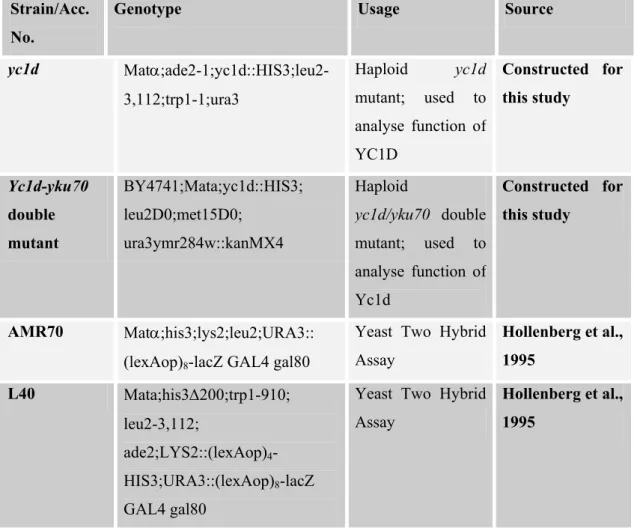
yc1d Matα;ade2-1;yc1d::HIS3;leu2-3,112;trp1-1;ura3 Haploid yc1d mutant; used to analyse function of YC1D Constructed for this study Yc1d-yku70 double mutant BY4741;Mata;yc1d::HIS3; leu2D0;met15D0; ura3ymr284w::kanMX4 Haploid yc1d/yku70 double mutant; used to analyse function of Yc1d Constructed for this study AMR70 Matα;his3;lys2;leu2;URA3:: (lexAop)8-lacZ GAL4 gal80
Yeast Two Hybrid Assay Hollenberg et al., 1995 L40 Mata;his3∆200;trp1-910; leu2-3,112; ade2;LYS2::(lexAop)4 -HIS3;URA3::(lexAop)8-lacZ GAL4 gal80
Yeast Two Hybrid Assay
Hollenberg et al., 1995
Table 2: List of S.cerevisiae strains used during the course of this study
2.2.1. Growth And Maintenance Of Yeast
Yeast strains were stored in YPAD (10 g Yeast Extract, 20 g Peptone and 0.1g Adenine dissolved in 900 ml ddH2O, sterilized by autoclaving, cooled to ~ 550C and 100 ml of 20% filter-sterilized glucose was added) rich medium with 50% glycerol and kept at –700C for long-term storage. Transformed yeast strains were stored in the appropriate minimal YC (1.2 g Yeast Nitrogen Base without amino acids, 5 g Ammonium Sulfate, 10 g Succinic Acid, 6 g NaOH and 0.1 g of all amino acids apart from Histidine, Uracil, Tryptophane and Leucine dissolved in 900 ml ddH2O, and autoclaved) medium to keep selective pressure on plasmid. Yeast cells
were recovered by scraping a small amount of cells from the frozen stock with an inoculation loop and streaking them onto YPAD- or (appropriate YC) agar plates. Transformed yeast strains were plated on selective YC-agar (20 g agar was added to 900 ml YC medium and sterilized by autoclaving) plates lacking specific amino acids that were expressed by yeast plasmid. Yeast extract, Yeast Nitrogen Base and Peptone were purchased from DIFCO Laboratories Ltd. and the rest of the components of yeast media were purchased from SIGMA.
Liquid cultures of yeast were prepared by inoculating one large colony per 5-ml medium and incubating the culture at 300C for 16-18 hr with shaking at 230-270 rpm. For mid-log phase liquid culture, enough volume of overnight culture was inoculated into fresh medium to give an OD600 of 0.2-0.3 and the culture was incubated at 300C for 3-5 hours with shaking till it reached to an OD600 of 0.5-0.8. The density of cells in a culture was determined by measuring its optical density (OD) at 600 nm using spectrophotometer (Beckman Instruments Inc., CA, USA). 0.1 OD600 value corresponds to 0.5x10⁷ and 0.25x10⁷ cells/ml for haploid and diploids strains, respectively.
2.3. MAMMALIAN CELLS
COS7 cells derived from kidney of SV40 transformed African green monkey was used (ATCC CRL-1651). They were maintained at DMEM (SIGMA) supplemented with 10% Foetal Calf Serum, 1% Streptomycin/Penicillin (both from SIGMA) and 2% Fungiozone (GIBCO). The cells were kept in air supplemented with 5% CO2 in a humidified incubator at 370C.
2.4. OLIGONUCLEOTIDES
2.4.1. Synthesis of Oligonucleotides
The oligonucleotides used for cloning and sequencing purposes were synthesized at the Beckman Oligo 1000M DNA Synthesiser (Beckman Instruments Inc. CA, USA) at Bilkent University, Department of Molecular Biology and Genetics (Ankara, Turkey). The longer oligonucleotides used in yeast assays and knockout experiments were either purchased from IONTEK (Bursa, Turkey) or were provided by MCRI (Oxted, U.K.)
2.4.2. List of Oligonucleotides Used in This Study
• Sequencing Primers: TC187 pACT Forward Primer 5’ TATCTATTCGATGATGAAGAT 3’ TC 238 T7-myc-pLink Forward Primer 5′ CAG AAG CTG ATC TCC GAG 3′ TC239 pGEXTK2 Forward Primer 5’ CCAAAATCGGATCTGGTT 3’ TC241 pQE30 Forward Primer
5′ GAA TTC ATT AAA GAG GAG AAA 3′ TC240 pCMV-HA Forward Primer 5’ GACGTCCCTGATTATGCA 3’
• Primer pairs used for PCR and cloning
Full-Length TRAX
TC234 F 5′ AGACGTCGACAAGGATCC ATG AGC AAC AAA GAA GGA TCA 3′
TC235 R 5′AGACGTCGACGGATCC CTA AGA AAT GCC CTC TTC 3′
N-Terminal TRAX
UY29 R 5’AGACGGATCCCTCGAGTTAAGCTTCCACATATTCCTG 3’ SalI
SalI
BamHI BamHI
MutLZ TRAX
UY30 R 5’AGACGGATCCCTCGAGTTAAGATCTAATTTCTGATTCAGTAAATAT 3’
UY31 F 5’AGACAGATCTGATGGTGTCAGACAAAAGATA 3’
Full-Length Translin
TC236 F 5′AGACGTCGACAAGGATCC ATG TCT GTG AGC GAG ATC TTC 3′
TC237 R 5′AGACGTCGACGGATCC CTA TTT TTC AAC ACA AGC 3′
Full-Length Human C1D
UY48 F5’ AGACAGATCTATGGCAGGTGAAGAAATTAATGAAGAC 3’
UY49 R 5’AGACAGATCTTTAACTTTTACTTTTTCCTTTATTGGCAAC 3’
• Primers Used in Yeast Based In-vivo Recombination Assay
Oligonucleotide used for “GV256 Dead”
5’CTGACTGAGTGAAGATCTTCACTCAGTCAGGAGCT3’
Primer pair used to amplify 800 bp region of LacZ F 5’AGACGGATCCTCCTTTGCGAATACGCCCAC 3’
R 5’AGACGAATTCTGTGAAAGAAAGCCTGACTG 3’
SalI SalI BamHI BamHI Bgl II SacI BamHI XhoI Bgl II Bgl II Bgl II Bgl II
STOP STOP STOP
BamHI
• Primers Designed to Knockout YC1D YC1D Disruption Cassette and PCR confirmation
F5’CAAAGCGGCAACGTCATAACCTTGGTATTTATTGGGCAACGTTTTAAGAGCT TGGTGAGC3’
R5’CAAAAGTGTTCACTGCCAACTACAAGAATAGCATATACACATTCGAGTTCAA GAGAAAAA 3’
R 5’CGGATCCCGTAGAAATGCTTTTGCCAAGG3’
2.5. RECOMBINANT DNA TECHNIQUES
2.5.1. Polymerase Chain Reaction
Target DNA sequences between a pair of oligonucleotide primers were amplified by using either thermostable DNA Polymerase of T. aquaticus (Taq)(MBI) or Pfu DNA polymerase (recombinant enzyme expressed and purified in bacteria, was a gift from Emine Ercikan Abali). A typical 50 µl PCR reaction contained 10-100 ng of target DNA, 16 pmol of each primer, 2mM of each dNTP (200mM) (MBI Fermentes; Cat. No.R0181), 3 mM MgCl2 (25 mM) (SIGMA), 1X Taq Pol buffer (SIGMA), and 0.5-1 units Taq DNA polymerase (SIGMA; Cat. No. D-4545). In cases, where longer DNA segments (>1kb) were amplified, 1 µl Pfu DNA polymerase was used with 1X Pfu Buffer (10x; 20mM Tris-HCl (pH: 8.0, 100mM KCl, 60 mM (NH4)2SO4, 1 % Triton X-100, 100µg/ml nuclease free BSA). The reaction was preheated to 94oC for 4 minutes and then subjected to 25-30 cycles of denaturation (0.5 min. at 950C), annealing (1-2 min. at 50C below the estimated Tm of the primers, which is generally between 48-550C) and synthesis (1-2 min. at 720C), in an automated thermal cycler (Perkin Elmer Gene Amp PCR System 9600). The product was assessed by gel electrophoresis and EtBr staining.
The PCR product was purified either by phenol/chloroform extraction and ethanol precipitation (Ausubel et al., 1987) or by gel extraction kit (Nucleospin- Macherey-Nagel; Cat.No. 740590) according to manufacturer’s instructions. The PCR products were digested with the appropriate restriction enzymes overnight and ligated into plasmids accordingly.
2.5.2. DNA Purification From Agarose Gel
The desired DNA bands were cut from the agarose gel under UV illumination and DNA was extracted from agarose gel slices by Gel extraction kit, Nucleospin extract (MN 740590), according to the manufacturer’s instructions.
2.5.3. Quantification of DNA
Concentrations and purity of nucleic acids were determined by measuring absorbency at 260nm and 280nm in a spectrophotometer (Beckmann PU640 Instruments Inc., CA, USA). The ratio between absorbance values at OD260 and OD280 was taken. Nucleic acid samples displaying OD260 and OD280 values in the range of 1.8 to 2.0 are regarded as highly pure. The concentration of DNA was determined automatically by using oligo/DNA quantitation program of BECKMAN spectrophotometer.
2.5.4. DNA Sequencing
2.5.5. Enzymatic Manipulation of DNA
2.5.5.1. Restriction Enzyme Digestion of DNA
Restriction enzyme digests of DNA were performed in 20-50 µl reactions that contained 5-10 units of enzyme/1 µg of DNA with the appropriate buffer. The digests were conducted at 370C for 1-5 hours, depending to the amount of DNA to be restricted. Restriction enzymes were purchased from MBI, ROCHE and GIBCO.
2.5.5.2. Dephosphorylation of DNA Ends
Shrimp Alkaline phosphatase (SAP- Roche, Cat. No. 1 758 250) was used for dephosphorylation of either staggered or blunt-ended DNA ends to prevent religation of plasmid DNA. After digestion of vector DNA with appropriate restriction enzymes, up to 1pmol 5’-terminal phosphorylated DNA fragments (either 5’ protruding or 5’ recessive ends) were incubated with 1 unit of SAP at 37oC for 1 hr. SAP was inactivated at 65 oC for 15 min and used for ligation.
2.5.5.3. DNA Ligation
The DNA to be ligated was purified by phenol extraction, ethanol precipitated and resuspended in ddH2O. The vector was dephosphorylated with SAP as mentioned above. Prior to ligation, vector and insert concentrations were checked by agarose gel electrophoresis, and a 1:4 vector:insert ratio was maintained in the ligation reactions. Ligations were performed in 15 µl reaction volumes containing 0.1 µg plasmid DNA and a molar excess of insert DNA in the presence of 1-4 Weiss units of T4 DNA ligase (Roche: Cat. No. 481220) with standard ligation buffer supplied by the manufacturer. The reactions were incubated either at room temperature for 4-6 hours or at 16oC overnight.
2.5.5.4. Repairing of 5’-Overhanging Ends to Generate Blunt Ends
Klenow fragment was applied to convert 5’-extensions to blunt ends that were required in cloning experiments. 0.1-4 µg DNA was digested with restriction enzymes that produced 5’-overhangs, and was purified with phenol/chloroform
extraction followed by ethanol precipitation. A 20 µl reaction contained 2 µl dNTP (0.5 mM each), 2 µl Klenow 10x buffer, 2 µl Klenow enzyme (STRATAGENE; Cat. No. 600071), 0.5 µl DTT (1 M stock), 0.5 µl BSA (10 mg/ml). The reaction was incubated at 370C for 1 hour and stopped by heating at 750C for 15 minutes.
2.5.6. Preparation of Plasmid DNA
2.5.6.1. Introduction of Plasmid DNA into E.Coli Cells Using Cacl2
A single colony of appropriate E.Coli strain was inoculated into 5 ml LB and the culture was incubated at 370C, 250 rpm overnight. The saturated culture was diluted in 50 ml LB to an OD600 of 0.2-0.3 and grown at 370C incubator with shaking at 250 rpm until the OD600 reached to 0.6-0.7. The cells were pelleted by centrifugation at 2500 g, at 40C for 10 minutes, resuspended in 40 ml ice cold 100 mM CaCl2 and incubated on ice for 1 hour. The cells were pelleted as above and resuspended in 2 ml ice cold CaCl2 (Maniatis et al., 1982). The competent cells prepared by this method were either used immediately or stored at 40C overnight.
DNA (less than 100 ng) was added to 150 µl competent cells in round bottom tubes, and incubated on ice for 30 minutes with occasional flicking. The cells were then exposed to a 90 second heat shock at 420C, chilled on ice for 1-2 minutes, 800 µl LB was added and incubation continued at 370C for one hour. The culture was then pelleted, resuspended in 100 µl LB, and was spread on L-agar plates supplemented with 50 µg/ml ampicillin and/or 25 µg/ml kanamaycin and incubated overnight at 370C.
2.5.6.2. High Efficiency Transformation with Quick and Simple Method
resuspended in 80 ml of ice-cold TB (10 mM Pipes, 55 mM MnCl2, 15 mM CaCl2, 250 mM KCl, pH 6.7, sterilised by filtration through 0.45 µm filter, stored at 40C), and incubated on ice for 10 minutes. The mixture was pelleted as above, resuspended gently in 20 ml TB and 7% DMSO was added, mixed gently and incubated on ice for 10 minutes. Aliquots of this mixture were then immediately chilled in liquid nitrogen, and stored in liquid nitrogen for up to 6 months without a significant loss in transformation efficiency.
For transformation with supercompetent cells, an aliquot was thawed at room temperature. 200 µl of the cells was dispersed to round bottom tubes and mixed with plasmid DNA (less than 100 ng) and the mixture was incubated on ice for 30 minutes. The cells were then exposed to a 30-second heat shock at 42oC, chilled on ice for 1-2 minutes, 800 µl SOC (SOB with 20 mM glucose) was added and incubation continued at 37oC for one hour with vigorous shaking at 250 rpm. The culture was then pelleted, washed with 1 ml LB and centrifuged again, resuspended in 100 µl SOC, and was spread on L-agar plates supplemented with 50 µg/ml ampicillin and/or 25 µg/ml kanamaycin and incubated overnight at 37oC (Inoue et al., 1990).
2.5.7. Plasmid DNA Purification
2.5.7.1. Small Scale Isolation (Alkaline Lysis Miniprep)
Small-scale preparation of plasmid DNA was performed by standard methods based on NaOH/SDS cell lysis and potassium acetate precipitation of cellular debris (Maniatis et al., 1982).
Cells were harvested by centrifugation (13,000 rpm, 1 minutes, at room temperature) from a 1.5 ml overnight culture of bacteria carrying the plasmid of interest. Following resuspension of the bacterial pellet in 100 µl solution I (50 mM glucose, 25 mM Tris-Cl, pH 8.0, 10 mM EDTA), 200 µl solution II (200 mM NaOH, 1% SDS) and 150 µl solution III (3M potassium acetate, 11.5% acetic acid glacial) was added, mixed and then protein was extracted with an equal volume of
phenol:chloroform 1:1. The phases were separated by centrifugation (13,000 rpm, 5 minutes at room temperature) and the DNA was precipitated from the aqueous phase by addition of 1 ml cold absolute ethanol. After washing once with 70% ethanol, the pellet was dried and resuspended in 30 µl ddH2O including 10µg/ml DNase-free RNase A to destroy the RNA in the product.
2.5.7.2. Medium Scale Isolation
Cells were grown in 30-50 ml LB (containing appropriate antibiotic) overnight to saturation and plasmid DNA was isolated by using Nucleobond Ax100 Midi Prep kit (MN 740573), according to the manufacturer’s instructions.
2.5.8. Expression Vectors and Cloning Strategy
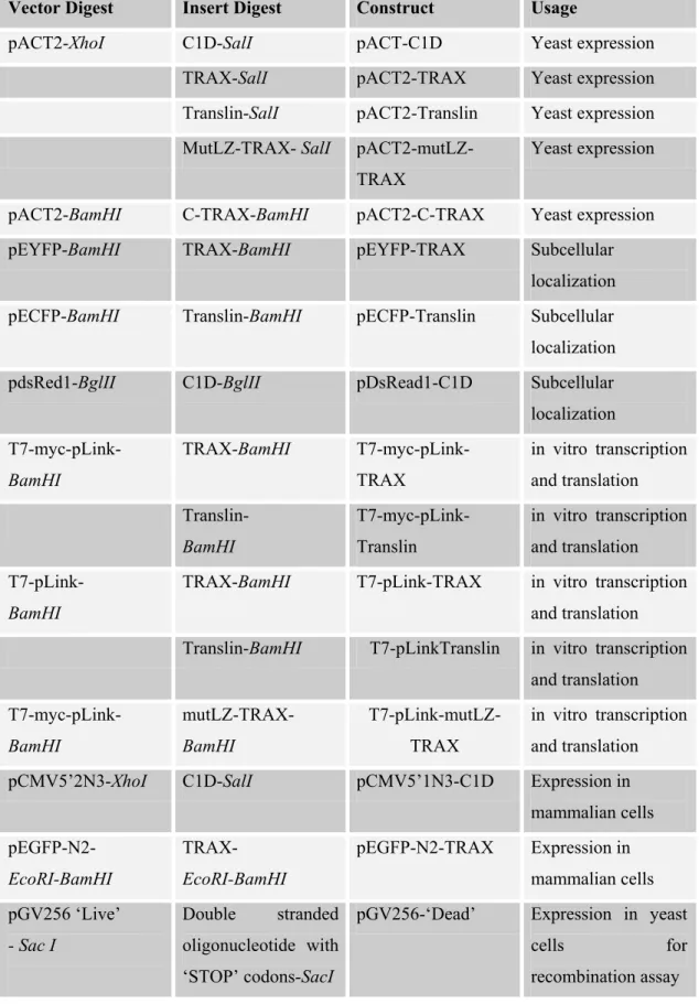
The way the constructs that were prepared and used during this study have been summarized in the table below. The maps of plasmids used in cloning are available in Appendix section.
Vector Digest Insert Digest Construct Usage
pQE30-XhoI C1D-SalI pQE30-C1D Bacterial expression, purification
pBTM116-
XhoI
C1D-SalI pBTM116-C1D Yeast expression
TRAX-SalI pBTM116-TRAX Yeast expression Translin-SalI pBTM116-Translin Yeast expression mutLZ-TRAX-SalI
pBTM116-mutLZ-TRAX
Yeast expression
Vector Digest Insert Digest Construct Usage
pACT2-XhoI C1D-SalI pACT-C1D Yeast expression TRAX-SalI pACT2-TRAX Yeast expression Translin-SalI pACT2-Translin Yeast expression MutLZ-TRAX- SalI
pACT2-mutLZ-TRAX
Yeast expression
pACT2-BamHI C-TRAX-BamHI pACT2-C-TRAX Yeast expression pEYFP-BamHI TRAX-BamHI pEYFP-TRAX Subcellular
localization pECFP-BamHI Translin-BamHI pECFP-Translin Subcellular localization pdsRed1-BglII C1D-BglII pDsRead1-C1D Subcellular localization T7-myc-pLink-BamHI TRAX-BamHI T7-myc-pLink-TRAX in vitro transcription and translation Translin- BamHI T7-myc-pLink-Translin in vitro transcription and translation T7-pLink- BamHI
TRAX-BamHI T7-pLink-TRAX in vitro transcription and translation Translin-BamHI T7-pLinkTranslin in vitro transcription
and translation T7-myc-pLink-BamHI mutLZ-TRAX- BamHI T7-pLink-mutLZ-TRAX in vitro transcription and translation pCMV5’2N3-XhoI C1D-SalI pCMV5’1N3-C1D Expression in
mammalian cells pEGFP-N2- EcoRI-BamHI TRAX- EcoRI-BamHI pEGFP-N2-TRAX Expression in mammalian cells pGV256 ‘Live’ - Sac I Double stranded oligonucleotide with ‘STOP’ codons-SacI
pGV256-‘Dead’ Expression in yeast
cells for recombination assay
2.5.8.1. Linker Scan and Deletion Mutagenesis
Linker Scan Mutagenesis method (Ausubel et al., 1987) was applied to introduce mutations into the putative leucine zipper motif of TRAX within an otherwise intact protein. MutLZ-TRAX was generated in two steps. First, the region between amino acids 1-194 was isolated by PCR with mismatch primers (TC234/ UY30) placing Sal I-Bam HI sites at the 5’ end and a Bgl II site at the 3’ end. The region between amino acids 194-272 was then amplifiedviaPCR, using primers (UY 31/TC235) that place a Bgl II site at the 5’ end and Sal I-Bam HI sites at the 3’ end. The two PCR fragments were digested with Sal I-Bgl II and cloned in a triple ligation into the Xho I site of pACT. This construct produced a mutant TRAX where Bgl II site is inserted, which changes the 194th amino acid leucine to arginine and preceding lysine to serine. The whole fragment was then cut out with BamHI and cloned into T7-Myc-plink, it was transcribed and translated in vitro using TnT Coupled Reticulocyte Lysate System (PROMEGA; Cat. No. L4610) to confirm that it gives a similar size product to wild type TRAX.
N- terminal region of TRAX between nucleotides 131-550 (including LZ region) was isolated by PCR with TC234 and UY29 primers placing Sal I to 5’ end and Xho I to 3’end. Then, this construct was cloned into XhoI site of pACT2.
2.5.8.2. Construction of “pGV256 Dead”
The oligonucleotide that contains a unique restriction enzyme site (Bgl II) in the middle, which is preceded by ‘STOP’ codons (“TGA” in three different reading frames, see section 2.4.2) was designed as a self-annealing primer and a Sac I restriction half-site was placed at the 3’-end. To obtain double-stranded oligonucleotide, 10 ng of primer was incubated at 950C for 2 minutes, 700C for 10 minutes, followed by at 370C for 10 minutes and finally, incubated at room temperature for an additional 10 minutes. As shown below, after hybridization the