The effect of nimodipine and prednisolone on traumatic
facial nerve injury treatment
Tolga Dölen1, ‹rfan Kaygusuz1, Nusret Akpolat2, Hayrettin Cengiz Alpay3, Erol Kelefl1, Turgut Karl›da¤1, fiinasi Yalç›n1, Koray Yüksel1
1
Department of ORL, Faculty of Medicine, F›rat University, Elaz›¤, Turkey 2
Department of Pathology, Faculty of Medicine, ‹nönü University, Malatya, Turkey 3
Department of ORL, Faculty of Medicine, Istanbul Kemerburgaz University, Istanbul, Turkey
Correspondence: Hayrettin Cengiz Alpay, MD. Department of ORL, Faculty of Medicine, Istanbul Kemerburgaz University, Istanbul, Turkey.
e-mail: [email protected]
Online available at: www.entupdates.org doi:10.2399/jmu.2017001011 QR code:
Özet: Nimodipin ve prednizolonun travmatik fasiyal
sinir hasar› üzerine etkisi
Amaç: Çal›flman›n amac› klempleme ile periferik fasiyal paralizi olufl-turulmufl hayvan modelinde nimodipin ve prednizolon tedavisinin histopatolojik etkisini araflt›rmakt›r.
Yöntem:Bukkal sinir dallar› klemplenerek fasiyal sinir felci olufltu-rulmufl 28 Yeni Zelanda orijinli tavflan, yediflerlik 4 gruba ayr›ld› ve her bir gruba 21 gün boyunca nimodipin, metilprednizolon ve nimo-dipin-metilprednizolon kombinasyonu uyguland›. Tedavi sonras›nda hasarl› nöral dokular histopatolojik olarak perinöral fibrozis, kollajen dejenerasyonu, aksonal dejenerasyon, miyelin dejenerasyonu, Schwann hücre proliferasyonu, normal miyelin yap›s› ve ödem aç›-s›ndan incelendi. Gruplar birbirleriyle ve kontrol grubuyla karfl›lafl-t›r›ld›.
Bulgular:Kollajen liflerde art›fl, miyelin dejenerasyonu, aksonal de-jenerasyon ve miyelin yap›s› aç›s›ndan nimodipin grubu ile kontrol grubu aras›nda; nimodipin grubu ile metilprednizolon grubu aras›nda ve nimodipin grubu ile nimodipin-metilprednizolon kombinasyon grubu aras›nda ise ödem oluflumu aç›s›ndan istatistiksel olarak anlam-l› farkanlam-l›anlam-l›k belirlendi (p<0.05). Metilprednizolon grubu ile kontrol grubu aras›nda kollajen liflerde art›fl, miyelin dejenerasyonu, aksonal dejenerasyon ve ödem, nimodipin-metilprednizolon kombinasyonu ile kontrol grubu aras›nda da kollajen liflerde art›fl, miyelin dejeneras-yonu, aksonal dejenerasyon ve normal miyelin yap›s› ve ödem aç›s›n-dan istatistiksel aç›aç›s›n-dan anlaml› veriler saptand› (p<0.05).
Sonuç: Hem nimodipin hem de metilprednizolon sinir bütünlü¤ü korunmufl travmatik sinir paralizisi üzerine olumlu etkilere sahiptir. Ancak nimodipinin metilprednizolona göre daha avantajl› oldu¤u ile-ri sürülemez.
Anahtar sözcükler:Fasiyal paralizi, nimodipin, metilprednizolon.
Abstract
Objective:To investigate the histopathological effect of nimodipine and prednisolone treatment on an animal model with peripheral facial nerve paralysis generated by clamping.
Methods:Twenty-eight New Zealand originated rabbits with facial nerve paralysis of the buccal branches generated by clamping were divided into four groups of seven each, administered with nimodipine, methylprednisolone and nimodipine-methylprednisolone combination throughout 21 days. The injured neural tissues were investigated histopathologically after treatment regarding perineural fibrosis, colla-gen decolla-generation, axonal decolla-generation, myelin decolla-generation, Schwann cell proliferation, normal myelin structure, and edema. The groups were compared with each other and with the control group.
Results: Statistically significant difference was determined between nimodipine and control groups regarding increased number of collagen fibers, myelin degeneration, axonal degeneration and myelin structure; between nimodipine and methylprednisolone groups, and between nimodipine and nimodipine-methylprednisolone combination groups regarding edema (p<0.05). Statistically significant data were also found between methylprednisolone and control groups in terms of increased number of collagen fibers, myelin degeneration, axonal degeneration and edema; between nimodipine-methylprednisolone combination and the control groups in terms of increased number of collagen fibers, myelin degeneration, axonal degeneration, normal myelin structure and edema (p<0.05).
Conclusion:Nimodipine and methylprednisolone both have positive effects on traumatic peripheral nerve paralysis with nerve integrity pre-served whereas advantage of nimodipine over methylprednisolone can-not be suggested.
The mimic muscles on our face reflect not only our genet-ic and physgenet-ical properties but also our mood expression, therefore, helping other people to understand our feelings. Patients with facial nerve dysfunction suffer from some functional and emotional problems.[1]
In order to prevent these issues and regain facial nerve functions, alternative methods of nerve recovery are needed to be developed in addition to surgical treatment.
Recently, there has been a remarkable achievement in the treatment of peripheral nerve lesions and constructing related defects after increased anatomic and histopatho-logical knowledge together with the improvement of sur-gical techniques to overcome nerve injury due to trauma, surgical intervention, tumors, compression and inflamma-tory processes.[2]
Markedly increased microsurgical tech-niques and improved histological and immunohistochem-ical methods have contributed to success in peripheral nerve injury treatment.[3]
In the treatment of traumatic peripheral facial nerve paralysis, substances such as neu-rotrophic factors, steroids, hormones and varying chemi-cals are also used for the purpose of nerve recovery in addition to many surgical techniques depending on the type of tissue damage.[4–6]
One of the chemical agents under investigation today is a 1,4 dihydropyridine derivative L-type voltage-depend-ent calcium channel antagonist, known as nimodipine. Calcium ions have key roles in depolarization, growth, excitability, aging, learning and cell proliferation, there-fore, maintaining neural plasticity.[7]
During peripheral nerve injury, permeability dysfunction of plasma mem-brane results in intracellular calcium accumulation due to electrochemical gradient difference.[8]
This intracellular calcium triggers a chain of periodic chemical reactions leading to cell death.[9]
In this study, we have developed a hypothesis of a chem-ical agent which prevents excessive calcium transition into the cell, with an assumption of having a role in diminishing mechanically induced nerve injury, therefore improving tis-sue recovery. For this purpose, we investigated the effects of nimodipine and methylprednisolone treatments in an ani-mal model of peripheral facial nerve paralysis.
Materials and Methods
This study is performed on 28 New Zealand originated rab-bits weighing 1200–1300 grams each, with the approval of Animal Experiments Ethic Committee, and the materials are supplied by Scientific Search Project Unit of our University
(Project No. TF05.11) All of the subjects were evaluated in terms of facial functions and those with no abnormalities were included. Symmetrical movements of mustache during chewing and presence of blinking reflex of the eyes during positive air pressure applied to the face by using a syringe were accepted as criteria of normal function.
All subjects underwent same surgical procedures by the same surgeon. They were given anesthesia by
administer-ing 10 mg/kg xylazine hydrochloride (Rompun®, Bayer
AG, Leverkusen, Germany) and 50 mg/kg ketamine
hydrochloride (Ketalar®, Eczac›bafl› Drug, Istanbul,
Turkey) and the area over facial nerve route on their faces were shaved before the intervention. A 2 cm long horizon-tal incision was made infraorbihorizon-tal parallel to the mandible. The skin and subcutaneous tissue were dissected, and the buccal branch of the facial nerve was identified with the help of nerve stimulator in each case. Marked area of the nerve was clipped by Yaflargil-Phynox Aneurysm Clips (Aesculap AG, Tuttlingen, Germany) with a standard
clo-sure presclo-sure of 188 g/cm2 and tolerance pressure of
162–198 g/cm2
lasting for one-minute compression (Fig. 1). Clipped area was pointed out by a suture applied to the underlying muscle tissue 5-0 silk (Ethicon Deutschland, Norderstedt, Germany) and the incision is closed at the end of clipping time by using 4-0 silk suture (Ethicon Deutschland, Norderstedt, Germany). 20–40 mg/kg of cefazoline sodium (Sefazol Flk®, Mustafa Nevzat, Istanbul,
Turkey) was administered to each subject prophylactically through intramuscular route, 1 hour before and after the surgical procedure.
28 subjects were divided into four randomized groups including seven of each and given medical treatment speci-fied below for 21 days:
• Group I (Nimodipine group): 0.5 mg/kg/day
nimodip-ine (Nimotop®, Bayer AG, Leverkusen, Germany),
intraperitoneally
• Group II (Methylprednisolone group): 1 mg/kg/day
methylprednisolone (Prednol-L®, Mustafa Nevzat,
Istanbul, Turkey), intramuscularly
• Group III (Nimodipine-methylprednisolone group): 0.5 mg/kg/day nimodipine intraperitoneally and 1 mg/kg/day methylprednisolone intramuscularly
• Group IV (Control group): 1 cc of saline solution intramuscularly
A re-incision was made over preexisting incision site on 21st day postoperatively. The destroyed segment of the buc-cal branch of the nerve was found as it was pointed out before, dissected from surrounding tissue and excised 5 mm proximally and 5 mm distally.
Excised specimens were fixed in 10% glutaraldehyde and cross-sectioned into vertical and horizontal slices with a 1.5 μ thickness of each by ultratome III glass knives (Shandon Finesse, Fisher Scientific, Leicester, UK). Slices were stained with Masson trichrome and hematoxylin-eosin (HE) and examined under the light microscope under ×40, 100, 200 and 1000 magnifications (Olympus, BX51, Tokyo, Japan).
Perineural fibrosis, increased number of collagen fibers, myelin degeneration, axonal degeneration, Schwann cell proliferation, normal structure of myelin and edema were studied on half thin tissue sections and graded as; none: -(0), mild: + (1), moderate: ++ (2), severe: +++ (3). Eyepiece graticule (ocular micrometer, 1x1 mm sized with 100 equal squares) attached to Olympus light microscope was used for counting. 4 areas of 4 cross-sections of each subject (×40, 100, 200, 1000 magnification) were counted and the aver-ages of 4 areas for each group were calculated.
Histopathological data were collected by subject follow-up forms, SPSS 11.5 (SPSS Inc., Chicago, IL, USA) pro-gram was used for statistical analysis and Mann-Whitney U test was used for group comparison. The statistical signifi-cance level was determined as 0.05.
Results
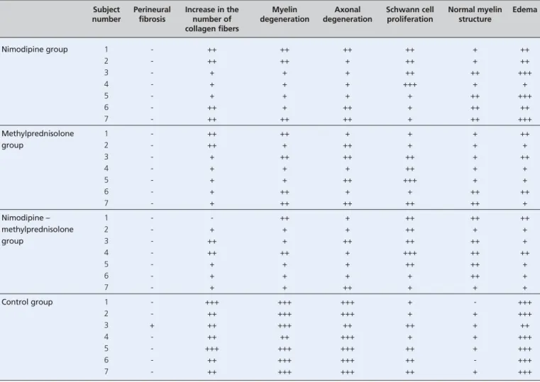
Histopathological grading of parameters for each group is shown in Table 1. As given in details, perineural fibrosis was not detected in any group except for 1 subject of the
control group. Therefore, no statistically significant dif-ference was found (p>0.05) (Table 2).
Increased number of collagen fibers was mostly seen in the control group (Group IV) followed by Groups I, II and III, respectively. Statistically significant data were found between Groups I, II, III, and the control group (p<0.05) whereas no significant data was found when Groups I, II and III were compared with each other (p>0.05) (Table 2; Fig. 2). Myelin and axonal degeneration were found mostly in control group and less in Group III. Statistically signifi-cant data were found between the control group (Group IV) and Groups I, II, III (p<0.05) and no significance detected when Groups I, II and III were compared between each other (p>0.05) (Table 2; Figs. 3 and 4).
In terms of Schwann cell proliferation, despite higher histopathological scores were detected in Groups I, II and III when compared with the control group (Group IV), no significance was found between the control group and Groups I, II, III and between Groups I, II and III when compared with each other (p>0.05) (Table 2; Fig. 5).
The normal myelin structure was mostly seen in Group III followed by Groups I, II and IV, respectively. Significant data were found between Groups I and III (p<0.05), whereas no significant data were found between Group II and the control group (Group IV) and between Groups I, II and III when compared with each other (p>0.05) (Table 2; Fig. 6).
Edema was found at least in Group III, followed by Groups II, I and IV, respectively. No statistically signifi-cant data were found between Groups II and III (p>0.05), whereas the data collected from comparisons between Groups II–III (together) and I and between Groups II–III (together) and IV were found to be significant (p<0.05). Also, no significance was found between Groups I and IV (p>0.05) (Table 2; Fig. 7).
Discussion
Facial paralysis is a clinical issue, which occurs due to a partial or complete dysfunction of the facial nerve, mostly seen as a result of trauma, surgical intervention, tumors, compression, inflammation or infection.[10]
Trauma is the second most common cause of peripher-al faciperipher-al nerve parperipher-alysis, following Bell’s pperipher-alsy.[2,11]
Facial nerve can be traumatized by temporal bone fractures, gun-shots, surgery (tympanomastoid surgery, acoustic neuro-ma surgery, parotid surgery) and penetrating laceration of the face.[12,13]
The treatment varies according to the type of the injury and clinical manifestation.[14] Many medical treat-ments have been suggested for years to improve neural function and shorten recovery period, and investigations still go on. Today, steroids are the most preferred agents as their anti-inflammatory and immunosuppressive effects are known to play a key role in the treatment of nerve injury, particularly in Bell’s palsy.[15]
Lieberman et al.[16] demonstrated a significant effect of low-dose steroids on the recovery of neural function by studying on rats with clamping induced nerve injury. Sekiya et al.[17]investigated the effect of methylprednisolone on cochlear nerve degen-eration created by clamping and suggested that it might prevent neural damage. Edema reducing the effect of methylprednisolone is believed to play an important role. In concordance with the literature, we observed
remark-able antiedema effect in groups of which methylpred-nisolone was administered.
Vita et al.[18] investigated neural regeneration rate in subjects with sciatic nerves clamped, examined from the point of injury to tibialis anterior and concluded that the group which received steroid had shown slightly signifi-cant improvement as compared to the control group. In our study, we observed less increase in the number of col-lagen fibers, less axonal and myelin degeneration and also less edema in the groups which were administered methyl-prednisolone (Groups II and III). In addition to those parameters, we found significant increment in a normal myelin structure in the nimodipine-methylprednisolone group (Group III). All these results suggest that steroids have a positive effect on traumatic nerve injury in those Subject Perineural Increase in the Myelin Axonal Schwann cell Normal myelin Edema number fibrosis number of degeneration degeneration proliferation structure
collagen fibers Nimodipine group 1 - ++ ++ ++ ++ + ++ 2 - ++ ++ + ++ + ++ 3 - + + + ++ ++ +++ 4 - + + + +++ + + 5 - + + + + ++ +++ 6 - ++ + ++ + ++ ++ 7 - ++ ++ ++ + ++ +++ Methylprednisolone 1 - ++ ++ + + + ++ group 2 - ++ + ++ + + + 3 - + ++ ++ ++ + ++ 4 - + + + ++ + + 5 - + + ++ +++ + + 6 - + ++ + + ++ ++ 7 - + ++ ++ ++ ++ + Nimodipine – 1 - - ++ + ++ ++ ++ methylprednisolone 2 - + + + ++ + + group 3 - ++ + ++ ++ ++ + 4 - ++ ++ + +++ ++ ++ 5 - + + + ++ ++ + 6 - + + + + ++ + 7 - + + ++ + + + Control group 1 - +++ +++ +++ + - +++ 2 - ++ +++ +++ + + +++ 3 + ++ +++ ++ ++ + ++ 4 - ++ ++ +++ + + +++ 5 - +++ +++ +++ ++ + +++ 6 - ++ +++ +++ ++ - +++ 7 - ++ +++ +++ ++ + +++
None: - (0), mild: + (1), moderate: ++ (2), severe: +++ (3)
with nerve integrity preserved, as in concordance with the literature.
Despite these facts, the effect of corticosteroids on trau-matic nerve injury recovery could not be thoroughly revealed. On the other hand, there are some reports about corticosteroids which emphasize their worsening effects on wound healing.[19–21]
Karl›da¤ et al.[22]
found no effect of methylprednisolone on recovery after cutting and suturing of the facial nerve. In our study, we observed no Schwann cell proliferation and no increase in the number of the nor-mal myelin structure, whereas we noticed an increase in myelin when administered in combination with nimodipine. For the first time, Van der Zee et al. investigated the effect of nimodipine on peripheral nerve injury and Groups compared Perineural Increase in Myelin Axonal Schwann Normal Edema
fibrosis the number degeneration degeneration cell myelin of collagens proliferation structure
fibers
Nimodipine group Control group .317 .030 .002 .002 .775 .015 .096
Nimodipine group Methylprednisolone group 1.000 .298 .606 .606 1.000 .298 .040
Nimodipine group Nimodipine- 1.000 .225 .591 .591 .674 .591 .020
methylprednisolone group
Methylprednisolone Control group .317 .006 .002 .002 .775 .054 .002
group
Methylprednisolone Nimodipine- 1.000 .705 .298 .298 .674 .122 ,591
group methylprednisolone group
Nimodipine- Control group .317 .007 .001 .001 .424 .006 .001
methylprednisolone group
Mann-Whitney U test
Table 2. The comparison of groups in terms of investigated parameters.
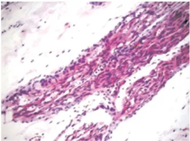
Fig. 2.The increase in the number of collagen fibers in nimodipine group (×200 HE).
reported a corrective effect on neuromuscular functions of rat sciatic nerve.[23]
Angelov et al.[24]
reported that nimodip-ine administration after cutting and suturing of facial nerve had improved the axonal regeneration, had stimu-lated nerve healing and had reduced the hyper-innerva-tion possibility. Our study supported the idea of reducing the effect of nimodipine on axonal and myelin degenera-tion.
Mattsson et al.[25]
underlined the importance of nerve surveillance on functional recovery after neural injury. For this purpose, they created facial nerve injury by cutting down subjects at the level of the middle cranial fossa and repaired them immediately, administered nimodipine and noticed that the number of surviving neurons in facial nerve motor nuclei was significantly high at the first month. In another study, Mattsson et al. created the intracranial nerve injury by crushing this time and report-ed that nimodipine had not much effect on motor nucleus cell loss (%13), but had stimulated axonal and myelin growth and improved functional recovery.[26]
In our study, we made a crushing type injury by compressing the nerve, and we detected decreased rates of axonal and myelin degeneration and increased normal myelin structure in nimodipine and nimodipine-methylprednisolone groups. Despite the fact that the increase in normal myelin struc-ture in nimodipine group compared to the control group suggests the efficacy of nimodipine on nerve regeneration, it has no superiority over methylprednisolone regarding statistically significant difference.
Scheller et al.[27] administered oral nimodipine in a group of patients with facial paralysis after maxillofacial surgery and graded the clinical manifestation by House-Brackmann scale. They indicated a lower duration of recovery period compared to the control group. In various studies, nimodipine used in the treatment of laryngeal nerve injury is shown to accelerate recovery period, and this is supported by objective measurements (EMG).[28–30]
Pointillart et al.[31]
defined the therapeutic effect of 1-week nimodipine administration on spinal cord injury by increasing blood flow. He stated significant improvement in neurologic signs after administering methylprednisolone and nimodipine to 100 patients with spinal cord injury in Fig. 5.Schwann cell proliferation in nimodipine group (×200 HE). Fig. 6.Normal myelin structure in nimodipine-methylprednisolone group
(×200 HE).
acute phase but found no difference between two groups.[32] Our study showed that nimodipine and methylprednisolone improved nerve regeneration, methylprednisolone had a superior effect on edema, and there was no significant dif-ference in other parameters. Regarding normal myelin structure, we found remarkable increment in nimodipine group, but there was no evidence on the superiority over methylprednisolone.
Conclusion
As a conclusion, evidence obtained from clinical trials sug-gests that nimodipine, a calcium-channel blocker, improves functional recovery after nerve injury. Our study also demonstrated the accelerating effect of nimodipine on neu-ral tissue healing, but we could not prove any superior effect of nimodipine over methylprednisolone. This result corre-lates with previous studies in the literature. Further studies should be performed to reinforce these results by using other objective methods such as EMG together with histopathological examination.
Conflict of Interest: No conflicts declared.
References
1. Proctor B, Nager GT. The facial canal: normal anatomy, varia-tions and anomalies. I. Normal anatomy of the facial canal. Ann Otol Rhinol Laryngol Suppl 1982;97:33–44.
2. Davis RE, Telischi FF. Traumatic facial nerve injuries: review of diagnosis and treatment. J Craniomaxillofac Trauma 1995;1:30–41. 3. Frostick SP, Yin Q, Kemp GJ. Schwann cells, neurotrophic factors, and peripheral nerve regeneration. Microsurgery 1998;18:397–405. 4. Makwana M, Raivich G. Molecular mechanisms in successful
peripheral regeneration. FEBS J 2005;272:2628–38.
5. Gordon T, Sulaiman O, Boyd GJ. Experimental strategies to pro-mote functional recovery after peripheral nerve injuries. J Peripher Nerv Syst 2003;8:236–50.
6. Mendonça AC, Barbieri HC, Mazzer N. Directly applied low intensity direct electric current enhances peripheral nerve regener-ation in rat. J Neurosci Methods 2003;129:183–90.
7. Gispen WH, Schuurman T, Traber J. Nimodipine and neural plasticity in the peripheral nervous system of adult and aged rats. In: Morad M, Nayler W, Kazda S, Schramm M, editors. The Ca2+ channel: structure, function and implications. Berlin: Springer; 1988. p. 491–502.
8. Borgens RB. Voltage gradients and ionic currents in injured and regenerating axons. Adv Neurol 1988;47:51–66.
9. Schanne F, Kant AB, Young EE, Farber JL. Calcium dependence of toxic cell death: a final common pathway. Science 1979;206: 700–2.
10. Gates GA. Facial paralysis. Otolaryngol Clin North Am 1987;20: 113–31.
11. Roob G, Fazekas F, Hartung HP. Peripheral facial palsy: etiology, diagnosis and treatment. Eur Neurol 1999;41:3–9.
12. Aky›ld›z N. Kulak Hastal›klar› ve mikrocerrahisi. Ankara: Bilimsel T›p Yay›nevi; 2002. p. 215–332.
13. May M. Trauma to the facial nerve. In. May M, editor. The facial nerve. New York, NY: Thieme; 1986. p. 192–224.
14. May M. Surgical rehabilitation of facial palsy: Total approach. In. May M, editor. The facial nerve. New York, NY: Thieme; 1986. p. 695–777.
15. fienel A, Kaya AH. Omurilik yaralanmas›nda farmakolojik tedavi. In: Kofral› E, Zileli M, editors. Temel nöroflirürji. Vol. 2. Ankara: Bulufl Tasar›m ve Matbaac›l›k; 2010. p. 1637–41.
16. Lieberman DM, Jan TA, Ahmad SO, Most SP. Effects of corticos-teroids on functional recovery and neuron survival after facial nerve injury in mice. Arch Facial Plast Surg 2011;13:117–24. 17. Sekiya T, Shimamura N, Suzuki S, Hatayama T. Methylprednisolone
ameliorates coclear nerve degeneration following mechanical injury. Hear Res 2001;151:125–32.
18. Vita G, Dattola R, Girlanda P, Oteri G, Lo Presti F, Messina C. Effects of steroid hormones on muscle reinnervation after nerve crush in rabbit. Exp Neurol 1983;80:279–87.
19. Wicke C, Halliday B, Allen D, et al. Effects of steroids and retinoids on wound healing. Arch Surg 2000;135:1265–70. 20. Pessoa ESI, Melhado RM, Theodoro LH, Garcia VG. A
histolog-ic assessment of the influence of low-intensity laser therapy on wound healing in steroid-treated animals. Photomed Laser Surg 2004;22:199–204.
21. Sharma N, Moeller CW, Marzo SJ, Jones KJ, Foecking EM. Combinatorial treatments enhance recovery following facial nerve crush. Laryngoscope 2010;120:1523–30.
22. Karlidag T, Yildiz M, Yalcin S, Colakoglu N, Kaygusuz I, Sapmaz E. Evaluation of the effect methylprednisolone and N-acetylcys-tein on anastomotic degeneration and regeneration of the facial nerve. Auris Nasus Larynx 2012;39:145–50.
23. van der Zee CE, Schuurman T, Traber J, Gispen WH. Oral admin-istration of nimodipine accelerates functional recovery following peripheral nerve damage in the rat. Neurosci Lett 1987;83:143–8. 24. Angelov DN, Neiss WF, Streppel M, Andermahr J, Mader K,
Stennert E. Nimodipine accelerates axonal sprouting after surgical repair of rat facial nerve. J Neurosci 1996;16:1041–8.
25. Mattsson P, Aldskogius H, Svensson M. Nimodipine-induced improved survival rate of facial motor neurons following intracra-nial transection of the facial nerve in the adult rat. J Neurosurg 1999;90:760–5.
26. Mattsson P, Janson AM, Aldskogius H, Svensson M. Nimodipine promotes regeneration and functional recovery after intracranial facial nerve crush. J Comp Neurol 2001;437:106–17.
27. Scheller K, Scheller C. Nimodipine promotes regeneration of peripheral facial nerve function after traumatic injury following maxillofacial surgery: An off label pilot-study. J Craniomaxillofac Surg 2012;40:427–34.
28. Mattsson P, Björck G, Remahl S, et al. Nimodipine and micro-surgery induced recovery of the vocal cord after recurrent laryn-geal nerve resection. Laryngoscope 2005;115:1863–5.
29. Nishimoto K, Kumai Y, Minoda R, Yumoto E. Nimodipine acceler-ates reinnervation of denervated rat thyroarytenoid muscle following nerve-muscle pedicle implantation. Laryngoscope 2012;122:606–13. 30. Hydman J, Björck G, Persson JKE, Zedenius J, Mattsson P.
Diagnosis and prognosis of iatrogenic injury of the recurrent laryngeal nerve. Ann Otol Rhinol Laryngol 2009;118:506–11.
31. Pointillart V, Gense D, Gross C, et al. Effect of nimodipine on posttraumatic spinal cord ischemia in baboons. J Neurotrauma 1993;10:201–13.
32. Pointillart V, Petitjean ME, Wiart L, et al. Pharmacological thera-py of spinal cord injury during the acute phase. Spinal Cord 2000; 38:71–6.
This is an open access article distributed under the terms of the Creative Commons Attribution-NonCommercial-NoDerivs 3.0 Unported (CC BY-NC-ND3.0) Licence (http://creativecommons.org/licenses/by-nc-nd/3.0/) which permits unrestricted noncommercial use, distribution, and reproduc-tion in any medium, provided the original work is properly cited.
Please cite this article as: Dölen T, Kaygusuz ‹, Akpolat N, Alpay HC, Kelefl E, Karl›da¤ T, Yalç›n fi, Yüksel K. The effect of nimodipine and