https://doi.org/10.1007/s00595-018-1690-3 ORIGINAL ARTICLE
Magnetic resonance-based pelvimetry and tumor volumetry can
predict surgical difficulty and oncologic outcome in locally advanced
mid–low rectal cancer
Gulsen Atasoy1 · Naciye Cigdem Arslan2,5 · Funda Dinc Elibol3 · Ozgul Sagol4 · Funda Obuz3 · Selman Sokmen1
Received: 15 March 2018 / Accepted: 24 June 2018 / Published online: 30 June 2018 © Springer Nature Singapore Pte Ltd. 2018
Abstract
Purpose To investigate the impact of the pelvic dimensions and tumor volume on surgery in locally advanced rectal cancer.
Methods Patients who underwent open surgery after neoadjuvant long-course chemoradiation for primary rectal cancer
were included. The predictive value of magnetic resonance-based pelvic measurements and tumor volume on the surgical difficulty and oncologic outcome were analyzed.
Results 125 patients were included. The independent risk factors related to the circumferential resection margin status were the pT stage [odds ratio (OR) 3.64, confidence interval (CI) 1.409–7.327] and tumor volume after neoadjuvant chemoradio-therapy (OR 1.59, CI 1.018–2.767). The operative time (p = 0.014, OR 1.453) and pelvic depth (p = 0.023, OR 1.116) were independent predictive factors for anastomotic leak. The median follow-up was 72 (2–113) months. Local recurrence was seen in 17 (14.1%) patients. Anastomotic leak (OR 1.799, CI 0.978–3.277), the circumferential resection margin status (OR 3.217, CI 1.262–7.870) and the relative tumor volume rate (OR 1.260, CI 1.004–1.912) were independent prognosticators of local recurrence. The 5-year overall survival was 66.7%. The circumferential resection margin status (hazard ratio: 4.739, CI 2.276–9.317), pN stage (OR 3.267, CI 1.195–8.930) and relative tumor volume rate (OR 2.628, CI 1.042–6.631) were independent prognostic factors for the overall survival.
Conclusions Relative dimensions of the tumor in the pelvis influence the local recurrence and overall survival rates.
Mag-netic resonance-based measurements can predict the difficulty of surgery and allow surgeons to consider the appropriate surgical approach.
Keywords Pelvimetry · Magnetic resonance · Rectal cancer · Survival · Local recurrence
* Naciye Cigdem Arslan cigdemarslan@hotmail.it Gulsen Atasoy glsenster@gmail.com Funda Dinc Elibol funda.elibol@deu.edu.tr Ozgul Sagol ozgul.sagol@deu.edu.tr Funda Obuz funda.obuz@deu.edu.tr Selman Sokmen selman.sokmen@deu.edu.tr
1 Department of Colorectal Surgery, Dokuz Eylul University
Medical Faculty, 35340 Izmir, Turkey
2 Department of Colorectal Surgery, Istanbul Medipol
University Medical Faculty, 34320 Istanbul, Turkey
3 Department of Radiology, Dokuz Eylul University Medical
Faculty, 35340 Izmir, Turkey
4 Department of Pathology, Dokuz Eylul University Medical
Faculty, 35340 Izmir, Turkey
5 Department of General Surgery, Istanbul Medipol University,
Introductıon
Prognostic risk categorization of rectal cancer is mainly based on TNM staging. The penetration of the tumor through the bowel wall, as well as the presence of meta-static lymph nodes have been well-known determinants of oncologic results. However, information on the utility of clinical data on the pelvic dimensions and tumor volume has been very scarce, with conflicting findings reported in the literature [1, 2].
The quality of total mesorectal excision (TME) is directly associated with the rates of local recurrence and overall survival (OS) in rectal cancer [3–5]. The mesorec-tal integrity is influenced by many variables, including the stage of the disease, tumor dimensions, histopathologic features, circumferential resection margin (CRM) status and surgeon-, center- and patient-related factors [5, 6]. Several studies have demonstrated that surgical limitations related to difficult pelvic anatomy can compromise the quality of TME and result in higher rates of an involved CRM, surgical complications and local recurrence [5,
7, 8]. The surgical difficulty in radical rectal resection depends on the location and size of the tumor, as well as the accessibility to the tumor within the anatomic con-straints of the bony pelvis. Easier dissection and a bet-ter quality of TME for a smaller tumor in a wider pelvis have been remarked by the majority of surgeons; however, objective evidence of the relationship between the pelvic anatomic measurements, tumor volume and outcome is lacking in the literature.
In this study, we assessed the impact of the tumor vol-ume, pelvic dimensions and relative tumor/pelvis dimen-sions on the surgical and oncologic outcome in locally advanced mid–low rectal cancer.
Methods
The study was approved by the local ethics committee of Dokuz Eylul University Faculty of Medicine (Approval number: 381-GOA). An institutional board-approved pro-spective rectal cancer database was reviewed. Patients who received neoadjuvant long-course chemoradiother-apy (CRT) and underwent open TME for locally advanced (T3-4 or node positive) primary rectal cancer < 10 cm from the anal verge were included in the study. The tumor level was determined by rigid sigmoidoscopy according to the distance from the anal verge as follows: distal (0–5 cm) and middle (6–10 cm). Pelvimetric and volumetric meas-urements were obtained from magnetic resonance (MR) images by two radiologists blinded to the design of the
study. The relationship between MR-based measurements and the surgical difficulty (operative time, blood loss, CRM status, anastomotic leak) and oncologic outcome (local recurrence and 5-year OS) were assessed.
Patients
Between October 2008 and February 2011, 267 patients with primary rectal cancer underwent radical resection with potential curative intent. The exclusion criteria were proximal tumors (> 10 cm) in 47 patients, incompletion of neoadjuvant treatment and/or protocol violation in 42 patients, laparoscopic surgery in 22 patients, synchronous distant/peritoneal metastasis in 21 patients, lack of proper MR imaging in 7 patients and poliposis coli syndrome in 3 patients.
All patients had colonoscopic biopsy-proven rectal cancer. Preoperative staging was performed with pelvic MR imaging and thoracoabdominal computed tomogra-phy (CT) before and after neoadjuvant therapy. Neoad-juvant long-course CRT consisted of 50.4-Gy irradiation and concomitant 225 mg/m2/day 5-fluorouracil infusion. The schedule of treatment and imaging was a strict inclu-sion criterion: neoadjuvant CRT had to be started within 1 week after the first MR scan was obtained; the second MR scan had to be performed at week 7 after completing neo-adjuvant CRT, with TME performed at week 8. Patients who did not meet this schedule were excluded.
MR imaging, pelvic measurements and tumor volumetry
Pelvic MR examinations were performed using a Philips Gyroscan NT15 Intera 1.5 T scanner (Philips Medical Sys-tems, Boston MA, USA). Sagittal and axial T2-weighted sequences were downloaded onto a digital workstation (Philips Medical Systems), and the pelvic dimensions and tumor volume were measured using hand-drawn irregu-lar ‘regions of interest’ for every section, as described by Curvo-Semedo et al. [9]. All measurements were made by the two blinded radiologists (FO, FDE), and pelvimetric measurements were repeated if the difference between the assessments of the two radiologists exceeded 10%.
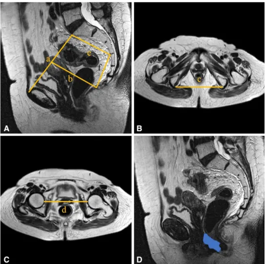
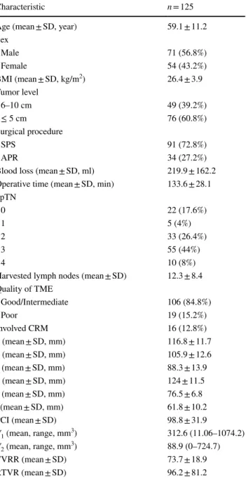
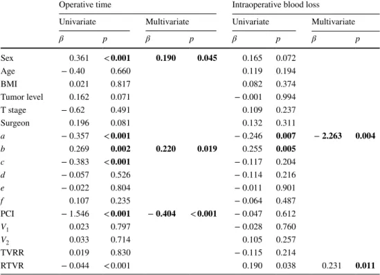
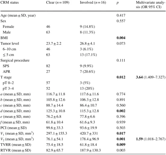
Six pelvic diameters were measured (Fig. 1). The tumor volume was calculated before (V1) and after (V2) neoadju-vant CRT. The pelvic cavity index (PCI) was calculated by the formula [pelvic inlet (a) × interspinous distance/pelvic depth (b)]. The tumor volume regression rate (TVVR) was expressed as [(V1 − V2) × 100/V1], and the relative tumor volume rate (RTVR) was expressed as [(V2/PCI) × 100].
Surgical technique
Radical open resection regarding TME principals was car-ried out by one of the three senior colorectal surgeons. Sphincter-preserving surgery was performed if a distal margin of 1 cm could be achieved, otherwise abdominop-erineal resection was carried out. Double-stapled colorectal anastomosis was performed in all sphincter-preserving pro-cedures. Diverting ileostomy was performed routinely. The duration between skin incision and closure was recorded as the operative time. Clinically or radiologically detected gastrointestinal extralumination or a pelvic abscess in the proximity of the anastomosis was considered indicative of an anastomotic leak [10].
Pathologic examinations and local recurrence
All specimens were examined in our institution by a patholo-gist blinded to the study (OS). Mesorectum integrity was determined as described by Quirke [5]: complete TME, nearly-complete TME and incomplete TME. Viable tumor cells within 1 mm of the resection plane were deemed to
indicate a positive CRM. Tumor staging was recorded according to 7th TNM. Any anastomotic, pelvic or perineal recurrence was accepted as local recurrence. Recurrent dis-ease was verified either radiologically with positron emis-sion tomography/CT or histopathologically.
Statistical analyses
Statistical analyses were performed using the SPSS software program, ver. 20.0, for Windows. Continuous variables were expressed as means ± standard deviation and categorical variables as the frequency and percentages. Linear regres-sion was performed to determine variables associated with the operative time and blood loss. Associations between categorical variables were tested with the Chi-square test. Associations between continuous variables were tested by an independent samples t-test.
Multivariate analyses of factors associated with the CRM status and local recurrence were assessed by logistic regres-sion. The OS rates were calculated using the Kaplan–Meier estimator, and the log-rank test was used to identify differ-ences among the survival curves. Cox’s proportional hazard
Fig. 1 Pelvic dimensions and volumetric measure-ments estimated by MR-based pelvimetry. A Pelvic inlet (a): distance between promontory and superior edge of sym-physis pubis. Pelvic depth (b): distance between the center of inlet and coccyx. (e): Distance between sacral promontory and interdiscal space of S3–S4. (f): Distance between S3 and coc-cyx. B Intertuberous distance (c): distance between tuberosi-ties of the ischium. C Interac-etabular distance (d): distance between medial aspects of the femur heads. D Manual tracing of free-hand region of interests in one sectional area for calcula-tion of tumor volume
model was used in univariate and multivariate analyses to assess the survival. p values < 0.05 were defined as statisti-cally significant.
Results
Of 267 patients, 125 met the inclusion criteria. 71 (56.8%) patients were male. The median age was 59 (19–83) years. The tumor site was the low rectum in 76 (60.8%) patients. Sphincter-preserving surgery was performed in 91 (72.8%) patients. The demographic and clinical characteristics of the patients are summarized in Table 1.
Surgical outcome
The mean operative time was 133.6 ± 28.1 min. Male gender (p = 0.045), a deeper pelvis (b) (p = 0.019) and a smaller PCI (p < 0.001) were associated with longer operative times in a multivariate analysis. The mean intraoperative blood loss was 219.9 ± 162.2 ml. Pelvic inlet (a) (p = 0.004) and TVRR (p = 0.011) were independent risk factors for blood loss (Table 2).
Clear CRM was achieved in 87.2% of the patients. Patients with a higher body mass index (BMI; p = 0.004), advanced T stage (p = 0.012), narrower interacetabular dis-tance (d) (p = 0.002), larger V1 (p = 0.017) and V2 (p = 0.001), lower TVRR (p = 0.009) and higher RTVR (p = 0.003) were more likely to have involved CRM than others. The inde-pendent risk factors associated with an involved CRM were pT stage [odds ratio (OR) 3.64, confidence interval (CI) 1.409–7.327] and V2 (OR 1.59, CI 1.018–2.767) (Table 3). The cut-off value for V2 to predict CRM involvement was 67.4 [area under the curve (AUC) 0.794] (Fig. 2).
Among 91 (72.8%) patients who underwent sphincter-preserving surgery, 9 (9.8%) had anastomotic leak. Four of those were managed with conservative approach, and the other five required relaparotomy (one died due to septic shock). The operative time (p = 0.032), blood loss (p = 0.016), pelvic depth (p = 0.039), distance between the sacral promontory and the interdiscal space of S3–S4 (e) (p = 0.049) and V2 (p = 0.030) were significantly associated with anastomotic leak, while the operative time (p = 0.014, OR 1.453) and pelvic depth (p = 0.023, OR 1.116) were independent risk factors.
Perioperative mortality was seen in 5 (4%) patients: 2 pulmonary infection, 1 stroke, 1 urinary sepsis and 1 anas-tomotic leak.
Oncologic outcomes
The median follow-up was 72 (2–113) months. Local recur-rence was seen in 17 (14.1%) patients. The median interval
between surgery and local recurrence was 13 (5–29) months. Four (50%) out of 8 patients with anastomotic leak had local recurrence (p = 0.009). The local recurrence rate was sig-nificantly higher in patients with involved CRM than in those without it (8.6 vs. 53.3%, p < 0.001). The operative
Table 1 Demographic, surgical, and pathologic characteristics and pelvimetric measurements of the patients
BMI body mass index, SPS sphincter-preserving surgery, APR abdominoperineal resection, ypTN pathologic TN stage in irradiated tumor, TME total mesorectal excision, CRM circumferential resec-tion margin, a pelvic inlet, b pelvic depth, c intertuberous distance, d interacetabular distance, e distance between sacral promontorium and interdiscal space of S3–S4, f distance between S3 and coccyx, PCI pelvic cavity index, V1 tumor volume before neoadjuvant therapy, V2
tumor volume after neoadjuvant therapy, TVRR tumor volume regres-sion rate, RTVR relative tumor volume rate, SD standard deviation
Characteristic n = 125
Age (mean ± SD, year) 59.1 ± 11.2
Sex Male 71 (56.8%) Female 54 (43.2%) BMI (mean ± SD, kg/m2) 26.4 ± 3.9 Tumor level 6–10 cm 49 (39.2%) ≤ 5 cm 76 (60.8%) Surgical procedure SPS 91 (72.8%) APR 34 (27.2%)
Blood loss (mean ± SD, ml) 219.9 ± 162.2 Operative time (mean ± SD, min) 133.6 ± 28.1 ypTN 0 22 (17.6%) 1 5 (4%) 2 33 (26.4%) 3 55 (44%) 4 10 (8%)
Harvested lymph nodes (mean ± SD) 12.3 ± 8.4 Quality of TME Good/Intermediate 106 (84.8%) Poor 19 (15.2%) Involved CRM 16 (12.8%) a (mean ± SD, mm) 116.8 ± 11.7 b (mean ± SD, mm) 105.9 ± 12.6 c (mean ± SD, mm) 88.3 ± 13.9 d (mean ± SD, mm) 124 ± 11.5 e (mean ± SD, mm) 76.5 ± 6.8 f (mean ± SD, mm) 61.8 ± 10.2 PCI (mean ± SD) 98.8 ± 31.9 V1 (mean, range, mm3) 312.6 (11.06–1074.2) V2 (mean, range, mm3) 88.9 (0–724.7) TVRR (mean ± SD) 73.7 ± 18.9 RTVR (mean ± SD) 96.2 ± 81.2
Table 2 Linear regression analysis of factors associated with operative time and intraoperative blood loss
a pelvic inlet, b pelvic depth, c intertuberous distance, d interacetabular distance, e distance between sacral promontorium and interdiscal space of S3–S4, f distance between S3 and coccyx, PCI pelvic cavity index, V1 tumor volume before neoadjuvant therapy, V2 tumor volume after neoadjuvant therapy, TVRR tumor
volume regression rate, RTVR relative tumor volume rate Bold values indicate better results than other filtering methods
Operative time Intraoperative blood loss
Univariate Multivariate Univariate Multivariate
β p β p β p β p Sex 0.361 < 0.001 0.190 0.045 0.165 0.072 Age − 0.40 0.660 0.119 0.194 BMI 0.021 0.817 0.082 0.374 Tumor level 0.162 0.071 − 0.001 0.994 T stage − 0.62 0.491 0.109 0.237 Surgeon 0.196 0.081 0.132 0.311 a − 0.357 < 0.001 − 0.246 0.007 − 2.263 0.004 b 0.269 0.002 0.220 0.019 0.255 0.005 c − 0.383 < 0.001 − 0.117 0.204 d − 0.057 0.526 − 0.114 0.216 e − 0.022 0.804 − 0.011 0.901 f 0.107 0.235 − 0.064 0.487 PCI − 1.546 < 0.001 − 0.404 < 0.001 − 0.047 0.612 V1 0.023 0.797 − 0.028 0.760 V2 0.033 0.714 0.105 0.257 TVRR 0.019 0.830 − 0.115 0.214 RTVR − 0.044 < 0.001 0.190 0.038 0.231 0.011
Fig. 2 ROC curves and cut-off values of independent prognostic pelvimetric factors. a Predictivity of volume after neoadjuvant chemotherapy (V2) for involved circumferential resection margin. b Predictivity of relative tumor volume rate for local recurrence
time (p = 0.012), quality of TME (p = 0.001), pT stage (p = 0.039), pelvic depth (b) (p = 0.001), intertuberous dis-tance (c) (p = 0.027), V2 (p = 0.004), TVRR (p = 0.001) and RTVR (p < 0.001) were other significant factors associated with local recurrence. A multivariate analysis revealed that anastomotic leak (OR 1.799, CI 0.978–3.277), CRM sta-tus (OR 3.217, CI 1.262–7.870) and RTVR (OR 1.260, CI 1.004–1.912) were independent prognostic factors of local recurrence (Table 4). The cut-off value for RTVR to predict local recurrence was 91.03 (AUC 0.712) (Fig. 2).
The 5-year OS was 66.7%. The quality of TME (p < 0.001), CRM status (p < 0.001), pT stage (p = 0.002), pN stage (p = 0.001), pathologic complete response (p = 0.034), perineural invasion (p = 0.002), adjuvant chemotherapy (p = 0.001), pelvic depth (b) (p = 0.042), PCI (p = 0.018) and RTVR (p = 0.009) were associated with the 5-year OS in the Kaplan–Meier analysis. The following factors were independent prognostic factors according to the multivariate analysis: CRM status (hazard ratio: 4.739, CI 2.276–9.317),
pN stage (OR 3.267, CI 1.195–8.930) and RTVR (OR 2.628, CI 1.042–6.631) (Table 5).
Discussion
Adequate TME is a prerequisite for achieving success in the management of rectal cancer. Some patient- and/or tumor-related factors that cannot be altered by technology or man-aged with mastery of the learning curve have been proposed to be responsible for surgical failure. A high BMI, distal tumor, advanced T stage, neoadjuvant radiotherapy and his-tory of abdominal surgery are well-known factors associated with difficulties and postoperative complications of TME [11–13]. However, objective data assessing the relationship between the pelvic anatomy and operative results are very limited and inconsistent.
In this study, we evaluated the influence of the anatomy and tumor volume on the surgical and oncologic outcomes
Table 3 Factors associated with circumferential resection margin status
OR odds ratio, CI confidence interval, SD standard deviation, TME total mesorectal excision, CRM circum-ferential radial margin, a pelvic inlet, b pelvic depth, c intertuberous distance, d interacetabular distance, e distance between sacral promontorium and interdiscal space of S3–S4, f distance between S3 and coccyx, PCI pelvic cavity index, V1 tumor volume before neoadjuvant therapy, V2 tumor volume after neoadjuvant
therapy, TVRR tumor volume regression rate, RTVR relative tumor volume rate
CRM status Clear (n = 109) Involved (n = 16) p Multivariate
analy-sis (OR 95% CI)
Age (mean ± SD, year) 0.417
Sex 0.557 Female 46 9 (14.8%) Male 63 8 (11.3%) BMI 0.004 Tumor level 23.7 ± 2.2 26.8 ± 4.1 0.073 6–10 cm 46 3 (6.1%) ≤ 5 cm 63 13 (17.1%) Surgical procedure 0.111 SPS 82 9 (9.9%) APR 27 7 (20.6%) T stage 0.012 3.64 (1.409–7.327) pT 0–2 57 3 (5%) pT 3–4 52 13 (20%) a (mean ± SD, mm) 116.7 ± 11.8 117.6 ± 11.6 0.774 b (mean ± SD, mm) 105.8 ± 12.6 106.3 ± 12.8 0.891 c (mean ± SD, mm) 88.7 ± 14.4 86.4 ± 10.7 0.560 d (mean ± SD, mm) 125.3 ± 10.8 115.3 ± 12.8 0.002 e (mean ± SD, mm) 76.2 ± 6.8 77.8 ± 6.6 0.396 f (mean ± SD, mm) 61.8 ± 10.4 61.6 ± 9.3 0.939 PCI (mean ± SD) 99.6 ± 33.3 93.6 ± 19.9 0.503 V1 (mean ± SD, mm3) 297.1 ± 155.3 420.7 ± 331 0.017 V2 (mean ± SD, mm3) 76.1 ± 54.1 178.4 ± 96.9 0.001 1.59 (1.018–2.767) TVRR (mean ± SD) 75.4 ± 18.5 61.8 ± 18.4 0.009 RTVR (mean ± SD) 82.9 ± 65.7 187.9 ± 130.3 0.003
Table 4 Factors associated with
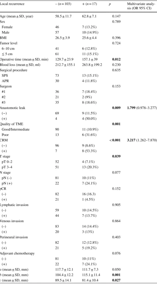
local recurrence Local recurrence – (n = 103) + (n = 17) p Multivariate analy-sis (OR 95% CI)
Age (mean ± SD, year) 58.5 ± 11.7 62.8 ± 7.1 0.147
Sex 0.789 Female 46 7 (13.2%) Male 57 10 (14.9%) BMI 26.5 ± 3.9 25.6 ± 4.4 0.396 Tumor level 0.724 6–10 cm 41 6 (12.8%) ≤ 5 cm 61 11 (15.1%)
Operative time (mean ± SD, min) 129.7 ± 23.9 157.1 ± 39 0.012 Blood loss (mean ± SD, ml) 212.7 ± 155.1 263.8 ± 199.2 0.230
Surgical procedure 0.635 SPS 73 13 (15.1%) APR 30 4 (11.8%) Surgeon 0.153 #1 36 7 (18.4%) #2 21 2 (9%) #3 35 8 (18.6%) Anastomotic leak 0.009 1.799 (0.978–3.277) (−) 69 9 (11.5%) (+) 4 4 (50.0%) Quality of TME 0.001 Good/Intermediate 90 11 (10.9%) Poor 13 6 (31.6%) CRM < 0.001 3.217 (1.262–7.870) (−) 96 9 (8.6%) (+) 7 8 (53.3%) T stage 0.039 pT 0–2 52 4 (7.1%) pT 3–4 51 13 (20.3%) N stage 0.077 pN (–) 81 10 (11%) pN (+) 22 7 (24.1%) pCR 0.152 (–) 82 16 (16.3) (+) 21 1 (4.5%) Lymphatic invasion 0.905 (–) 59 10 (14.5%) (+) 44 7 (13.7%) Venous invasion 0.864 (–) 83 14 (14.4%) (+) 20 3 (13%) Perineural invasion 0.403 (–) 82 12 (12.8%) (+) 21 5 (19.2%) Adjuvant chemotherapy 0.076 (–) 81 10 (11%) (+) 22 7 (24.1%) a (mean ± SD, mm) 117.7 ± 12.1 111.7 ± 7.3 0.050 b (mean ± SD, mm) 104.4 ± 12.2 115.1 ± 11.4 0.001 c (mean ± SD, mm) 89.5 ± 14.1 81.4 ± 10.4 0.027
of open TME. We analyzed not only the pelvic dimensions but the tumor volume and tumor/pelvis volume relativity. Our results indicated that the pelvic anatomy, tumor volume and tumor/pelvis relationship influence the surgical difficulty and rates of local recurrence and survival.
When deciding on neoadjuvant treatment for rectal can-cer, the accurate prediction of the CRM status and involved lymph nodes as well as distant metastasis are essential. Guidelines recommend thoracoabdominal CT for systemic screening and MR or endorectal ultrasound for local staging of the tumor [14]. Recently, there has been a trend toward using CT alone for initial staging, as CT is more widely available and enables screening of the thorax, liver, perito-neum and pelvis all at once for a lower cost than CT with MR imaging [15]. However, the predictive value of CT for involved CRM is decreased in low-lying tumors. MR imag-ing is more accurate than CT for detectimag-ing subcentimetric lymph nodes and mesorectal fascia status in distal tumors [16]. The reproducibility and a better understanding of the levators are other advantages associated with MR imaging [17]. A recent study including 559 patients assessed the quality of CT and MR imaging for the preoperative stag-ing of rectal cancer and concluded that MR imagstag-ing was underutilized, CT reporting of the mesorectal fascia status was low, and the accuracy of T and N staging were similar. This study included patients from several institutions in a wide region of Ontario and highlighted the importance of standardized technics and reporting of imaging modalities. At our institution, we have a colorectal team including a radiologist specializing in rectal cancer with the ability to perform MR routinely with standardized techniques and reporting. Our study includes mid–low-rectal tumors. We, therefore, preferred to use MR imaging for local staging and pelvimetric/volumetric measurements.
A reliable indicator of the difficulty of pelvic dissec-tion may help deciding the surgical procedure (open,
laparoscopic, robotic or transanal) as well as stoma and/ or anastomosis formation, and (neo-)adjuvant therapies. Surgical difficulty was demonstrated by the operative time, intraoperative blood loss, CRM involvement and rate of anastomotic leak in our study. The surgery was performed by one of three senior colorectal surgeons. Surgical diffi-culty may depend on the surgeons’ skills, but this was not a significant factor in our series. All of the surgeons had similar results regarding the operative time, blood loss and rates of local recurrence and 5-year OS. The definition of operative time was a limitation, as the pure pelvic dissec-tion time was lacking in our records, and abdominoperineal resections were included in the study. In their study includ-ing 79 laparoscopic low anterior resections, Akiyoshi et al. [18] represented surgical difficulty as the pelvic dissection time and showed that pelvic outlet as well as the BMI, tumor distance from anal verge and tumor depth were independent predictive factors of the operative time. They did not find any association between the pelvic measurements and post-operative complications [18]. In our study, the pelvic depth and operative time were independent risk factors for anasto-motic leak. Another study focusing on pelvic dimensions by Killeen et al. [19] showed that a less acutely curved sacrum and wider pelvic outlet were associated with a longer opera-tive time. In contrast, Ogiso et al. [20] suggested no correla-tion between the pelvic dimensions and operative time in 50 patients who underwent laparoscopic TME. The tumor size, BMI and tumor location were also independent predictors of the operative time in their study. In our study, male gender, a deeper pelvis and a smaller peritoneal cavity independently predicted a longer operative time. The pelvic inlet and rela-tive tumor/pelvis volume were independently correlated with the intraoperative blood loss. In contrast with the findings of other studies, the BMI was not significantly associated with surgical difficulty, probably because our series consisted of open TME procedures.
OR odds ratio, CI confidence interval, SD standard deviation, TME total mesorectal excision, CRM circum-ferential radial margin, a pelvic inlet, b pelvic depth, c intertuberous distance, d interacetabular distance, e distance between sacral promontorium and interdiscal space of S3–S4, f distance between S3 and coccyx, PCI pelvic cavity index, V1 tumor volume before neoadjuvant therapy, V2 tumor volume after neoadjuvant
therapy, TVRR tumor volume regression rate, RTVR relative tumor volume rate
Table 4 (continued) Local recurrence – (n = 103) + (n = 17) p Multivariate
analy-sis (OR 95% CI)
d (mean ± SD, mm) 124.8 ± 11.2 119.4 ± 12.7 0.078 e (mean ± SD, mm) 76.1 ± 6.7 78.7 ± 7.6 0.142 f (mean ± SD, mm) 61.2 ± 10.4 65.7 ± 8.5 0.091 PCI (mean ± SD) 101.7 ± 33.4 81.4 ± 10.1 0.015 V1 (mean ± SD, mm3) 291.1 ± 147.5 509.4 ± 312.4 0.535 V2 (mean ± SD, mm3) 77.3 ± 90.7 163.9 ± 201.8 0.004 TVRR (mean ± SD) 76.6 ± 16.6 61.2 ± 18.5 0.001 RTVR (mean ± SD) 77.8 ± 86.3 206.6 ± 254.4 < 0.001 1.260 (1.004–1.912)
Table 5 Factors associated with
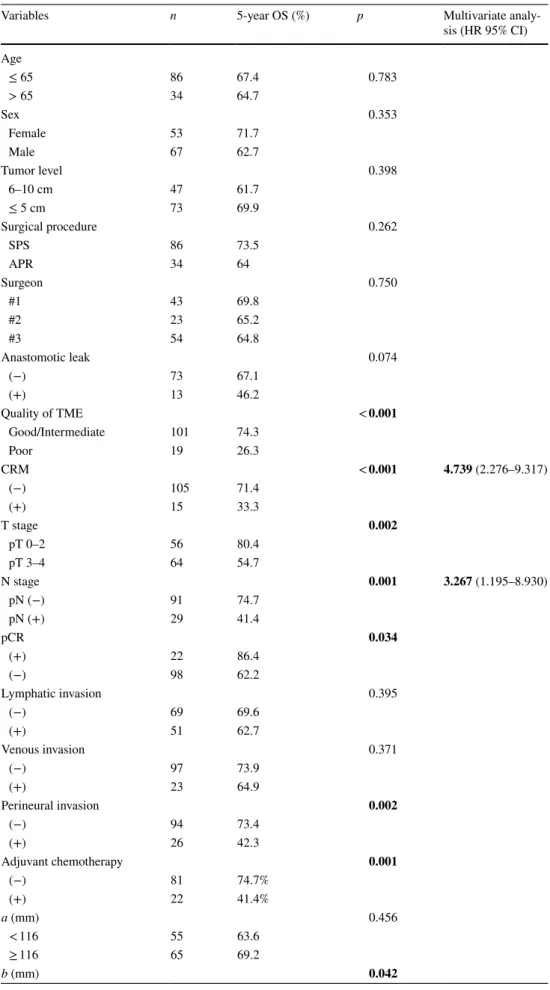
5-year overall survival Variables n 5-year OS (%) p Multivariate analy-sis (HR 95% CI)
Age ≤ 65 86 67.4 0.783 > 65 34 64.7 Sex 0.353 Female 53 71.7 Male 67 62.7 Tumor level 0.398 6–10 cm 47 61.7 ≤ 5 cm 73 69.9 Surgical procedure 0.262 SPS 86 73.5 APR 34 64 Surgeon 0.750 #1 43 69.8 #2 23 65.2 #3 54 64.8 Anastomotic leak 0.074 (−) 73 67.1 (+) 13 46.2 Quality of TME < 0.001 Good/Intermediate 101 74.3 Poor 19 26.3 CRM < 0.001 4.739 (2.276–9.317) (−) 105 71.4 (+) 15 33.3 T stage 0.002 pT 0–2 56 80.4 pT 3–4 64 54.7 N stage 0.001 3.267 (1.195–8.930) pN (−) 91 74.7 pN (+) 29 41.4 pCR 0.034 (+) 22 86.4 (−) 98 62.2 Lymphatic invasion 0.395 (−) 69 69.6 (+) 51 62.7 Venous invasion 0.371 (−) 97 73.9 (+) 23 64.9 Perineural invasion 0.002 (−) 94 73.4 (+) 26 42.3 Adjuvant chemotherapy 0.001 (−) 81 74.7% (+) 22 41.4% a (mm) 0.456 < 116 55 63.6 ≥ 116 65 69.2 b (mm) 0.042
In patients who received long-course neoadjuvant CRT, the CRM status is an indicator of adequate surgery and the major prognostic factor for local recurrence [3, 7]. Previous studies reported that a narrow pelvis is associated with poor operative outcomes, including an increased rate of anasto-motic leak and an involved CRM [4, 12, 21]. In 2004, Boyle et al. showed the significance of preoperative MR imaging on predicting CRM positivity in female patients [12]. In their study of 126 patients, they concluded that preoperative MR imaging can be used to estimate surgical resectability and help proper patient selection for neoadjuvant CRT. In contrast, Salerno et al. stated that the only predictive factor for CRM positivity was the tumor height; however, MR-pel-vimetry and CRM positivity had no significant relationship. In their cohort of 186 patients, 18.8% of those who received
neoadjuvant long-course CRT had rectal cancer [11]. In our series patients were homogenous regarding neoadjuvant treatment. In distal tumors, the rate of involved CRM was higher than in mid-rectal tumors (6.1 vs. 17%), but the dif-ference was not statistically significant, presumably due to a type-2 error (false negative). The initial tumor volume, tumor response to neoadjuvant treatment and relative tumor/ pelvis volume relationship were significantly associated with the CRM status. The pT stage and tumor volume after neo-adjuvant CRT were independent risk factors.
The tumor volumetry and tumor volume regression meas-ured by MR imaging have been proposed to be correlated with the pathologic tumor response to neoadjuvant CRT and the survival in several studies [21–26]; however, the results are conflicting. Most of those studies include both colon and
OR odds ratio, CI confidence interval, SD standard deviation, TME total mesorectal excision, CRM circum-ferential radial margin, a pelvic inlet, b pelvic depth, c intertuberous distance, d interacetabular distance, e distance between sacral promontorium and interdiscal space of S3–S4, f distance between S3 and coccyx, PCI pelvic cavity index, V1 tumor volume before neoadjuvant therapy, V2 tumor volume after neoadjuvant
therapy, TVRR tumor volume regression rate, RTVR relative tumor volume rate
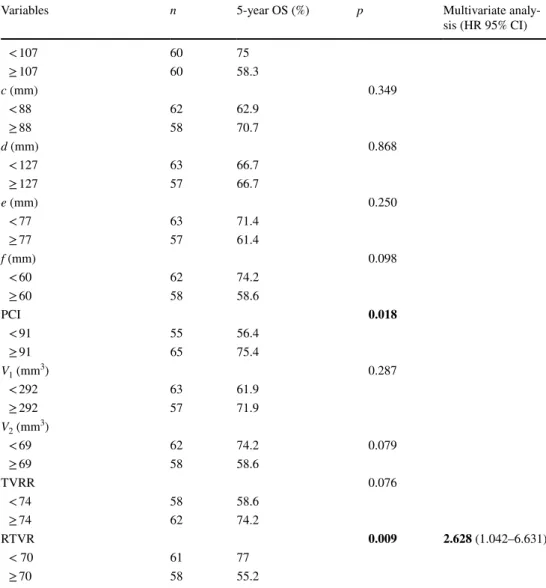
Table 5 (continued) Variables n 5-year OS (%) p Multivariate
analy-sis (HR 95% CI) < 107 60 75 ≥ 107 60 58.3 c (mm) 0.349 < 88 62 62.9 ≥ 88 58 70.7 d (mm) 0.868 < 127 63 66.7 ≥ 127 57 66.7 e (mm) 0.250 < 77 63 71.4 ≥ 77 57 61.4 f (mm) 0.098 < 60 62 74.2 ≥ 60 58 58.6 PCI 0.018 < 91 55 56.4 ≥ 91 65 75.4 V1 (mm3) 0.287 < 292 63 61.9 ≥ 292 57 71.9 V2 (mm3) < 69 62 74.2 0.079 ≥ 69 58 58.6 TVRR 0.076 < 74 58 58.6 ≥ 74 62 74.2 RTVR 0.009 2.628 (1.042–6.631) < 70 61 77 ≥ 70 58 55.2
rectal tumors, and the analyses were performed with regard to either pelvic diameters or tumor size [1, 26, 27]. Nougaret et al. [26] showed that a TVRR of at least 70% was associ-ated with a better disease-free survival in 51 patients with mostly low-lying tumors. In another study, Ferko et al. [28] assessed the association between the pelvimetry parameter A5 (the angle between the longitudinal axis of the symphy-sis, and the lines between the symphysis and the promon-tory) and the quality of TME. They concluded that pelvic measurements can be useful for determining the risk of poor mesorectum integrity and aid in the consideration of adopt-ing a transanal approach. In a study performed by Yeo et al., 430 patients with locally advanced rectal cancer underwent pre-CRT and post-CRT MR-pelvimetry. The tumor volume regression rate was found to be an independent prognostic factor for both disease-free survival and OS [22]. In contrast to other studies, we evaluated the relationship between the size of the tumor and the size of the pelvis. The relative tumor volume regression rate was an independent predicator of local recurrence and the 5-year OS in our series.
A few studies have focused on laparoscopic or robotic resections that were suggested to overcome anatomic chal-lenges. In our study, we did not include laparoscopic proce-dures, since the number was too small to perform an analy-sis. In a study of 74 laparoscopic procedures, Kim et al. [29] evaluated the anatomical difficulty with pelvic dissection time and found that a long sacrum, shallow sacral angle, narrow pelvis and large tumor were associated with pro-longed dissection. They categorized the patients into three groups according to the MR pelvimetric measurements: easy group, moderate group and difficult group. The intraopera-tive complication rate was significantly higher in the difficult group than in the other groups. In 2013, the same group published the results of robotic resections, indicating that a high BMI, neoadjuvant CRT and lower tumor location were significantly associated with a longer operative time, while the pelvimetric parameters were not [28]. They concluded that the outcome of robotic surgery was not influenced by pelvic anatomy. In addition to these reports, the drawbacks of laparoscopic TME in a narrow pelvis have been demon-strated in several studies [19, 30–32]. Transanal TME is a promising technique for high-risk distal tumors regarding CRM positivity. A recent meta-analysis of 209 transanal and 257 laparoscopic TME procedures in mid- and low-rectal cancer concluded that a transanal approach is superior to laparoscopy in terms of the CRM involvement, quality of TME and operative time [33]. Randomized studies are needed to identify the most favorable technique for manag-ing difficult TME.
The tumor size, pelvic dimensions and tumor/pelvis rela-tionship predict the surgical difficulty, local recurrence and survival in locally advanced mid- and low-rectal cancer. MR-based measurements may aid in the planning of rectal
surgery, contribute to the TME quality and positively influ-ence surgeons’ intuitive choices in high-risk patients. Based on the preoperative MR-pelvimetry, alternative approaches, such as robotic or transanal TME, may be considered.
Compliance with ethical standards
Conflict of interest The authors declare that they have no conflicts of interest.
References
1. Kornprat P, Pollheimer MJ, Lindtner RA, Schlemmer A, Rehak P, Langner C. Value of tumor size as a prognostic variable in colorectal cancer: a critical reappraisal. Am J Clin Oncol. 2011;34(1):43–9.
2. Crozier JE, McMillan DC, McArdle CS, Angerson WJ, Ander-son JH, Horgan PG, et al. Tumor size is associated with the sys-temic inflammatory response but not survival in patients with primary operable colorectal cancer. J Gastroenterol Hepatol. 2007;22(12):2288–91.
3. Trakarnsanga A, Gonen M, Shia J, Goodman KA, Nash GM, Temple LK, et al. What is the significance of the circumferential margin in locally advanced rectal cancer after neoadjuvant chemo-radiotherapy? Ann Surg Oncol. 2013;20(4):1179–84.
4. Baik SH, Kim NK, Lee KY, Sohn SK, Cho CH, et al. Factors influencing pathologic results after total mesorectal excision for rectal cancer: analysis of consecutive 100 cases. Ann Surg Oncol. 2008;15(3):721–8.
5. Quirke P, Durdey P, Dixon M, Williams N. Local recurrence of rectal adenocarcinoma due to inadequate surgical resection. Lan-cet. 1986;2(8514):996–9.
6. Leonard D, Penninckx F, Fieuws S, Jouret-Mourin A, Sempoux C, Jehaes C, et al. Factors predicting the quality of total mesorectal excision for rectal cancer. Ann Surg. 2010;252(6):982–8. 7. Quirke P, Dixon MF. The prediction of local recurrence in rectal
adenocarcinoma by histopathological examination. Int J Colorec-tal Dis. 1988;3(2):127–31. https ://doi.org/10.1007/BF016 45318 . 8. Wibe A, Rendedal P. Prognostic significance of the circumferen-tial resection margin following total mesorectal excision for rectal cancer. Br J Surg. 2002;98(8):327–34.
9. Curvo-Semedo L, Lambregts DM, Maas M, Thywissen T, Mehsen RT, Lammering G, et al. Rectal cancer: assessment of complete response to preoperative combined radiation therapy with chemo-therapy–conventional MR volumetry versus diffusion-weighted MR imaging. Radiology. 2011;260(3):734–43.
10. Rahbari NN, Weitz J, Hohenberger W, Heald RJ, Moran B, Ulrich A, et al. Definition and grading of anastomotic leakage following anterior resection of the rectum: a proposal by the International Study Group of Rectal Cancer. Surgery. 2010;147(3):339–51. 11. Salerno G, Daniels IR, Brown G, Heald RJ, Moran BJ. Magnetic
resonance imaging pelvimetry in 186 patients with rectal cancer confirms an overlap in pelvic size between males and females. Colorectal Dis. 2006;8(9):772–6.
12. Boyle KM, Petty D, Chalmers G, Quirke P, Cairns A, Finan PJ, et al. MRI assessment of the bony pelvis may help predict resect-ability of rectal cancer. Colorectal Dis. 2005;7(3):232–40. 13. Kim CW, Baek SJ, Hur H, Min BS, Baik SH, Kim NK.
Anas-tomotic leakage after low anterior resection for rectal cancer is different between minimally invasive surgery and open surgery. Ann Surg. 2006;263(1):130–7.
14. National Comprehensive CancerNetwork (2017) Rectal Cancer. (Version 3. 2017). https ://www.nccn.org/profe ssion als/physi cian_ gls/pdf/recta l_block s.pdf.
15. Bogach J, Tsai S, Zbuk K, Wong R, Grubac V, Coates A, et al. Quality of preoperative pelvic computed tomography (CT) and magnetic resonance imaging (MRI) for rectal cancer in a region in Ontario: a retrospective population-based study. J Surg Oncol. 2018;117(5):1038–42.
16. Can CT. Replace MRI in preoperative assessment of the circum-ferential resection margin in rectal cancer? Dis Colon Rectum. 2010;53(3) 308–14.
17. Nasseri Y, Langenfeld SJ. Imaging for colorectal cancer. Surg Clin North Am. 2017;97(3):503–13.
18. Akiyoshi T, Watanabe T, Ueno M. Pelvic dimensions as a predic-tor of difficulty in laparoscopic surgery for rectal cancer. Surg Endosc. 2011;25(9):3122–3.
19. Killeen T, Banerjee S, Vijay V, Al-Dabbagh Z, Francis D, Warren S. Pelvic dimensions as a predictor of difficulty in laparoscopic surgery for rectal cancer. Surg Endosc. 2012;26(1):277. 20. Ogiso S, Yamaguchi T, Hata H, Fukuda M, Ikai I, Yamato T.
Evaluation of factors affecting the difficulty of laparoscopic ante-rior resection for rectal cancer: “narrow pelvis” is not a contrain-dication. Surg Endosc. 2011;25(6):1907–12.
21. Bertani E, Chiappa A, Della Vigna P, Radice D, Papis D, Cossu L, Biffi R, et al. The Impact of pelvimetry on anastomotic leak-age in a consecutive series of open, laparoscopic and robotic low anterior resections with total mesorectal excision for rectal cancer. Hepatogastroenterology. 2014;61(134):1574–81.
22. Yeo SG, Kim DY, Kim TH, Jung KH, Hong YS, Chang HJ, Park JW, et al. Tumor volume reduction rate measured by magnetic resonance volumetry correlated with pathologic tumor response of preoperative chemoradiotherapy for rectal cancer. Int J Radiat Oncol Biol Phys. 2010;78(1):164–71.
23. Young HK, Dae YK, Tae HK, Jung KH, Chang HJ, Jeong SY, et al. Usefulness of magnetic resonance volumetric evaluation in predicting response to preoperative concurrent chemoradiotherapy in patients with resectable rectal cancer. Int J Radiat Oncol Biol Phys. 2005;62(3):761–8.
24. Lambrecht M, Vandecaveye V, De Keyzer F, Roels S, Penninckx F, Van Cutsem E, et al. Value of diffusion-weighted magnetic resonance imaging for prediction and early assessment of response to neoadjuvant radiochemotherapy in rectal cancer: preliminary results. Int J Radiat Oncol Biol Phys. 2012;82(2):863–70. 25. Lee Y-C, Hsieh C-C, Chuang J-P. Prognostic significance
of partial tumor regression after preoperative chemoradio-therapy for rectal cancer: a meta-analysis. Dis Colon Rectum. 2013;56(9):1093–101.
26. Nougaret S, Rouanet P, Molinari N, Pierredon MA, Bibeau F, Azria D, et al. MR volumetric measurement of low rectal cancer helps predict tumor response and outcome after combined chemo-therapy and radiation chemo-therapy. Radiology. 2012;263(2):409–18. 27. Poritz LS, Sehgal R, Hartnett K, Berg A, Koltun WA. Tumor
volume and percent positive lymph nodes as a predictor of 5-year survival in colorectal cancer. Surgery. 2011;150(4):649–55. 28. Ferko A, Malý O, Örhalmi J, Dolejš J. CT/MRI pelvimetry as a
useful tool when selecting patients with rectal cancer for transanal total mesorectal excision. Surg Endosc. 2016;30(3):1164–71. 29. Kim JY, Kim YW, Kim NK, Hur H, Lee K, Min BS, et al. Pelvic
anatomy as a factor in laparoscopic rectal surgery: a prospective study. Surg Laparosc Endosc Percutan Tech. 2011;21(5):334–9. 30. Baek SJ, Kim CH, Cho MS, Bae SU, Hur H, Min BS, et al.
Robotic surgery for rectal cancer can overcome difficulties asso-ciated with pelvic anatomy. Surg Endosc. 2015;29(6):1419–24. 31. Yang Y, Wang F, Zhang P, Shi C, Zou Y, Qin H. Robot-assisted
versus conventional laparoscopic surgery for colorectal disease, focusing on rectal cancer: a meta-analysis. Ann Surg Oncol. 2012;19(12):3727–36.
32. Zur Hausen G, Gröne J, Kaufmann D, Niehues SM, Aschenbren-ner K, Stroux A, et al. Influence of pelvic volume on surgical out-come after low anterior resection for rectal cancer. Int J Colorectal Dis. https ://doi.org/10.1007/s0038 4-017-2793-9 (Epub 2017 Mar 18)
33. Xu W, Xu Z, Cheng H, Ying J, Cheng F, Xu W, et al. Comparison of short-term clinical outcomes between transanal and laparo-scopic total mesorectal excision for the treatment of mid and low rectal cancer: a meta-analysis. 2016;42(12):1841–50.