Chemopreventive Effects of Hydatid Disease on Experimental Breast Cancer
Asian Pac J Cancer Prev, 16 (4), 1391-1395
Introduction
Breast cancer is the most common of all cancers and is the leading cause of cancer deaths in women worldwide. Breast cancer can be treated using a multimodality approach of surgery, chemotherapy, radiotherapy and targeted therapy (Swamy et al., 2014).
The immune system has evolved in order to detect and eliminate pathogens that may do harm to the organism. Moreover, it serves as a watchdog against transformed cells that may lead to cancer (Smyth et al., 2001). Numerous tumor antigens have been identified that can be recognized by T cells (Rosenberg et al., 2001). Probaby the key to understand complex connection between immune system and cancer is Human leukocyte antigen complex (HLA) genes. HLA genes are located on the short arm of chromosome 6 and the most polymorphic loci within the human genome. The primary function of HLA is to allow the immune system to identify infectious pathogens and eliminate them. HLA alleles play a significant role in immune responses and immunologic tolerance. It has
1Department of Pharmacology, 2Department of Histology and Embryology, 7Department of Anesthesiology, Faculty of Medicine, Cumhuriyet University, 3Department of General Surgery, SIVAS Hospital, Sivas, 4Department of General Surgery, Numune Education and Research Hospital, Ankara, 5Department of General Surgery, Medipol University, Faculty of Medicine, Istanbul, 7Department of Genetics, Faculty of Science, Bartın University, Bartın, Turkey *For correspondence: [email protected]
Abstract
Breast cancer is one of the most common and letal cancers in all over the world. Since there have been significant improvements in treatment of breast cancer, there is still a big need for alternative approaches. In this study, we aimed to investigate protective role of hydatid disease against breast cancer. Twenty Wistar rats were divided into two groups of 10 rats each Group I (control) and Group II. In Group II intraperitoneal hydatidosis was performed. Then DMBA was applied to mammary tissues of all rats. Immunohistochemistry studies for Ki-67 and S-100 in the tumoral tissue sections of DMBA induced mammary tumor in rats were performed. TUNEL Assay was used to detect apoptotic cells of tumoral tissue. In vivo anticancer activity testing was carried out by preventing the tumorigenesis by DMBA in mammary tissue of rats. The expressions of the Ki-67 and S-100 protein decreased in rats who had Hydatid Disease (HD) (Group II), compared with the control rats (Group I). TUNEL positive cells were higher in rats with HD (Group II), compared with the control rats (Group I). In vivo studies showed that HD prevented the tumorigenesis by DMBA in mammary tissue of rats with 50 percent.In the light of the evidence the present study showed that HD may have chemopreventive effects on DMBA induced breast cancer.
Keywords: Apoptosis - breast cancer - chemoprevention - hydatid disease
RESEARCH ARTICLE
Chemopreventive Effects of Hydatid Disease on Experimental
Breast Cancer
Ahmet Altun
1*, Serpil Unver Saraydin
2, Sinan Soylu
3, Deniz Sahin Inan
2, Cinar
Yasti
4, Yasar Ozdenkaya
5, Binnur Koksal
6, Cevdet Duger
7, Cemil Isbir
7, Mustafa
Turan
7been recently shown that certain type of HLA genes are strictly related to certain type of cancers (An et al., 2014). Cystic echinococcosis (CE) is a cosmopolitan zoonosis caused by the larval cystic stage of the tapeworm
Echinococcus granulosus. It is a frequently encountered
parasitic infection in endemic areas of the world, such as the Mediterranean countries (Akgul et al., 2003). The clinical evolution of these cysts is silent for several months, and the symptoms are not specific. Since it is long term chronic infection source the immune system is activated and different kinds of many cytokines including IL-6, IL-12 and IFN-γ become a part of the equation (Mezioglu et al., 2009).
In Turkey, clinicians have noticed an exceedingly low rate of incidental diagnoses of hydatid disease (HD) among patients undergoing surgery for various solid tumors. Akgul et al. evaluated 2086 patients with various solid tumors according to the accompanying HD (Akgul et al., 2003). Although the prevalence rate of the HD in Turkey is reported to be approximately 1-2%, prevalence rate of the cancer is 0.64%, the coexistance of both
conditions (cancer and HD) had been observed in a rate of 0.09 % (Kalyoncu et al., 1989; Akgul et al., 2003)
In an attempt to evaluate the presence of cancer at the time of diagnosis of Echinococcus granulosus, Akgul et al. screened the medical reports of a total of 1000 patients who were treated for HD (Akgul et al., 2003). In the group of patients diagnosed with HD, they identified only 1 patient who underwent surgery for esophageal carcinoma and this also occurred 8 years prior to the diagnosis of HD.
Similar observations among surgeons in Turkey, brought a legend that hydatid disease may have protective role against cancer”. In this study we aimed to investigate if hidatid disease has a preventive effect on development of breast cancer.
Materials and Methods
Preparation of the hydatid ‘‘sand’’ (scolices)
We used the fertile cysts of sheep livers supplied from the local slaughterhouse in Sivas. The aspirated portion of the hydatid liquid was collected into sterile tubes and centrifuged at 4000 g for 10 minutes; then the supernatant was discarded. The remaining sediment (hydatid sand) contained 1500 scolices/ml, 95% to 99% of which were viable as determined from their motility characteristics seen with 0,1% Eosin staining under light microscopy.
Preparation of intraperitoneal hydatidosis model
A total of 20 female albino Wistar rats (160±10 g body weight) 50-59 days old maintained in Animal Laboratory of Cumhuriyet Universty Faculty of Medicine, at temperature 24±2ºC with a 12 hour light/dark cycle and 60%±5% humidity. They were provided with standard pellet diet and water ad libitum. The experiment was carried out as per the guidelines of Ethical Committee for the Purpose of Control and Supervision of Experiments on Animals, of Cumhuriyet Universty Faculty of Medicine.
All rats were divided into two groups of 10 rats each Group I (control) and Group II (rats with hydatidosis). All of the manipulations described below were performed with strict compliance to antiseptic principles. In Group II, the rats were anesthetized with 3 mg/kg Xylasine (Rhompun®, Bayer) I.P., and 90 mg/kg Ketamin Hydrochloride (Ketalar®, Eczacibasi, Istanbul, Turkey) I.M. Then those rats were subjected to a median laparotomy of 1 cm length performed with a scalpel. The peritoneum was exposed and opened to reach the intraperitoneal cavity, into which 1 ml of concentrated hydatid “sand” (scolices), was instilled with a pipette. Laparotomy incision was sutured with a continuous 2/0 polyglactin suture. None of the rats received any medication before this operation. Rats received analgesia in the postoperative period.
The animals in the study were kept for 3 months. During this period, all animals were fed ad libitum, given tap water and kept at room temperature (18-20 C0) in the separate cages. Then, rats in Group II were again anesthetized with 3 mg/kg Xylasine (Rhompun®, Bayer) I.P., and 90 mg/kg Ketamin Hydrochloride (Ketalar®, Eczacibasi, Istanbul, Turkey) I.M. underwent to a median relaparotomy of 2 cm, for confirmation of hydatidosis.
Swabs and aspirate were obtained from intrabdominal cysts for confirmation of hydatidosis. Then, laparotomy incision was sutured with a continuous 2/0 polyglactin suture. None of the rats received any medication before this operation. Rats received analgesia in the postoperative period.
Swabs and aspirate were prepared and inspected with 0.1% Eosin staining under light microscopy. After confirmation of hydatidosis of rats in Group II, mammary carcinogenesis induction of rats in was planned in Group I (control) and Group II (rats with hydatidosis).
In vivo chemopreventive studies
20 days later, rats in Groups I and II were induced mammary carcinogenesis by providing single subcutaneous injection in right pectoral area of 25 mg Dimethyl Benzanthracene (DMBA) in 1 mL emulsion of sunflower oil (0.75 mL) and physiological saline (0.25 mL) to each rat. During the experimental period, animals were weighed weekly. Animals were observed daily to assess their general health.
After DMBA administration, right pectoral area of all rats was followed up for the tumoral development. Palpation of mammary nodules began 4 week after animals received DMBA. 8 weeks after DMBA administration rats were sacrificed and injected pectoral areas of rats were removed from the animals. Histopathological examination was performed from that resected tissue for each nodule.
Histopathology and morphological observations
Each tumor tissues were sampled macroscopically and fixed in 10% formaldehyde solution for 24 hours. Tumor tissues then processed in autotechnicon device later embedded in parafin blocks and cut sections with 3-5 μm thickness were obtained and stained with Haemotoxylen-Eosin stain for routine histopathological examination by light microscopy.
Immunohistochemistry
Ki -67 and S-100: For immunohistochemical staining, the deparaffinized and redehydrated tissue sections were inactivated the endogenous peroxidase by an incubation with 3% H2O2 for 10 minutes. To recover antigen, these
sections were put into EDTA solution (pH 8.5) and heated in the microwave oven twice. The slides were then washed with PBS (pH 7.2-7.6) twice. Non-specific binding sites were blocked with Ultra V Block (ScyTek Laboratories, Logan, Utah, USA) for 20 minutes. After the redundant liquid was discarded, the sections were incubated with primary antibody Ki-67 and S-100 (ScyTek Laboratories, Logan, Utah, USA) at room temperature for 1,5 hour and washed with PBS. Then the slides were incubated with biotinylated secondary antibody (ScyTek Laboratories, Logan, Utah, USA) for 20 minutes and washed with PBS, followed by incubation with streptavidin-HRP (ScyTek Laboratories, Logan, Utah, USA) for 20 minutes and washed with PBS. The S100 antibody binding sites were visualized by incubation with a AEC kromogen (Scy Tec Laboratories, Logan, Utah, USA) solution. The Ki-67 antibody binding sites were visualized by incubation with
Chemopreventive Effects of Hydatid Disease on Experimental Breast Cancer
USA) solution. The slides were counterstained for 1 min. with hematoxylin and then dehydrated with sequential ethanol for sealing and microscope observation.
In immunohistochemistry studies the semiquantitative scoring system was used in considering the staining intensity and area extent, which has been widely accepted and used in previous studies [3]. Every tumor was given a score according to the intensity of the nuclear or cytoplasmic staining (no staining=0; weak staining=1; moderate staining=2; strong staining=3) and the extent of stained cells (0%=0; 1-10%=1; 11-50%=2; 51-80%=3; 81-100%=4).
The percentage of proliferating neoplastic cells was evaluated directly by light microscopy. Quantification of the proliferation was performed by counting, Ki-67 positive cells in 4-6 random fields per slide. S-100 activity was evaluated semi quantativelly in the cytoplasm of the tumor cells either in living or in the necrotic tumor cell areas.
In vivo apoptosis assay
Apoptosis was evaluated by using terminal deoxynucleotydil transferase dUTP nick and labelling (TUNEL) method. In Situ Cell Death Detection Kit, POD (Roche, Germany) is used for apoptosis. Tumor sections were deparaffinized and dehydrated according to standard protocols. Tissue sections were incubated with Proteinase K working solution for 30 min at 21-37oC. The slides were
then washed with PBS (pH 7.2-7.6) twice. Positive control was incubated with DNase I recombinant for 10 min at 15-25oC. Negative control was incubated with Label solution
(without terminal transferase) instead of TUNEL reaction mixture. The slides were then washed with PBS (pH 7.2-7.6) three times. Converter-POD was added on slides and incubated in a humidified chamber for 30 min at 37oC.
The slides were then washed with PBS (pH 7.2-7.6) three times. DAB Substrate was added on slides and incubated for 10 min at 15-25oC. The slides were then washed with
PBS (pH 7.2-7.6) three times. The slides were mounted and analysed by fluorescence microscope (Olympus DP 70, Melville, NY, USA).
Statistical analysis
Results are reported as mean±standard error (SE). We tested the data from the experiments for statistical significance using the Mann-Whitney U Test. A p-value less than 0.05 were considered significant.
Results
Histopathology and morphological results
Mammary tissue from DMBA-treated group (Group I) showed hyperplasia of the lobules characterized by a
typical arborization. Atypical epithelial hyperplasia was observed. Epithelial cells showed variation in nuclear size with irregular chromatin and nucleoli (Figure 1a, b). In the tissues of the rats with hydatid disease (Group II), the normal lobular structure of the breast was lost, and the border between intralobular and dense interlobular connective tissue was obscured (Figure 1c).
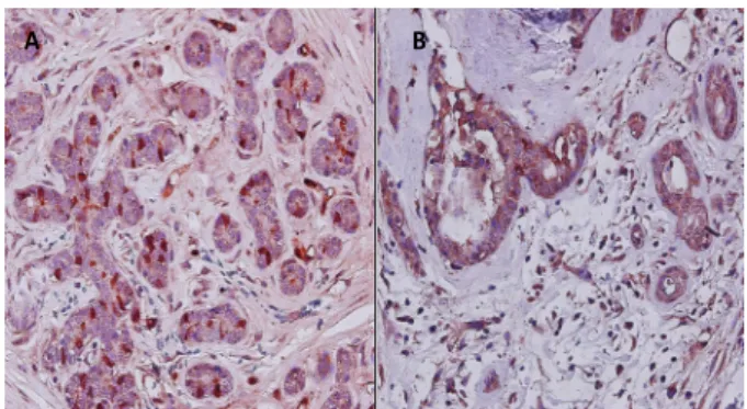
Whereas S-100 expression was high in the Group I, it was low in the Group II (Figure 2, a, b) (Table 1). Ki-67 activity was most intensive in the group I (Table 1). TUNEL-Positive cells has been detected rarely in the Group I. (Figure 3a). TUNEL-Positive cells were increased in the Group II (Figure 3 b).
Table 1. Results of Immunohistochemistry Studies According to Semiquantitative Scoring System. Statistical Significance was set at p<0.05.
Immunohistochemical examination of tumoral tissues Ki-67 S-100
In control rats (Group I) 2.44±0.09 2.27±0.23
In rats with hydatid disease (Group II) 1.25±0.23 1.23±0.18
Statististical results ( p=0.003) (p=0.004)
Figure 1. 1a) Intraductal carcinoma. Group I, H-E, 10X.
1b) Intraductal carcinoma. Group I, H-E, 40X. In both the ducts
filled completely with malignant cells. 1c) Fibrocystic change. The normal lobular structure of the breast is lost, and the border between intralobular and dense interlobular connective tissue is obscured. Group II, H-E, 20X
A
B
C
Figure 2. S-100 Expression was high in the Group I, it was Low in the Group II 40X (Figure 2, a,b)
Tumorigenesis and volume inhibition studies
After 8 weeks of DMBA administration histopathological examination was performed from that resected pectoral tissue for each nodule. While tumoral tissues were detected in 8 of 10 rats (80%) in Group I, they were detected in 4 of 10 rats (40%) in Group II. The mean tumorigenesis inhibition ratio of hydatid disease was calculated as 50 %.
Discussion
In Turkey anectodal reports indicate that the prevalence of hydatid disease is significantly lower than expected in cancer patients (Kalyoncu et al., 1989; Akgul et al., 2003). Based on these observations, we propose that
Echinococcus granulosus may elicit a protective effect
against the development of cancer through common antigenic properties with cancerous cells.
Antigen KI-67 is a protein that in humans is encoded by the MKI67 gene (Scholzen et al., 2000). It is a nuclear protein and associated with cell proliferation and it is an excellent marker to determine the growth fraction of a given cell population (Scholzen et al., 2000). In this study, we studied the Ki-67 levels of the DMBA induced tumoral tissues to determine the HD effect on tumoral growth. In-vivo immunohistochemistry experiments of this study showed that HD decreased the Ki-67 activity in tumoral tissues of rats.
S-100 protein is a family of low molecular weight protein found in vertebrates. S100 proteins are involved in regulation of protein phosphorylation, transcription factors, enzyme activities, cell growth and differentiation, and the inflammatory response (Donato et al., 2003). Several members of the S-100 protein family are useful as markers for certain tumors and epidermal differentiation (Nonaka et al., 2008). In this study, we studied the S-100 levels of the DMBA induced tumoral tissues to determine the HD effect on tumoral growth. In-vivo immunohistochemistry experiments of this study showed that HD decreased the S-100 activity in tumoral tissues of rats.
During apoptosis a group of proteases are activated which cause DNA fragmentation, cytoplasmic shrinkage and membrane blebbing. Here, we therefore examined apoptosis in tumoral tissues by using terminal
(TUNEL) method. TUNEL Assay results of our in-vivo studies demonstrated that HD increased the apoptotic cells in tumoral tissues of rats.
After 8 weeks of DMBA administration histopathological examination showed that tumoral tissues were detected in 8 of 10 rats (80%) in Group I and they were detected in 4 of 10 rats (40%) in Group II. These results showed that HD caused 50 percent chemoprevention.
Suppression of neoplastic growth via infectious agents has been observed for bacterial (Listeria monocytogenes, Corynebacterium parvum) and protozoan (Toxoplasma gondii, Besnoitia jellisoni) pathogens (Hunter et al., 2001). Similar cross-reactions between bacteria and tumour cells have been reported. For example, Mycobacterium bovis (BCG) and guinea-pig line 10 hepatocarcinoma cells as well as human melanoma cells share surface antigens (Hunter et al., 2001). Stimulation of the immune response (such as nonspecific macrophage activation to kill tumor cells or systemic inhibition of angiogenesis) has been put forward to explain the biologic basis of these observations (Springer et al., 1997; Hunter et al., 2001).
It is interesting to note that an antigenic similarity between various tumors and cysts of Echinococcus granulosus infection was reported for cancer-associated O-glycosylated Tn antigen (Van Knapen et al., 1980; Springer et al., 1997; Alvarez Errico et al., 2001; Hunter et al., 2001). Tn antigen is a glycoprotein that is expressed during the early phases of various malignancies, including carcinomas of the breast, pancreas, lung, gastrointestinal tract, upper aerodigestive tract, and genitourinary tract (Springer et al., 1997). Yong et al. also reported a possible antigenic similarity between pulmonary carcinoma and cysts of Echinococcus granulosus (Yong et al., 1979). Pfister M, et al reported the elevated carbohydrate antigen 19-9 (CA 19-9) in patients with Echinococcus infection (Pfister et al., 2001).
Karadayi et al reported that sera of patients with hydatid diseases had cytotoxic effects on human small cell lung cancer cells, but they did not have cytotoxic effects on fibroblast cells. Sera from healthy subjects did not have a cytotoxic effect on the tumor cell line or control fibroblasts (Karadayi et al., 2013). As the evidence for an antigenic similarity between E granulosus and some tumour types increases we should keep in mind that the presence of specific antibodies in the serum may have immunotherapy and immunoprophylaxis in malignancies. This may influence the antitumoral effect of HD in this experimental tumour model.
Furthermore, beside with antigenic similarty hypothesis, it should be kept in mind that choronic parasitic infections like hidatid disease can induce immune system which is one of the main systems keep tumorigenic development under control. It is believed that one of the reasons why cancer develops and spreads so rapidly is because of the weak immunosurveillance (Reiman et al., 2007). Although alterations in biological systems, which upregulate immune system, increase the risk of autoimmune diseases, these alterations decrease the risk of cancer development (Ghaderi et al., 2011). Cytotoxic Figure 3. Amount of TUNEL-Positive Apoptotic Cells
in Group I (a), Increased TUNEL-Positive Apoptotic Cells in Group II (b)
Chemopreventive Effects of Hydatid Disease on Experimental Breast Cancer protein involved in the downregulation of immune system
(Ghaderi et al., 2011). Since CTLA-4 downregulates T-cell production individuals with GG genotype (less CTLA-4 production) have higher T-cell proliferation than those with the AA genotype (Maurer et al., 2002). It is due to this reason that the risk allele for susceptibility to cancer (allele A) is the opposite of that found for susceptibility to autoimmune diseases (allele G) (Sun et al., 2009). It was found to be positively associated with breast cancer by Sun et al. and Wang et al. in Chinese and by Ghaderi et al. in Iranian breast cancer patients (Ghaderi et al., 2004; Wang et al., 2007; Sun et al., 2008). These results are consistent with our findings that suggest more active immune system is resistant to the tumor devolepment. On the other hand, in a similar study to Maurer and Sun, Minhas et al., found that CTLA-4 +49 G/A polymorphism was not found to be associated with breast cancer risk in our North Indian population
In conclusion, these results showed that hydatid disease may have preventive properties against breast cancer. It may be related to antigenic similarities of the parasite with certain type of cancer cells or induction of the immune sytem against cancer development. Further studies needed to reach the main mechanism of these effects. .
Acknowledgements
The study has been planned and conducted by Ahmet Altun and Mustafa Turan. Anesthesia of animals during the operations has been applied by Cevdet Duger and Cemil Isbir. Histopathologic examinations have been performed by Sepil Unver Sariaydin and Deniz Sahin. Operations and tissue collection have been done by Sinan Soylu, Çinar Yasti and Yasar Ozdenkaya. Statistical analysis has been done by Binnur Koksal.
References
Akgul H, Tez M, Unal AE, et al (2003). Echinococcus against cancer: why not? Cancer, 98, 1999-2000.
Alvarez Errico D, Medeiros A, Míguez M, et al (2001). O-glycosylation in Echinococcus granulosus: identification and characterization of the carcinoma-associated Tn antigen. Exp Parasitol, 98, 100-9.
An WX, Fan YX, Liang XH, Liu H (2014). Changes in median ages at death from selected cancer types in relation to HLA-DRB1/DQB1. Asian Pac J Cancer Prev, 15, 4125-8. Donato R (2003). Intracellular and extracellular roles of S100
proteins. Microsc Res Tech, 60, 540-51.
Ghaderi A, Yeganeh F, Kalantari T, et al (2004). Cytotoxic T lymphocyte antigen-4 gene in breast cancer. Breast Cancer Res Treat, 86, 1-7.
Ghaderi A (2011). CTLA4 gene variants in autoimmunity and cancer: a comparative review. Iran J Immunol, 8,127-49. Han CP, Kok LF, Wang PH, et al (2009). Scoring of p16INK4a
immunohistochemistry based on independent nuclear staining alone can sufficiently distinguish between endocervical and endometrial adenocarcinomas in a tissue microarray study. Modern Pathology, 22, 797-806. Hunter CA, Yu D, Gee M, et al (2001). Cutting edge: systemic
inhibition of angiogenesis underlies resistance to tumors during acute toxoplasmosis. J Immunol, 166, 5878-81.
Kalyoncu AF, Emri AS, Akhan O, et al (1989). Asağiesence koyu Beysehir Konya’da hidatik kist hastaliği prevalans calismasi. In: Baris I˙, Sahin A, editors. Hidatik kist hastaliği ve Turkiye’deki konumu Ankara: Turkiye Akciğer Hastaliklari Vakfi Yayini, 90-8.
Karadayi S, Arslan S, Sumer Z, et al (2013). Does hydatid disease have protective effects against lung cancer? Mol Biol Rep, 40, 4701-4.
Mäurer M, Loserth S, Kolb-Mäurer A, et al (2002). A polymorphism in the human cytotoxic T-lymphocyte antigen 4 (CTLA4) gene (exon 1 +49) alters T-cell activation. Immunogenetics, 54: 1-8.
Mezioug D, Touil-Boukoffa C (2009). Cytokine profile in human hydatidosis: Possible role in the immunosurveillance of patients infected with Echinococcus granulosus. Parasite,
16, 57.
Minhas S, Bhalla S, Shokeen Y, et al (2014). Lack of any association of the CTLA-4 +49 G/A polymorphism with breast cancer risk in a North Indian population. Asian Pac J Cancer Prev, 15, 2035-8.
Nonaka D, Chiriboga L, Rubin BP (2008). Differential expression of S100 protein subtypes in malignant melanoma, and benign and malignant peripheral nerve sheath tumors. J Cutan Pathol, 35, 1014-9.
Pfister M, Gottstein B, Kretschmer R, Cerny T, Cerny A (2001). Elevated carbohydrate antigen 19-9 (CA 19-9) in patients with Echinococcus infection. Clin Chem Lab Med, 39, 527-30.
Reiman JM, Kmieciak M, Manjili MH, Knutson KL (2007). Tumor immunoediting and immunosculpting pathways to cancer progression. Semin Cancer Biol, 17, 275-8. Rosenberg SA (2001). Progress in human tumour immunology
and immunotherapy. Nature, 411, 380-4.
Scholzen T, Gerdes J (2000). The Ki-67 protein: from the known and the unknown. J Cell Physiol, 182, 311-22.
Smyth MJ, Godfrey DI, Trapani JA (2001). A fresh look at tumor immunosurveillance and immunotherapy. Nat Immunol, 2, 293-99
Springer GF (1997). Immunoreactive T and Tn epitopes in cancer diagnosis, prognosis, and immunotherapy. J Mol Med, 75, 594-602.
Sun T, Hu Z, Shen H, Lin D (2009). Genetic polymorphisms in cytotoxic T-lymphocyte antigen 4 and cancer: the dialectical nature of subtle human immune dysregulation. Cancer Res,
69, 6011-4.
Sun T, Zhou Y, Yang M, et al (2008). Functional genetic variations in cytotoxic T-lymphocyte antigen 4 and susceptibility to multiple types of cancer. Cancer Res, 68, 7025-34. Swamy ST, Radha CA, Kathirvel M, Arun G, Subramanian S
(2014). Feasibility study of deep inspiration breath-hold based volumetric modulated arc therapy for locally advanced left sided breast cancer patients. Asian Pac J Cancer Prev,
15, 9033-8.
Van Knapen F (1980). Echinococcus granulosus infection and malignancy. BMJ, 281, 195-96.
Wang L, Li D, Fu Z, et al (2007). Association of CTLA-4 gene polymorphisms with sporadic breast cancer in Chinese Han population. BMC Cancer, 7, 173.
Yong WK, Heath DD, Savage T (1979). Possible antigenic similarity between pulmonary carcinoma and cysts of Echinococcus granulosus. Br Med J, 1463-4.