© 2016 Wiley Periodicals, Inc. R E S E A R C H A R T I C L E
The importance of serum biglycan levels as a fibrosis marker in
patients with chronic hepatitis B
Rafiye Ciftciler
1| Seren Ozenirler
2| Aysegul Atak Yucel
3| Mustafa Cengiz
4|
Gulbanu Erkan
5| Erkan Buyukdemirci
6| Cemile Sönmez
7| Guldal Yılmaz Esendaglı
81Department of Internal Medicine, Gazi
University Faculty of Medicine, Ankara, Turkey
2Department of Gastroenterology, Gazi
University Faculty of Medicine, Ankara, Turkey
3Department of Immunology, Gazi University
Faculty of Medicine, Ankara, Turkey
4Department of Gastroenterology, Ankara
Oncology Education and Research Hospital, Ankara, Turkey
5Department of Gastroenterology, Istanbul
Medipol University Faculty of Medicine, Istanbul, Turkey
6Department of Public Health, Gazi University
Faculty of Medicine, Ankara, Turkey
7Microbiology Specialist, Vaccine preventable
Bacterial Diseases Research Laboratory, Public Health Institution of Turkey, Ankara, Turkey
8Department of Pathology, Gazi University
Faculty of Medicine, Ankara, Turkey Correspondence
Rafiye Ciftciler, Departments of Internal Medicine, Gazi University Faculty of Medicine, Ankara, Turkey.
Email: [email protected] Funding
The whole financial cost of the study was paid by the authors; no grants or financial support was taken from any other third parties.
Background: Liver biopsy is recommended in the majority of patients with chronic viral hepatitis for fibrosis evaluation. Because of the potential risks of liver biopsy, many studies related to non- invasive biomarkers of hepatic fibrosis have been per-formed. We aimed to assess the diagnostic value of serum biglycan as a non- invasive fibrosis marker in chronic hepatitis B patients.
Methods: This study included 120 patients with biopsy- proven hepatitis B patients and 60 healthy controls. Fibrosis stage and necroinflammatory activity were assessed in liver biopsy specimens. Biglycan level was measured using an ELISA assay.
Results: Serum biglycan levels of chronic hepatitis B patients were found to be signifi-cantly higher than those of healthy controls (337.3±363.0 pg/mL vs 189.1±61.9 pg/ mL, respectively, P<.001). There was a statistically significant positive correlation be-tween serum biglycan level and fibrosis stage (P=.004; r=.213). Besides, a statistically significant positive correlation was found between serum biglycan level and necroin-flammatory activity (P<.001; r=.271). The AUROC of BGN levels was 0.702 for fibrosis stage, differentiating patients from healthy controls with statistical significance (P<.001). The AUROC of BGN levels was 0.632 for necroinflammatory activity score, differentiating patients from healthy controls with statistical significance (P=.004). Conclusions: Serum biglycan might be used as a non- invasive marker of liver fibrosis. Further studies are needed to evaluate the usefulness of this marker.
K E Y W O R D S
biglycan, chronic hepatitis B, liver biopsy, liver fibrosis, non-invasive marker
1 | BACKGROUND
Chronic hepatitis B infection, chronic hepatitis C infection, and non- alcoholic steatohepatitis are the most common chronic liver diseases all over the world.1 Hepatitis B virus infection is a global public health
problem. It is estimated that there are 350 million HBV carriers and nearly 1 million patients die from HBV- related liver disease annually.2 HBV infection can cause acute or chronic infection, liver cirrhosis, or hepatocellular carcinoma.3 HBV infection is the most common cause of hepatocellular carcinoma. Liver biopsy is the gold standard method for the diagnosis of many liver diseases such as the non- alcoholic ste-atohepatitis, assessment of liver fibrosis, and for determining the prog-nosis of chronic hepatitis infection. However, liver biopsy has some disadvantages: It is an invasive procedure and is prone to variation in the length and size of tissue specimen. Therefore, the development of non- invasive biomarkers of fibrogenesis is important for the staging of fibrosis and monitoring of chronic liver disease. Many tests have been evaluated in specific populations.4,5
Institutional review board statement: The study was approved by the ethics committee of
Gazi University Faculty of Medicine, Ankara, Turkey.
Informed consent statement: All patients gave informed consent.
Abbreviations: ALT, alanine aminotransferase; AST, aspartate aminotransferase; AUROC,
area under the receiver operator curve; BGN, biglycan; ECM, extracelullar matrix; ECMR, extracellular matrix remodeling; GGT, gamma-glutamyl transpeptidase; HBV, hepatitis B virus; HBsAg, hepatitis B surface antigen; HCV, hepatitis C virus; HIV, human immunodefi-ciency virus; INR, international normalized ratio; NIA, necroinflammatory activity index; SD, standard deviation; SLRP, small leucine-rich proteoglycan family.
J Clin Lab Anal. 2017;31:e22109. wileyonlinelibrary.com/journal/jcla
|
1 of 6Fibrosis occurs in region where injury is most severe, par-ticularly in chronic liver diseases due to alcohol or viral infection. Extracellular matrix (ECM) proteins play major roles in fibrotic dis-eases, metastasis, and tumor progression.6 ECM represents a group of macromolecules, including collagen, non- collagen glycoprotein, glycosaminoglycans, proteoglycans, and matrix proteins. Biglycan (BGN) is a secreted proteoglycan; it is involved in collagen fibril as-sembly while its fragmentation is likely to be associated with col-lagen turnover during the pathogenesis of diseases which involve dysregulated extracellular matrix remodeling (ECMR), such as liver fibrosis.7,8 BGN has a role in fibrogenesis. It belongs to a small leucine- rich proteoglycan (SLRP) family. BGN regulates cytokine activity in the pathogenesis of fibrosis. Based on its capacity to bind TGF- β and TNFα, BGN is known to be involved in regulating cyto-kine activity.9 Numerous attempts have been made to identify the non- invasive markers that are capable of providing accurate infor-mation about the extent of fibrosis in liver. There are some studies in rats about BGN and the extent of liver fibrosis. To the best of our knowledge, there is no human study about the association of serum BGN level and the extent of liver fibrosis.
Therefore, this study is the first one to investigate any correlation between serum BGN level and the extent of liver fibrosis in chronic hepatitis B patients. We aimed to assess the diagnostic value of serum BGN as a non- invasive fibrosis marker to predict liver fibrosis in treatment- naive chronic hepatitis B patients.
2 | OBJECTIVES
We aimed to assess the diagnostic value of serum biglycan as a non- invasive fibrosis marker in chronic hepatitis B patients.
3 | MATERIALS AND METHODS
3.1 | Study subjects
This prospective study involved 120 patients diagnosed with chronic hepatitis B infection, as defined by positive hepatitis B surface anti-gen (HBsAg) for at least 6 months, and 60 age- and anti-gender- matched healthy controls. Peripheral venous blood samples were evaluated for the presence of HBV surface antigen; as well as levels of ALT, AST, hemoglobin, and albumin. Platelet count and prothrombin time were also determined. The patients’ liver biopsies were performed in the Department of Gastroenterology at Gazi University Hospital between December 2012 and January 2015. Liver biopsy was per-formed for the assessment of the severity of liver fibrosis and inflam-mation prior to treatment of the underlying liver disease. Exclusion criteria included co- infection with hepatitis C, hepatitis D virus, and HIV, chronic liver disease due to other causes, non- alcoholic steato-hepatitis, and alcoholic liver disease. As for 60 healthy controls, there was no indication for liver biopsy; their liver and renal function tests were normal, they had negative serology for HBV, HCV, and HIV, their abdominal ultrasonography examinations were within normal limits. These healthy controls had never suffered from any form of
hepatitis, had not been exposed to hepatotoxic drugs, and had no history of alcohol abuse.
Laboratory tests, including hemoglobin, platelet count, ALT, AST, prothrombin time, and albumin were evaluated in all patients on the day of the liver biopsy. The markers of hepatitis virus including HBsAg and HBV DNA concentration were recorded.
The study protocol was in accordance with the ethical guidelines of the 1975 Declaration of Helsinki. All patients provided written in-formed consents for their participation in the study.
3.2 | Measurement of serum biglycan levels
For each participant, 5 mL of peripheral venous blood sample was drawn using Vacutainer system and centrifuged at 1600 g for 15 min-utes in order to separate serum. All serum samples were kept at −80°C until the assay date.
In order to measure the BGN level in serum samples, a com-mercially available enzyme- linked immunosorbent assay (ELISA) kit (Human Biglycan ELISA Kit, Catalog number: EK1357, Boster Immunoleader, Boster Biological Technology Co., Ltd., Pleasanton, CA, USA) was used and the assay was performed according to manu-facturer’s instructions. The test principle for biglycan ELISA assay can be summarized as follows: A monoclonal antibody from mouse spe-cific for Biglycan has been precoated onto 96- well plates. Standards and test samples were added to the wells; a biotinylated detection polyclonal antibody from goat specific for Biglycan was added sub-sequently and then followed by washing with PBS or TBS buffer. Avidin- biotin- peroxidase complex was added and unbound con-jugates were washed away with PBS or TBS buffer. HRP substrate TMB was used to visualize HRP enzymatic reaction. TMB was cat-alyzed by HRP to produce a blue color product that changed into yellow after adding acidic stop solution. The assay range is 156 pg/ mL- 10 000 pg/mL. The specificity of the assay has been defined as natural and recombinant human biglycan, whereas its sensitivity is <10 pg/mL. No detectable cross- reactivity with other relevant pro-teins has been defined. The average coefficient of variation (CV) re-garding intra- assay precision is 4.66%; and the average CV rere-garding inter- assay precision is 5.36%.
After the steps of ELISA assay were completed, the optical density (OD) values were read spectrophotometrically at 450 nm. BGN lev-els in serum samples were calculated using OD values of standards with known concentrations by regression–correlation analysis using CurveExpert Basic (Version 1.4; CurveExpert, Hixson, TN, USA) statis-tical software package. Producer of the software program is Mr. Daniel Hyams of 1698 Chadwick Court, Hixson, TN 37343. CurveExpert is validated against the Statistical Reference Datasets Project of the National Institute of Standards and Technology. http://www.curveex-pert.net/products/curveexpert-basic/
3.3 | Histological assessment
Liver biopsies were performed employing ultrasonography guidance with 16 F true- cut biopsy needles. Specimens of minimum 10 mm
length were immediately fixed in %10 formalin and routinely embed-ded in paraffin. The tissue sections were stained with hematoxylin- eosin, Masson trichrome and reticular fiber staining. All specimens were at least 20 mm in length with a minimum of 11 portal tracts. All biopsies were assessed by experienced pathologists who were blinded to the results of serum BGN levels, and were scored according to the Ishak’s scoring (10): F0, no fibrosis; F1, fibrous expansion of some por-tal areas, with or without short fibrous septa; F2, fibrous expansion of most portal areas, with or without short fibrous septa; F3, fibrous expansion of most portal areas, with occasional portal to portal bridg-ing; F4, fibrous expansion of portal areas with marked bridging (portal to portal) as well as portal to central; F5, marked bridging (portal to portal and/or portal to central) with occasional nodules (incomplete cirrhosis); F6, cirrhosis, probable or definite.
3.4 | Statistics
Baseline characteristics were presented as mean ± standard deviation, median (interquartile range), and categorical variables as number (per-centage). Statistical analyses were carried out using SPSS v.15 (SPSS, Chicago, IL, USA) software statistical package. Normality of distribu-tion was assessed using Kolmogorov- Smirnov test. Statistical analyses were performed using t test and Mann- Whitney U test for compari-sons between patient and healthy control groups. Correlations were calculated using Spearman rank test and Kendall’s Tau- b test. A value of P<.05 was considered statistically significant. The predictive ac-curacy of each non- invasive index was assessed by calculating the AUROC. The sensitivity, specificity, positive predictive value (PPV), and negative predictive value (NPV) of biglycan was calculated using the receiver operator curves (ROC).
4 | RESULTS
4.1 | Patient characteristics
One hundred and twenty patients with chronic hepatitis B and 60 healthy controls were enrolled in this study. Their age and gender characteristics are given in Table 1. The mean age of chronic hepatitis B patients was 46.8±13.3 years, and 56 patients were female while 64 were male. The mean age of 60 healthy controls was 45.4±13.0 years, and 32 were female while 28 were male. There was no significant dif-ference between the patient group and healthy controls either in age (P=.233) or in sex distribution (P=.312).
The fibrosis stages were identified pathologically upon liver bi-opsy: Fibrosis stage was F0 in 22 patients (%18.3), F 1- 2- 3 in 84 pa-tients (%70), and F4- 5- 6 in 14 papa-tients (%11.7) as seen in Table 1.
The mean levels of AST and ALT were significantly higher in the pa-tient group than in healthy controls (P<.001). The mean platelet count was significantly lower in the patient group than in healthy controls (P<.001; Table 1). There were no statistically significant differences in the mean values of other parameters between the two groups. The mean level of serum BGN was significantly higher in the patient group when compared to that in healthy controls (337.3±363.0 pg/mL vs 189.1±61.9 pg/mL, respectively, P<.001; Table 1).
4.2 | Correlation between laboratory tests and
stages of liver fibrosis
The mean levels of serum AST (r=.332; P<.001) and ALT (r=.262; P=.004) increase as the fibrosis score increases from F0 to F4 or higher: The differences were statistically significant (Table 2). T A B L E 1 The baseline characteristics of patients and healthy controls
Patient group (n:120) Healthy controls (n:60)
P
Mean±SD Median (min–max) Mean±SD Median (min–max)
Sex (female/male) 56 (46.7%)/64 (53.3%) 32 (53.3)/28 (46.7) .399 Age (y) 46.8±13.3 50 (18- 79) 45.4±13.0 43 (24- 75) .233 Hb (g/dL) 14.1±1.6 14.3 (9.6- 17.2) 13.6±1.1 13.7 (11.3- 17.8) .021 Plt (e3/UL) 215.1±61.3 246.0 (146.0- 457.0) 262.7±61.3 264.0 (146- 457.0) <.001 AST (U/L) 33.1±26.6 20.0 (12- 221) 20.9±5.9 19.5 <.001 ALT (U/L) 44±38 26 (5- 257) 23.0±7.8 19.5 (9- 40) <.001 Albumin (g/dL) 4.2±0.3 4.3 (3.4- 5.3) 4.3±0.3 4.3 (3.4- 5.3) .704 INR 0.9±0.08 0.9 (0.8- 1.4) 0.9±0.06 0.9 (0.8- 1.05) .320 Biglycan (pg/mL) 337.3±363.0 222.0 (110.8- 2330.3) 189.1±61.9 182.4 (95.9- 427.4) <.001 Fibrosis stage (n/%) F0 22 (18.3%) patients F1- 3 84 (70.0%) patients F4- 6 14 (11.7%) patients
Hb, hemoglobin; plt, platelet number; AST, aspartate aminotransferase; ALT, alanine aminotransferase; GGT, gamma- glutamyl transpeptidase; INR, inter-national normalized ratio.
Data are expressed as mean±standard deviation. Bolded text indicates statistical significance at P<.05.
Moreover, there was a statistically significant positive correla-tion between necroinflammatory activity and fibrosis score (r=.406; P<.001), and there was a statistically significant negative correlation between platelet count and fibrosis stage (P=.010; r=−.235; Table 2).
There was a statistically significant positive correlation between serum BGN level and fibrosis stage (P=.004; r=.213). The mean level of serum BGN was significantly higher in patients with F4- 6 than in patients with F1- 3 (2294.1±2094.4 vs 243.1±110.7 pg/mL, respec-tively, P<.001). Besides, a statistically significant positive correlation was found between serum BGN level and necroinflammatory activity (P<.001; r=.271). The mean level of serum BGN was significantly higher in patients with NIA 9- 12 than in patients with NIA 5- 8 (1569.9±2050.6 vs 300.2±183.1 pg/mL, respectively, P=.004; Table 3).
There was no significant correlation between serum BGN level and HBV DNA level (P=.320; r=−.092).
4.3 | ROC
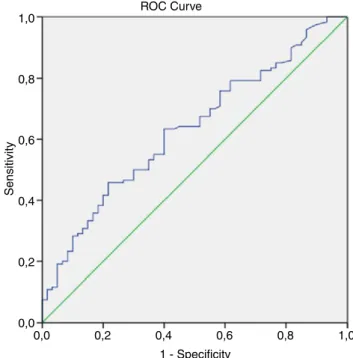
We assessed the diagnostic performance of serum BGN levels and fibrosis stages using a ROC. The AUROC of BGN levels was 0.702
for fibrosis stage, differentiating patients from healthy controls with statistical significance (P<.001). The AUROC of BGN levels was 0.632 for necroinflammatory activity score, differentiating pa-tients from healthy controls with statistical significance (P=.004; Figure 1). The AUROC of BGN levels was 0.524 for F0- F3 vs F4- F6 (Figure 2).
T A B L E 2 Correlation between laboratory studies and stage of fibrosis Fibrosis r P Hb (g/dL) .100 .277 Plt (e3/UL) −.235 .010 AST (U/L) .332 <.001 ALT (U/L) .262 .004 INR −.056 .548 Alb (g/dL) −.59 .432 NIA .406 <.001 Biglycan (pg/mL) .213 .004 HBV DNA copies/ML .320 −.092
Hb, hemoglobin; plt, platelet number; AST, aspartate aminotransferase; ALT, alanine aminotransferase; GGT, gamma- glutamyl transpeptidase; INR, international normalized ratio; NIA, necroinflammatory activity index; SD, standard deviation; r, correlation coefficient.
T A B L E 3 Serum biglycan levels according to the stage of fibrosis and necroinflammatory activity index
Mean±SD Median (min–max)
F0 200.1±90.7 180.7 (95.9- 687.3) F1- 3 243.1±110.7 214.1 (110.3- 869.7) F4- 6 2294.1±2094.4 853.9 (141.0- 4982.0) NIA0 208.1±101.6 186.0 (95.9- 687.3) NIA (1- 4) 208.5±67.2 196.7 (110.1- 428.1) NIA (5- 8) 300.2±183.1 224.3 (110.8- 953.9) NIA (9- 12) 1569.9±2050.6 278.4 (151.2- 4982.0) NIA (13- 16) — —
NIA, necroinflammatory activity index; SD, standard deviation.
F I G U R E 1 Receiver operator curves for necroinflammatory activity ROC Curve Sensitivity 1,0 0,8 0,6 0,4 0,2 0,0 0,0 0,2 0,4 1 - Specificity 0,6 0,8 1,0
Diagonal segments are produced by ties.
F I G U R E 2 Receiver operator curves for having clinical significant fibrosis vs not (F0- F3 vs F4- F6) ROC Curve Sensitivity 1,0 0,8 0,6 0,4 0,2 0,0 0,0 0,2 0,4 1 - Specificity 0,6 0,8 1,0
5 | DISCUSSION
Liver fibrosis is a serious medical condition that occurs as a result of chronic viral hepatitis infection, alcoholic liver disease, non- alcoholic steatohepatitis, drug abuse, and autoimmune hepatitis.11 Liver fibrosis is the final common pathway of most chronic liver diseases, and it is characterized by increasing accumulation of collagen in the matrix result-ing in scar tissue.12 Liver biopsy is the gold standard diagnostic method for grading fibrosis. Nevertheless, it is an invasive procedure and has some disadvantages.13 Therefore, many studies have been performed in order to find out new non- invasive biomarkers of liver fibrosis, and to in-vestigate the roles of these biomarkers to predict liver fibrosis. BGN be-longs to the small leucine- rich family of proteoglycans.9 Decorin, BGN, lumican, and fibromodulin are the key regulators of collagen fibers.14
Aim of this study was to determine whether serum BGN level could be a potential serological marker of pathological extracellular matrix remodeling in chronic HBV infection. Previously, some animal studies have sought the answer to this question; however, ours is the first human study on this subject.
In a study of Leeming et al., C3M, BGM, C4M, P4NP- 7S, ELM were studied in 249 HIV- infected patients and 55 controls. The serum levels of MMP- degraded collagen type III (C3M), biglycan (BGM), elastin (ELM), as well as the formation marker 7S (P4NP- 7S), and MMP- degraded collagen type IV (C4M) were determined using spe-cific ELISAs. Sixty- eight patients underwent a follow- up visit 3 years later including assessment of ECM markers and fibrosis using transient elastography (Fibroscan). These markers were found to be significantly higher in HIV- infected patients than in the control group; and in pa-tients with high viral load compared to those with low viral load. A negative correlation was found between the duration of antiretroviral therapy and the levels of these markers. This study has shown that BGM level measurements are effective in monitoring the hepatic ef-fects of antiretroviral therapy.15
In another study that involved 94 alcoholic cirrhosis patients and 20 control subjects, the levels of type 1 collagen, C3M, PROC3, C6M, P4NP- 7S, C5M, biglycan, and elastin denoted the degree of hepatic dysfunction. A significant correlation was found between these mark-ers and both hepatic venous pressure gradient and portal hyperten-sion. By this study, new non- invasive markers were found to spot clinically important portal hypertension.16
Genovese et al., established a hepatic fibrosis model in rats by em-ploying CCL4 and bile duct ligation. Biglycan that is cleaved by matrix metalloproteinases was found to be correlated with pathological ex-tracellular matrix remodeling. Hence, a significant positive correlation was shown between the degree of hepatic fibrosis and serum biglycan level in an animal study.17
In a study performed by Jia et al., 30 rats were employed. In 20 of them, hepatocellular carcinoma (HCC) model was established by administering N- diethylnitrosamine. Aggrecan (chondroitin sulfate proteoglycan 1), biglycan, and decorin (proteoglycan core protein) lev-els were found to be significantly higher in hepatic tissues of these rats.18 It is not surprising that decorin and biglycan levels were high
in hepatic fibrosis because they are members of the small leucine- rich proteoglycan (SLRP) family.11 Interestingly, in HCC tissue, aggrecan, biglycan, and decorin spread into cytoplasm, pericellular matrix, and cellular membrane in contrast to the control group in which they are found exclusively on hepatocyte membrane and pericellular matrix.19
In another study, decorin, biglycan, proteoglycan 100, and pro-teoglycan colony stimulating factor were studied in both normal and fibrotic human livers. In HBsAg- positive chronic active hepatitis pa-tients, decorin and biglycan showed strong immunoreactivity in fi-brotic areas while they are clearly seen in the space of Disse in normal hepatic tissue.20
In an animal study performed on 52 rats, a hepatic fibrosis model was established by administering CCL4. Biomarkers including type 1 collagen, type 3 collagen, type 4 collagen, type 6 collagen, citrulline, vi-mentin, biglycan, PN3P, P4NP- 7S, and PCP5 were shown to correlate with the degree of hepatic fibrosis induced by CCL4.21
In a study by Nielsen et al., Pro- C3 and C3M levels were measured by ELISA in plasma obtained from CHC patients (n=194) who had been enrolled in a prior phase II antifibrotic trial. Pro- C3 levels were sig-nificantly higher in CHC patients in Ishak stage 4 compared to those in stage 2 (P<.001) or in stage 3 (P<.01). Pro- C3 could significantly distinguish moderate fibrosis (stage 4) from mild fibrosis (stage 2/3) (AUC=0.72, P<.001). Pro- C3 was a useful test to predict fibrogenesis and monitor disease progression. Moreover, it could differentiate mild from moderate disease.22
In this study, we aimed to investigate any association between serum biglycan levels and fibrotic stage and necroinflammatory activ-ity in chronic HBV infection, because there was no previously pub-lished human study to show this association. Serum biglycan levels of 120 chronic hepatitis B patients were compared with 60 control sub-jects. In chronic hepatitis B patients, mean serum biglycan level was found to be significantly higher than that in the control group. When biglycan levels were assessed according to histopathological fibrotic stages in liver biopsy specimens, a positive correlation was shown, and histopathologically determined necroinflammatory activity increased as serum biglycan levels increased.
Significance of level is evaluated with ROC curve. Area under the ROC curve (AUROC), can be between values of 0.5 and 1.0 according to effectiveness. Significance is shown by the levels near 1 in chronic hepatitis B patients; serum biglycan proved to be a promising diagnostic test for the evaluation of hepatic fibrosis (0.702). Serum biglycan level was statistically meaningful to evaluate the presence of fibrosis. It also performed fairly as a diagnostic test to predict the liver necroinflamma-tory activity (0.632) and it was statistically significant also. The aim of our study was to investigate an alternative new screening method, re-garding liver biopsy. In chronic hepatitis B patients, a significant correla-tion was found between serum biglycan level and the stage of fibrosis.
6 | CONCLUSION
As a result, we have shown a correlation between serum biglycan level and fibrotic and necroinflammatory stage in chronic hepatitis
B patients. Nevertheless, it should be noted that this correlation is modest. It was also found that serum biglycan levels of the patients and healthy control group showed statistically significant differences. Validity of this non- invasive method needs to be verified by perform-ing further studies. On the other hand, liver biopsy is still the gold standard diagnostic method in the evaluation of hepatic fibrosis. This non- invasive marker may be beneficial in the circumstances when liver biopsy needs to be repeated or when it is contraindicated.
AUTHORS’ CONTRIBUTIONS
Rafiye Ciftciler was responsible for collecting blood samples, inform-ing volunteers, data collection, performed the majority of experi-ments, and wrote the article; Seren Ozenirler organized research and corrected the article; Aysegul Atak Yucel performed immunological analysis of the samples and corrected the article; Mustafa Cengiz and Erkan Buyukdemirci performed statistical analysis of data; Gulbanu Erkan served as scientific adviser, drafted the article, and revised it critically for important intellectual content; performed statistical anal-ysis of data; Cemile Sönmez performed immunological analanal-ysis of the samples; Guldal Yılmaz Esendaglı evaluated liver tissue samples and determined fibrosis stage.
REFERENCES
1. Schuppan D, Afdal NH. Liver cirrhosis. Lancet. 2008;371:838–851. 2. Maynard JE. Hepatitis B: global importance and need for control.
Vaccine. 1990;8(Suppl):S18.
3. Parkin DM, Bray F, Ferlay J, Pisani P. Global cancer statistics. CA
Cancer J Clin. 2002;55:74–108.
4. Maharaj B, Maharaj RJ, Leary WP, Cooppan RM, Naran AD, Pirie D. Localisation and semiquantitative assessment of hepatic procollagen mRNA in primary biliary cirrhosis. Gut. 1998;43:433–440.
5. Metavir F, Cooperative Study Group. Intraobserver and interob-server variations in liver biopsies in patients with chronic hepatitis C.
Hepatology. 1994;20:15–20.
6. Karsdal MA, Nielsen MJ, Sand JM. Extracellular matrix remodeling: the common denominator in connective tissue diseases possibilities for evaluation and current understanding of the matrix as more than a passive architecture, but a key player in tissue failure. Assay Drug Dev
Technol. 2012;11:70–92.
7. Wiberg C, Hedbom E, Khairullina A, et al. Biglycan and decorin bind close to the n- terminal region of the collagen VI triple helix. J Biol
Chem. 2001;276:18947–18952.
8. Melchior-Becker A, Dai G, Ding Z. Deficiency of biglycan causes car-diac fibroblasts to differentiate into a myofibroblast phenotype. J Biol
Chem. 2011;286:17365–17375.
9. Schaefer L, Babelova A, Kiss E. The matrix component biglycan is proinflammatory and signals through Toll- like receptors 4 and 2 in macrophages. J Clin Invest. 2005;115:2223–2233.
10. Ishak K, Baptista A, Bianchi L, Callea F, De Groote J, Gudai F. Histological grading and staging of chronic hepatitis. J Hepatol. 1995;22:696–699.
11. Friedman SL. Liver fibrosis- from bench to bedside. J Hepatol. 2003;38(Suppl. 1):S38–S53.
12. Liu C, Gaça MD, Swenson ES, Velluci VF, Reiss M, Wells RG. Smads 2 and 3 are differentially activated by transforming growth factor beta in quiescent and activated hepatic stellate cells. Constitutive nuclear localization of smads in activated cells is TGF- beta- independent. J Biol
Chem. 2003;278:11721–11728.
13. Guido M, Rugge M. Liver biopsy sampling in chronic viral hepatitis.
Semin Liver Dis. 2004;24:89–97.
14. Bataller R, Brenner DA. Liver fibrosis. J Clin Invest. 2005;115:209–218. 15. Leeming DJ, Anadol E, Schierwagen R, et al. Combined antiretrovi-ral therapy attenuates hepatic extracellular matrix remodeling in HIV patients assessed by novel protein fingerprint markers. AIDS. 2014;28:2081–2090.
16. Leeming DJ, Karsdal MA, Byrjalsen I, et al. Novel serological neo- epitope markers of extracellular matrix proteins for the detection of portal hypertension. Aliment Pharmacol Ther. 2013;38:1086–1096. 17. Genovese F, Barascuk N, Larsen L, et al. Biglycan fragmentation in pathologies associated with extracellular matrix remodeling by matrix metalloproteinases. Fibrogenesis Tissue Repair. 2013;6:9.
18. Jia X, Li S, Dang S, et al. Increased expression of chondroitin sul-phate proteoglycans in rat hepatocellular carcinoma tissues. World J
Gastroenterol. 2012;18:3962–3976.
19. Meyer DH, Krull N, Dreher KL, Gressner AM. Biglycan and decorin gene expression in normal and fibrotic rat liver: cellular localization and regulatory factors. Hepatology. 1992;16:204–216.
20. Högemann B, Edel G, Schwarz K, Krech R, Kresse H. Expression of biglycan, decorin and proteoglycan- 100/CSF- 1 in normal and fibrotic human liver. Pathol Res Pract. 1997;193:747–751.
21. Leeming DJ, Byrjalsen I, Jiménez W, Christiansen C, Karsdal MA. Protein fingerprinting of the extracellular matrix remodelling in a rat model of liver fibrosis- a serological evaluation. Liver Int. 2013;33:439–447.
22. Nielsen MJ, Veidal SS, Karsdal MA, et al. Plasma Pro- C3 (N- terminal type III collagen propeptide) predicts fibrosis progression in patients with chronic hepatitis C. Liver Int. 2015;35:429–437.