increasing activity of antioxidant enzymes in rat brain
Burak Dik
1*, Devran Coskun
2, Emre Bahcivan
3and Ayşe Er
11
Department of Pharmacology and Toxicology, Veterinary Faculty, Selcuk University, Konya, Turkey 2
Department of Pharmacology and Toxicology, Veterinary Faculty, Siirt University, Siirt, Turkey 3
Department of Pharmacology and Toxicology, Veterinary Faculty, Kafkas University, Kars, Turkey
Abstract: The aim of this study is to determine the effects of alone or combined usage of doxycycline and meloxicam on
brain superoxide dismutase (SOD), catalase (CAT), malondialdehyde (MDA), and matrix metalloproteinase (MMP)-9 levels of lipopolysaccharide (LPS)-induced brain inflammation. Totally 78 rats were divided into 5 groups; Healthy control (n=6), LPS (n=18, 0.05µg/µL/rat, intracranially), LPS+D (n=18, LPS 0.05µg/µL/rat, intracranially and doxycycline 40 mg/kg, intraperitoneally), LPS+M (n=18, LPS 0.05 µg/µL/rat, intracranially and meloxicam 2 mg/kg, intraperitoneally), LPS+Combination (n=18, LPS 0.05 µg/µL/rat, intracranially and simultaneously both drug combination) groups. Animals were euthanized at 1, 3 and 6 hours following injections and the brains were removed. Brain SOD, CAT, MDA and MMP-9 levels were determined by ELISA reader. Parameters of LPS groups generally different from Healthy control group. When compared to LPS group, increased SOD level of LPS+D at 3 hours and CAT levels of LPS+M and LPS+D groups were determined (P<0.05) at 3 and 6 hours, respectively. In addition, all treatments statistically significantly (P<0.05) decreased MMP-9 levels at 6 hours. In conclusion, doxycycline and meloxicam may show antioxidant effect via increasing antioxidant enzyme production in the brain; however combined usage of drugs may show more beneficial effect for neuroinflammation.
Keywords: Antioxidants, doxycycline, meloxicam, neuroinflammation.
INTRODUCTION
Neuroinflammation is important in the healing and progression of nervous system diseases. It is the key for many neurodegenerative diseases (Lima et al., 2012). Neuroinflammatory diseases are modeled on experimental animals for the diagnosis and treatment of neurodegenerative diseases such as Parkinson and Alzheimer's diseases (Amor et al., 2010). Adaptive and innate immune systems in the central nervous system (CNS) limit the tumor and neurotrophic viral infections, bacterial diseases, and regenerate the brain tissue after inflammation. Bacterial and viral infections also play an important role in the development of neuroinflammation. Neuroinflammation is necessary for to promote regeneration and repair (Amor et al., 2014). In the treatment of neuroinflammation, the main target is the reduction of cytokines, matrix metalloproteinase (MMP)-9 levels and oxidative stress (Boulamery et al., 2017). In general, inflammation, oxidative stress and mitochondrial dysfunction are the main cause of neurodegenerative diseases. Free radicals are one of the most important factors that cause death of neurons (Buendia et al., 2016). Reactive oxygen radicals interact with carbonhydrates, proteins, lipids and nucleic acids, leading to cellular dysfunction. The brain tissue is more susceptible to oxidative damage due to its high polyunsaturated fat content, high oxygen consumption,
and inadequate antioxidants (Valko et al., 2007). Astrocytes produce antioxidants and free radical scavengers that protect the brain from oxidative stress. However, different signal molecules, mainly cytokines and neurotransmitters, trigger reactive astrogliosis. If inflammation cannot be controlled with anti-inflammatory agents, activated astrocytes release several inflammatory factors. Increased neuronal inflammatory apoptosis lead to increased oxidative damage markers. As a result, in the development of various neurodegenerative diseases, amyloid deposits and glial scarring occurs when astrocytes lose their function (Agostinho et al., 2010). Superoxide dismutase (SOD), catalase (CAT), glutathione peroxidases and glutathione reductase have antioxidant effects in the body. Hence they may inhibit neuronal inflammation (Mosley et al., 2006). Various anti-inflammatory and antioxidant agents prevent the progression of neuroinflammations with these mechanisms (Agostinho et al., 2010; Valko et al., 2007). MMP-9 releases from microglia, macrophages, and infiltrating neutrophils MMP-9 is activated by the formation of free radicals in neuroinflammation. MMP has a multitude of roles in inflammation and tissue repair. It contributes to the damage of the tissue and the later recovery phases (Yang et al., 2015).
Doxycycline is a broad-spectrum tetracycline antibiotic. The tetracycline group antibiotics have anti-inflammatory effect and they function by decreasing NF-κB activity
(Bastos et al., 2012). Doxycycline inhibits microglial activation by decreasing interleukin-1β (IL-1β) and tumor necrosis factor-α (TNF-α). However, doxycycline may inhibit MMP depending in a dose dependent manner (Jantzie et al., 2010). Doxycycline inhibits neuroinflammatory diseases by modulating lipopolysaccharide (LPS)-induced p38 MAP kinase and NF-κB pathways (Santa-Cecilia et al., 2016). Doxycycline chelates the zinc ion present in the catalytic site of MMPs and inhibits MMP activity. It also has antioxidant properties (Antonio et al., 2014). While it reduces malondialdehyde (MDA) and total oxidative status (TOS) levels, it increases total antioxidant status level (Yagan et al., 2014). Doxycycline antagonizes free radicals, lipid peroxidation and glutamate excitotoxicity and prevents neuronal degeneration (Nogueira et al., 2011).
Meloxicam is a preferential COX-2 inhibitor and protects the brain from damage (Jun-Qing et al., 2006). Oxidative stress in the brain triggers inflammation by COX-2 enzyme. The effect of meloxicam on COX-2 reduces inflammation. Also, this effect can increase the antioxidant effect by reducing oxidative stress (Hakan et
al., 2010). It is recommended as an neuroprotective agent
by reducing lipid peroxidation and increasing endogenous antioxidant enzymes (Goverdhan et al., 2012). Meloxicam inhibits neuronal death by regulating increased MDA and decreased SOD levels in neuroinflammation (Yu et al., 2014). It has been reported that this effect alone is not enough, despite the antioxidant activity of meloxicam. It can change the level of glutathione and superoxide enzymes (Khan et al., 2017). However, oxidative stress and antioxidant balance may vary depending on the duration of meloxicam administration (Khan et al., 2014). In the acute phase of the disease and with the treatment of meloxicam, it can limit the enhanced activity of various MMPs and prevent the progression of inflammatory diseases (de Grauw et
al., 2009).
The aim of this study was to determine the effects of doxycycline and meloxicam treatment on activities of SOD, CAT and levels of MDA and MMP-9 in lipopolysaccharide (LPS)-induced brain inflammation.
MATERIALS AND METHODS
Experimental design
Seventy-eight male Wistar Albino rats (10-12 weeks old, 250-300 g) were housed and allowed free access to food and water. All procedures were approved by Selcuk University Experimental Medical Practice and Research Center Ethic Committee.
Doxycycline (Doksimis 100 ml, Mistav, Ankara, Turkey) and meloxicam (Maxicam X4, Sanovel, Istanbul, Turkey) were supplied. Lyophilized Lipopolysaccharide (LPS,
Escherichia coli 0111: B4, Sigma-Aldrich Chemie, USA)
was diluted with 0.9% saline for experimental neuroinflammation model.
The rats were divided in the following groups for experiment. Control Group (C) (Control, n=6) were used as healthy controls. The healthy control group received 10 µL 0.09 % saline solution, intracranially. LPS Group (n=18) animals received 0.5 µg/rat LPS within 10 µL, intracranially. The microinjection was applied intracranially through a 10 µL Hamilton syringeby stereotaxic surgery (Tsai et al., 2003). The application was performed according to the coordinates (0.8 mm posterior to bregma, 1.5 mm lateral to saggital suture by Hamilton microsyringe). LPS + D Group (n=18) animals received 0.5 µg/rat LPS within 10 µL, intracranially and simultaneously 40 mg/kg doxycycline intraperitoneally. LPS + M Group (n=18) animals received 0.5 µg/rat LPS within 10 µL, intracranially and simultaneously 2 mg/kg meloxicam intraperitoneally. The last, LPS+ Combination Group (n=18) animals received 0.5 µg/rat LPS within 10 µL, intracranially and simultaneously 40 mg/kg doxycycline combination with 2 mg/kg meloxicam intraperitoneally. The rats were anesthetized [20 mg/kg thiopental sodium, intraperitoneally (Pental 1 g; Ulagay, Istanbul, Turkey)] before the drug application and later they were euthanized. The brain tissues were immediately removed at 1, 3 and 6 hours after the last administration in all experiment groups. The brain tissues were separately put on ice.
Analysis of parameters
The brain of rats were homogenized in PBS and activities of SOD (Superoxide Dismutase Assay Kit, Item No. 706002, Cayman Chemical Company, USA), CAT (Catalase Assay Kit, Item No. 707002, Cayman Chemical Company, USA) and levels of MDA (Malondialdehyde ELISA kit, Catalog No: E-EL-0060, Elabscience Biotechnology Co. Ltd., China), MMP-9 (Rat matrix metalloproteinase 9 ELISA kit, Catalog No: YLA0585RA, YL Biotech Co., Ltd. China) were measured by ELISA reader (Bio-Tek Instruments Inc., MWGt Lambda Scan 200).
STATISTICAL ANALYSIS
All data obtained from brain tissues are defined as mean ± standard error of the mean (SEM). The data were analyzed using ANOVA and Duncan test as a post hoc test (SPSS 22.0). In all parameters, p < 0.05 was the criterion for statistical significance
RESULTS
The effects of doxycycline, meloxicam and combination of both on levels of MDA, MMP-9 and activities of SOD, CAT at different times of neuroinflammation were presented in figs. 1, 2, 3 and 4.
While MDA levels didn’t change in any groups, SOD activity of LPS+D was statistically significantly higher from LPS+ Combination and Control groups at 1 hour. Additionally, SOD activity of LPS+ Combination group was statistically significant higher than Control group at 1 hour. The SOD activity of LPS+D group was statistically higher than all experiment group and LPS+M and LPS+ Combination groups were statistically significant higher than Control group at 3 hours. SOD activities of LPS+D and LPS+M groups were statistically higher than Control group.
Fig. 1: MDA levels of the brain at different sampling
times (mean ± SEM) No statistical difference was found (P > 0.05).
Fig. 2: SOD activity of the brain at different sampling
times (mean ± SEM) Different letters are statistically significant (P<0.05).
Higher CAT activities were determined in LPS+M and LPS+ Combination groups than Control group at 1 and 6 hours, whereas CAT activity of LPS+M group was higher than all groups except LPS+ Combination group at 3 hours (p<0.05). CAT activity of LPS+D group was higher than in LPS and Control groups at 6 hours.
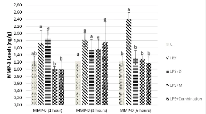
MMP-9 levels statistically significantly decreased in LPS+M and LPS+ Combination groups when compared to LPS group at 1 hour, while its level was highest in LPS group from all other groups at 6 hours (p<0.05).
DISCUSSION
Many neuroinflammatory diseases have proved to be the underlying cause of oxidative stress associated with
inflammation (Gustaw-Rothenberg et al., 2010; Negi et
al., 2011). Although MDA is considered a potential
biomarker for neuroinflammatory diseases such as dementia and Alzheimer (Gustaw-Rothenberg et al., 2010), some authors think that this situation is controversial (Sekler et al., 2008). Although there is no difference at the MDA level in some periods of the disease (Sekler et al., 2008; Zafrilla et al., 2006), there is a difference in the advance phase. Researchers have reported different neurotoxic reactions and oxidative responses to aldehydes in different regions of the brain such as the frontal and occipital lobes (Zafrilla et al., 2006). In the present study, MDA level was not significantly different among the groups at any sampling times (fig. 1), and this result may be derived from sampling time.
Fig. 3: CAT activity of the brain at different sampling
times (mean ± SEM) Different letters are statistically significant (P<0.05).
Fig. 4: MMP-9 levels of the brain at different sampling
times (mean ± SEM) Different letters are statistically significant (P<0.05).
SOD is the first protective defensive line against oxidative stress. Increased SOD level in neurons is related to various oxidative stress-associated neurodegenerative and neuroinflammatory diseases (Schreibelt et al., 2007). Oxidized and misfolded SOD causes mitochondrial dysfunction and disease progression in neuronal diseases (Shvil et al., 2018). In the current study, the SOD level increased with neuroinflammation in the first 3 hours but the endogenous SOD increases in the neuroinflammatory (LPS) group was not enough and decreased at 3 hours. On the contrary, it increased at 6 hours in the treatment
groups and the LPS + D group at 3 hours were statistically higher than the LPS group (fig. 2). In the present study, doxycycline may have prevented oxidative stress via inhibiting SOD degradation and protected against apoptosis from oxidative stress or inflammation (Rhieu et al., 2014), hence this effect increases the activity of SOD as microglia and astrocytes cells protect against apoptosis. Although meloxicam does not pass enough amounts to the brain. Nanoparticle forms of meloxicam may pass to the brain and show anti-inflammatory and antioxidant effects via inhibiting COX-2 (Ianiski et al., COX-201COX-2). In the current study, insufficient antioxidant effects of meloxicam may not have passed enough to the brain.
CAT activity decreases in systemic LPS induced neuroinflammation. However, brain function can improve by reducing inflammation and triggering antioxidant enzymes (Salmani et al., 2018). Increased catalase activity protects against oxidative stress and neuroinflammation (Godinho et al., 2018). In the neuroinflammation induced by LPS for 1 week, the oxidative stress occurred and the level and activity of antioxidant enzymes such as catalase decreased (Khajevand-Khazaei et al., 2018). In the present study, the increase in CAT activity of the LPS-administered group may be an antioxidant response against inflammation (fig. 3). The level and activity of antioxidant enzymes may not have decreased because endogenous antioxidant enzymes do not cause exhaustion due to LPS dose. Doxycycline is thought to be effective in the treatment of neuropsychiatric disorders such as schizophrenia by increasing SOD and CAT activity (Ben-Azu et al., 2018). Meloxicam does not cause a change in CAT activity in human erythrocytes (Burak Cimen et al., 2003), while it induces the activity in the renal injury (Hassan et al., 2014). The findings of the present study showed that the antioxidant effects (CAT activity) of doxycycline at 6 hours and meloxicam at 3 hours can prevent neurodegenerative diseases by reducing oxidative and inflammatory damage.
LPS induce oxidative stress, matrix metalloproteinase-9 (MMP-9), cytokines and chemokines by damaging microglia cells (Lee et al., 2015; Mayer et al., 2016). Excessive increase in MMP level accompanies pathological, neuroinflammatory brain disorders (Yang et
al., 2017). The reduction of the MMP-9 level modulates
neuroinflammation and it plays a neuroprotective role (Lee et al., 2015). Doxycycline inhibits cellular damage by suppressing MMP-9 in ischemia and neuronal damage, at the same time it repairs neurovascular matrix degradation, hemorrhage, edema, blood-brain barrier breakdown and neuroinflammation (Lee et al., 2009). Meloxicam diminishes the level of MMP-9 by inhibiting the enzyme COX-2. It has also important role for extracellular matrix remodeling (Kim et al., 2008). In the
present study, MMP-9 level was induced by LPS administration. Especially at the beginning of neuronal damage, the level of MMP-9 increased later. COX-2 inhibitor meloxicam treatment, early MMP-9 levels may have regulated the healing process by decreasing MMP-9 levels in the early stages. In the later stages of neuroinflammation, anti-inflammatory and antioxidant doxycycline may have inhibited by neuronal degeneration and MMP-9 expression.
CONCLUSION
The combination of doxycycline and meloxicam are not significantly different when applied alone on oxidative status. However, the combination of drugs may be effective for anti-inflammatory purposes and their different doses could be suitable for the treatment of neuronal diseases.
ACKNOWLEDGMENTS
The abstract of the study was presented at the 14th International Congress of the European Association for Veterinary Pharmacology and Toxicology (EAVPT), in Wroclaw, Poland.
REFERENCES
Agostinho P, Cunha RA and Oliveira C (2010). Neuroinflammation, oxidative stress and the Pathogenesis of Alzheimer's disease. Curr. Pharm.
Design, 16(25): 2766-2778.
Amor S, Peferoen LA, Vogel DY, Breur M, van der Valk P, Baker D and van Noort JM (2014). Inflammation in neurodegenerative diseases: An update. Immunology,
142(2): 151-166.
Amor S, Puentes F, Baker D and van der Valk P (2010). Inflammation in neurodegenerative diseases.
Immunology, 129(2): 154-169.
Antonio RC, Ceron CS, Rizzi E, Coelho EB, Tanus-Santos JE and Gerlach RF (2014). Antioxidant effect of doxycycline decreases MMP activity and blood pressure in SHR. Mol Cell Biochem, 386(1-2): 99-105. Bastos LF, de Oliveira AC, Watkins LR, Moraes MF and
Coelho MM (2012). Tetracyclines and pain. Naunyn
Schmiedebergs Arch Pharmacol., 385(3): 225-241.
Ben-Azu B, Omogbiya IA, Aderibigbe AO, Umukoro S, Ajayi AM and Iwalewa EO (2018). Doxycycline prevents and reverses schizophrenic-like behaviors induced by ketamine in mice via modulation of oxidative, nitrergic and cholinergic pathways. Brain
Res. Bull., 139: 114-124.
Boulamery A and Desplat-Jego S (2017). Regulation of neuroinflammation: What role for the tumor necrosis factor-like weak inducer of apoptosis/Fn14 Pathway?
Front Immunol, 8: 1534.
and Leon R (2016). Nrf2-ARE pathway: An emerging target against oxidative stress and neuroinflammation in neurodegenerative diseases. Pharmacol Ther, 157: 84-104.
Burak Cimen MY, Cimen OB, Eskandari G, Sahin G, Erdogan C and Atik U (2003). In vivo effects of meloxicam, celecoxib and ibuprofen on free radical metabolism in human erythrocytes. Drug Chem
Toxicol., 26(3): 169-176.
de Grauw JC, van de Lest CH, Brama PA, Rambags BP and van Weeren PR (2009). In vivo effects of meloxicam on inflammatory mediators, MMP activity and cartilage biomarkers in equine joints with acute synovitis. Equine Vet. J., 41(7): 693-699.
Godinho J, de Sa-Nakanishi AB, Moreira LS, de Oliveira RMW, Huzita CH, Mello JCP, da Silva AOF, Nakamura CV, Previdelli IS, Ribeiro M and Milani H (2018). Ethyl-acetate fraction of Trichilia catigua protects against oxidative stress and neuroinflammation after cerebral ischemia/reperfusion. J. Ethno-pharmacol., 221: 109-118.
Goverdhan P, Sravanthi A and Mamatha T (2012). Neuroprotective effects of meloxicam and selegiline in scopolamine-induced cognitive impairment and oxidative stress. Int. J. Alzheimers Dis., 2012: 974013. Gustaw-Rothenberg K, Kowalczuk K and
Stryjecka-Zimmer M (2010). Lipids' peroxidation markers in Alzheimer's disease and vascular dementia. Geriatr
Gerontol Int., 10(2): 161-166.
Hakan T, Toklu HZ, Biber N, Ozevren H, Solakoglu S, Demirturk P and Aker FV (2010). Effect of COX-2 inhibitor meloxicam against traumatic brain injury-induced biochemical, histopathological changes and blood-brain barrier permeability. Neurol Res., 32(6): 629-635.
Hassan MH, Ghobara M and Abd-Allah GM (2014). Modulator effects of meloxicam against doxorubicin-induced nephrotoxicity in mice. J. Biochem. Mol.
Toxicol., 28(8): 337-346.
Ianiski FR, Alves CB, Souza AC, Pinton S, Roman SS, Rhoden CR, Alves MP and Luchese C (2012). Protective effect of meloxicam-loaded nanocapsules against amyloid-beta peptide-induced damage in mice.
Behav. Brain Res., 230(1): 100-107.
Jantzie LL and Todd KG (2010). Doxycycline inhibits proinflammatory cytokines but not acute cerebral cytogenesis after hypoxia-ischemia in neonatal rats. J
Psychiatry Neurosci., 35(1): 20-32.
Jun-Qing Y, Bei-Zhong L, Bai-Cheng H and Qi-Qin Z (2006). Protective effects of meloxicam on aluminum overload-induced cerebral damage in mice. Eur. J.
Pharmacol., 547(1-3): 52-58.
Khajevand-Khazaei MR, Ziaee P, Motevalizadeh SA, Rohani M, Afshin-Majd S, Baluchnejadmojarad T and Roghani M (2018). Naringenin ameliorates learning and memory impairment following systemic lipopolysaccharide challenge in the rat. Eur. J.
Pharmacol., 826: 114-122.
Khan AM and Rampal S (2014). Effects of repeated oral administration of pazufloxacin mesylate and meloxicam on the antioxidant status in rabbits. J. Am.
Assoc. Lab. Anim. Sci., 53(4): 399-403.
Khan AM, Rampal S and Sood NK (2017). Effect of repeated oral administration of levofloxacin, enrofloxacin, and meloxicam on antioxidant parameters and lipid peroxidation in rabbits. Hum. Exp.
Toxicol., 36(1): 42-50.
Kim SM, Park KC, Kim HG and Han SJ (2008). Effect of selective cyclooxygenase-2 inhibitor meloxicam on liver fibrosis in rats with ligated common bile ducts.
Hepatol. Res., 38(8): 800-809.
Lee EJ, Ko HM, Jeong YH, Park EM and Kim HS (2015). beta-Lapachone suppresses neuroinflammation by modulating the expression of cytokines and matrix metalloproteinases in activated microglia. J. Neuroinflammation, 12: 133.
Lee H, Park JW, Kim SP, Lo EH and Lee SR (2009). Doxycycline inhibits matrix metalloproteinase-9 and laminin degradation after transient global cerebral ischemia. Neurobiol Dis., 34(2): 189-198.
Lima IV, Bastos LF, Limborco-Filho M, Fiebich BL and de Oliveira AC (2012). Role of prostaglandins in neuroinflammatory and neurodegenerative diseases.
Mediators Inflamm, 2012: 946813.
Mayer AM, Murphy J, MacAdam D, Osterbauer C, Baseer I, Hall ML, Feher D and Williams P (2016). Classical and Alternative Activation of Cyanobacterium Oscillatoria sp. Lipopolysaccharide-Treated Rat Microglia in vitro. Toxicol Sci., 149(2): 484-495.
Mosley RL, Benner EJ, Kadiu I, Thomas M, Boska MD, Hasan K, Laurie C and Gendelman HE (2006). Neuroinflammation, oxidative stress and the pathogenesis of Parkinson's disease. Clin. Neurosci.
Res., 6(5): 261-281.
Negi G, Kumar A and Sharma SS (2011). Melatonin modulates neuroinflammation and oxidative stress in experimental diabetic neuropathy: Effects on NF-kappaB and Nrf2 cascades. J. Pineal. Res., 50(2): 124-131.
Nogueira CR, Damasceno FM, de Aquino-Neto MR, de Andrade GM, Fontenele JB, de Medeiros TA and Viana GS (2011). Doxycycline protects against pilocarpine-induced convulsions in rats, through its antioxidant effect and modulation of brain amino acids.
Pharmacol. Biochem. Behav., 98(4): 525-532.
Rhieu BH, Shinde A, Epperly MW, Dixon T, Wang H, Chaillet R and Greenberger JS (2014). Organ-specific responses of total body irradiated doxycycline-inducible manganese superoxide dismutase Tet/Tet mice. In vivo., 28(6): 1033-1043.
Salmani H, Hosseini M, Beheshti F, Baghcheghi Y, Sadeghnia HR, Soukhtanloo M, Shafei MN and Khazaei M (2018). Angiotensin receptor blocker,
losartan ameliorates neuroinflammation and behavioral consequences of lipopolysaccharide injection. Life Sci,
203: 161-170.
Santa-Cecilia FV, Socias B, Ouidja MO, Sepulveda-Diaz JE, Acuna L, Silva RL, Michel PP, Del-Bel E, Cunha TM and Raisman-Vozari R (2016). Doxycycline suppresses microglial activation by inhibiting the p38 MAPK and NF-kB signaling pathways. Neurotox Res,
29(4): 447-459.
Schreibelt G, van Horssen J, van Rossum S, Dijkstra CD, Drukarch B and de Vries HE (2007). Therapeutic potential and biological role of endogenous antioxidant enzymes in multiple sclerosis pathology. Brain Res.
Rev., 56(2): 322-330.
Sekler A, Jimenez JM, Rojo L, Pastene E, Fuentes P, Slachevsky A and Maccioni RB (2008). Cognitive impairment and Alzheimer's disease: Links with oxidative stress and cholesterol metabolism.
Neuropsychiatr. Dis. Treat., 4(4): 715-722.
Shvil N, Banerjee V, Zoltsman G, Shani T, Kahn J, Abu-Hamad S, Papo N, Engel S, Bernhagen J and Israelson A (2018). MIF inhibits the formation and toxicity of misfolded SOD1 amyloid aggregates: implications for familial ALS. Cell Death Dis., 9(2): 107.
Tsai SM, Lin MT, Wang JJ and Huang WT (2003). Pyrogens enhance β-endorphin release in hypothalamus and trigger fever that can be attenuated by buprenorphine. J. of Pharm. Sci., 93(2): 155-162.
Valko M, Leibfritz D, Moncol J, Cronin MT, Mazur M and Telser J (2007). Free radicals and antioxidants in normal physiological functions and human disease. Int.
J. Biochem. Cell Biol., 39(1): 44-84.
Yagan A, Kesim S and Liman N (2014). Effect of low-dose doxycycline on serum oxidative status, gingival antioxidant levels, and alveolar bone loss in experimental periodontitis in rats. J. Periodontol.,
85(3): 478-489.
Yang CC, Hsiao LD, Yang CM and Lin CC (2017). Thrombin Enhanced Matrix Metalloproteinase-9 Expression and Migration of SK-N-SH Cells via PAR-1, c-Src, PYK2, EGFR, Erk1/2 and AP-1. Mol
Neurobiol., 54(5): 3476-3491.
Yang Y and Rosenberg GA (2015). Matrix metalloproteinases as therapeutic targets for stroke.
Brain. Res., 1623: 30-38.
Yu L, Jiang R, Su Q, Yu H and Yang J (2014). Hippocampal neuronal metal ion imbalance related oxidative stress in a rat model of chronic aluminum exposure and neuroprotection of meloxicam. Behav.
Brain Funct, 10: 6.
Zafrilla P, Mulero J, Xandri JM, Santo E, Caravaca G and Morillas JM (2006). Oxidative stress in Alzheimer patients in different stages of the disease. Curr. Med.