Black Sea Journal of Health Science
doi: 10.19127/bshealthscience.844062
BSJ Health Sci / Feyza SÖNMEZ TOPCU et al.
124
This work is licensed under Creative Commons Attribution 4.0 International LicenseOpen Access Journal
e-ISSN: 2619 – 9041
COVID-19 AND STROKE
Feyza SÖNMEZ TOPCU
1*, Şirin YURTLU TEMEL
2, Yıldıray TUTPINAR
11İstinye University Bahçeşehir Liv Hospital, Department of Radiology, 34510, Esenyurt, İstanbul Turkey 2İstinye University Bahçeşehir Liv Hospital, Department of Chest Disease, 34510, Esenyurt, İstanbul Turkey
Abstract: Most patients with COVID-19 present with constitutional and respiratory symptoms and some with atypical gastrointestinal,
cardiovascular, or neurological manifestations. Recent studies suggest that there are neurologic manifestations of COVID-19, including acute cerebrovascular disease (CVD). The aim of this study is to find out any evidence of COVID-19 related stroke. Radiologic studies of the patients admitted to the emergency department (ED) of our center from March 11 Th to June 10 Th 2020, with acute stroke symptoms and of whom the acute cerebrovascular disease is confirmed by Computed Tomography (CT) and/or Magnetic Resonance Imaging (MRI) are searched retrospectively. CT Angiography (CTA) and MR Angiography (MRA) studies obtained for stroke management, searched for acute thromboembolism. We noticed some radiologic evidence of acute cerebrovascular disease in 56 patients of 528 patients admitted with immediate neurological symptoms. 11 (19.64 %) of these patients who were not diagnosed before, proved to be simultaneous COVID-19 infection with laboratory tests and/or thorax CT. It was noteworthy that these 11 patients presented with an acute cerebrovascular event supported by neurological and radiological findings instead of the well-known constitutional or respiratory symptoms of COVID 19 infection. 45 (80.35 %) patients were negative for COVID-19 infection. CT /or MR Angiography demonstrated carotid or intracranial major arterial thromboembolism in 5 (11.1 %) of the non-COVID-19 patients and 5 (45.4 %) of the simultaneous COVID-19 disease diagnosed ones. COVID19 positive 5 patients presented with acute internal carotid artery (ICA) or major ICA branch thrombosis at the first stage of COVID-19 infection, rather than a complication of the serious lung disease or a component of multiorgan disfunction related COVID-19. Acute cerebrovascular disease symptoms bringing patients to the ED instead of the respiratory symptoms, aroused high suspicion of the direct neuropathy and early coagulopathy effect of the virus. COVID- 19 disease, itself thought to be a great risk factor for stroke alone. Even in initial cases and in cases where COVID-19 infection do not show a severe and fatal course, stimulation in the coagulation cascade in the early stages, increased the risk of acute stroke.
Keywords: COVID- 19, SARS Cov-2, Acute cerebrovascular disease, Infection related stroke
*Corresponding author: İstinye University Bahçeşehir Liv Hospital, Department of Radiology, 34510, Esenyurt, İstanbul Turkey E mail: [email protected] (F. SÖNMEZ TOPCU)
Feyza SÖNMEZ TOPCU https://orcid.org/0000-0002-7450-2949 Received: December 23, 2020 Accepted: January 31, 2021 Published: May 01, 2021
Şirin YURTLU TEMEL https://orcid.org/0000-0003-3081-4577 Yıldıray TUTPINAR https://orcid.org/0000-0001-9445-6604
Cite as: Sönmez Topcu F, Yurtlu Temel Ş, Tutpinar Y. 2021. COVID-19 and stroke. BSJ Health Sci, 4(2): 124-128.
1. Introduction
Corona Virus Disease 2019 (COVID-19) caused by the novel severe acute respiratory syndrome coronavirus 2 (SARS-CoV-2) has become a worldwide pandemic since the first reports identified in December 2019 Wuhan, Hubei Province, China. On March 11 2020, the World Health Organization (WHO) declared the infection of SARS-CoV-2 as a pandemic (Phelan et al., 2020; Cucinotta and Vanelli, 2020). Symptoms of SARS-Cov2 infection range from asymptomatic disease to life-threatening acute respiratory distress syndrome (ARDS), severe pneumonia, acute kidney injury, myocarditis, multi-organ failure and death (Huang et al., 2020).
There is a growing body of published evidence that complications of COVID-19 include a wide range of neurologic manifestations, such as acute cerebral infarcts (Mao et al., 2020; Helms et al.,2020). However, multiple reports have raised concerns about the virus’ tendency to invade the central nervous system (CNS). Evidence of cerebrovascular complications associated with SARS-CoV-2 is limited, but previous reports from the SARS
epidemic in Asia in 2003 suggested a higher incidence of
thromboembolic complications, including stroke
(Umapathi et al., 2004).
The aim of our study is to answer questions 1) Is SARS-Cov2 a neuropathic virus same like the previous coronaviruses caused SARS and MERS epidemic? 2) Can SARS-Cov2 cause direct coagulopathy effect on CNS vessels without serious lung disease and multiorgan dysfunction?
2. Material and Methods
We searched the hospital (İstinye University Bahçeşehir Liv Hospital) database of three months of pandemic disease from March 11 Th to June 10 Th, for patients admitted to emergency department who presented with acute stroke symptoms. The laboratory test results, thorax CT images, cranial MR and/or CT images and carotid/cerebral CTA or MRA of the individuals reviewed from the database retrospectively.
All radiological images were evaluated independently by two radiologists and patients with consensus were
Research Article
BSJ Health Sci / Feyza SÖNMEZ TOPCU et al.
125
included in the study. Patients admitted to the ED with radiological evidences of acute cerebrovascular disease and concomitant features of COVID-19 infection included. Patients with acute cerebrovascular disease as a component of previously diagnosed COVID-19 infection related multiorgan dysfunction excluded from the study. Demographic data (age, gender) noted. With respect to the common risk factors for stroke, the coexistence of hypertension, diabetes mellitus, hypercholesterolemia, coronary heart disease and smoking had been investigated for all patients at admission and the data of these risk factors were collected from records. Clinical course of all individuals noted.
Patients were imaged at 1.5 T Philips Ingenia MRI system equipped with a head coil (Philips dS headneck coil 8 channel), for angiography studies a total amount of 14 ml Dotarem ® 0.5 mmol/ml (Gadoteric acid, Guerbet BP 57400, 95943 Roissy, CdG Cedex, France) admitted with 2 ml/ sec.
CT images performed at Philips Ingenuity 128 multi-slice CT. CTA contrast agent Kopaq ® 300mg I/ml (Ioheksol, Pharmavision San. ve Tic. A.Ş. Davutpaşa Cad. No: 145 34010 Topkapı/İstanbul Turkey) admitted totally 50 ml with 4ml/sec. Precautions (oxygen support unit, IV adrenalin, corticosteroids and antihistaminic agents) were already prepared before contrast media admitted MRA and CTA.
2.1. Ethical Consideration
All procedures were performed in accordance with the institutional ethics committee and the Declaration of Helsinki. İstinye University Ethics Committee has approved the study with the number 2017-KAEK-120 /2/2020.G-069.
3. Results
In our center, from March 11 th to June 10 th 2020, 526 patients admitted with symptoms suspected for stroke and 56 of them diagnosed as acute cerebrovascular disease proven with cerebral MRI or CT. Acute neurologic symptoms of the patients were altered mental status, slurred speech or hemiparesis. 11 (19.64%) of the 56 stroke patients were also with suspicious clinical and chest imaging findings for COVID-19 infection. As a result of the physical and laboratory examinations performed at the time of admission to the hospital, it was understood that these patients were also infected with SARS CoV-2 and were unaware of this situation. The reason that brought these 11 patients to the hospital was acute neurological findings rather than well-known respiratory symptoms for COVID- 19 infection. 45 (80.35%) of the radiologically proven stroke patients were negative for COVID-19 infection.
Focusing on these COVID-19 infected 11 patients, Polymerase Chain Reaction test (PCR) from nasal swap results were positive for 8, viral antibody IgM was positive in blood analysis for 1, IgG positive for 1 and one patient was negative for PCR but CT imaging and clinical manifestation were significant for the coronavirus
infection. The common risk factors of stroke for two group summarized in Table 1.
Table1. Common risk factors for stroke in COVID-19
positive and negative group
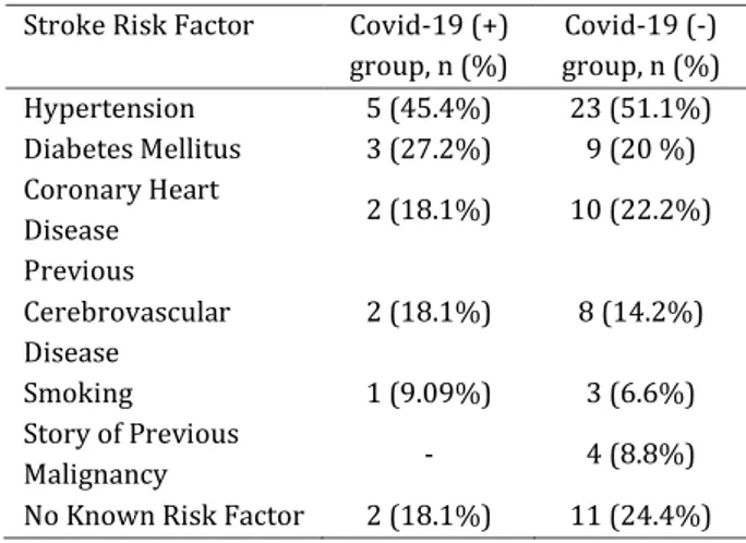
Stroke Risk Factor Covid-19 (+)
group, n (%) Covid-19 (-) group, n (%) Hypertension 5 (45.4%) 23 (51.1%) Diabetes Mellitus 3 (27.2%) 9 (20 %) Coronary Heart Disease 2 (18.1%) 10 (22.2%) Previous Cerebrovascular Disease 2 (18.1%) 8 (14.2%) Smoking 1 (9.09%) 3 (6.6%) Story of Previous Malignancy - 4 (8.8%)
No Known Risk Factor 2 (18.1%) 11 (24.4%)
Chest CT findings of 9 COVID-19 positive patients were pathognomonic with ground glass opacities of whom 2 with bilateral, 7 with unilateral lung involvement. 2 patients’ chest CT images were not typical for COVID-19 pneumonia. Chest CT findings of Covid-19 positive patients summarized in Table 2.
Table 2. Chest CT findings of 9 COVID-19 positive
patients
Chest CT Findings Number of
patients
%
Unilateral focal ground glass 3 27.27
Bilateral multiple ground glass 4 36.36
Consolidation 2 18.18
No specific finding 2 18.18
Total 11 99.9
We did not find a specific infarct pattern in the brain radiological images of COVID-19 positive patients. In the COVID-19 positive stroke group, all the patients had ischemic infarcts while none of them had hemorrhage. There was no evidence of specific area infarct pattern. Acute infarct findings observed in patients' CT and MRIs’ are as summarized in the Table 3.
Table 3. Cranial CT/MR findings of patients
Infarct patterns COVID-19 (+)
group n (%)
COVID-19 (-) group
n (%)
Single millimetric focus 2 (18.1%) 11 (24.4%)
Multiple millimetric
focus 5 (45.4%) 15 (33.3%)
Territorial 3 (27.2%) 17 (37.7%)
Cortical watershed zone 1 (9.09%) 1 (2.2 %)
Hemorrhagic - 1 (2.2 %)
BSJ Health Sci / Feyza SÖNMEZ TOPCU et al.
126
Overall, 52 stroke patients also underwent contrast enhanced carotid CT or MRI angiography for diagnostic purposes. 10 patients belonged to COVID-19 positive stroke group and 42 patients belonged to non-COVID-19 stroke group. Contrast enhanced CT or MR Angiography results of COVID-19 positive stroke group are summarized in Table 4.
Table 4. CTA / MRA findings of COVID-19 positive stroke
group
CTA/MRA findings Number of
patients
Percentage Total internal carotid
artery (ICA) occlusion 4 36.36
Total middle cerebral
artery (MCA) occlusion 1 9.09
Non-stenotic atheroma
plaque 1 9.09
Normal CTA/MRA 4 36.36
No CTA/MRA, normal vascular signal void in MRI
1 9.09
Total 11 100
Acute internal carotid artery (ICA) or major ICA branch thrombus was proven in totally 10 (19.23%) patients with CTA/MRA. Of these 10 patients, 5 were COVID-19 positive and 5 were not. In summary, acute ICA or major ICA branch thrombus was proven in 5 (10.6%) of 42 19 negative patients and 5 (45.4%) of 10 COVID-19 positive patients.
When we compared the radiologically proven ICA or major branch thromboembolism rate in COVID-19 positive patients with the rate of thromboembolism in negative patients, we found that COVID-19 positive patients had a remarkably higher rate. We used the Pearson chi-square test to answer the question of whether we can establish a significant statistical connection between COVID-19 infection and the development of carotid artery thromboembolism. Depending on the Pearson Chi-Square test, calculated P value: 0.0076 (P<0.05) which showed a significant relation between COVID-19 infection and carotid thrombus.
Some imaging samples of patients are shown in the following, Figures 1 to 3.
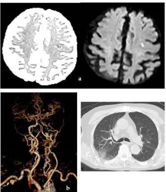
Figure 1. 83 years male, application with slurred speech.
PCR test of the nasal swap is positive. a) ADC map and Diffusion weighted MRI show millimeter-sized cortical acute infarct in the left frontal lobe, b) 3D Reformation of Contrast admitted CT angiography shows total occlusion of Left ICA from the origin, c) Chest CT image demonstrate localized ground-glass opacities in the upper lobe posterior of the right lung.
Figure 2. 46 years male, application with right
hemiparesis. PCR test of the nasal swap is positive. a) CT perfusion image, acute infarct in the left MCA territory, b) Chest CT, bilateral parenchymal consolidation.
BSJ Health Sci / Feyza SÖNMEZ TOPCU et al.
127
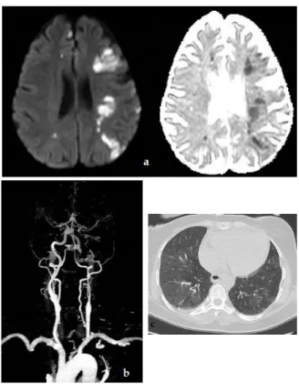
Figure 3. 55 years female, application with speech loss,
PCR test is positive. is shown in CTA. a) DWI and ADC images shows bilateral acute infarcts, b) CTA image shows left total ICA occlusion from the origin, c) Chest CT with non-specific findings.
4. Discussion
In a case study with 214 patients with confirmed COVID-19 infections, 36.4% showed neurological symptoms such as dizziness, headache and confusion, with 5.7% of these patients having cerebrovascular disease, including ischemic stroke and intracerebral hemorrhage (Mao et al., 2020).
In a study by Li et al. (2020) of 219 patients with COVID-19, 10 (4.6%) developed acute ischemic stroke and 1 (0.5%) had intracerebral hemorrhage. COVID-19 with new onset of CVD were significantly older, and were more likely to have cardiovascular risk factors, including hypertension, diabetes and medical history of CVD. Average time of onset of stroke after COVID-19 diagnosis was 12 days.
In our study, 11 patients did not have any diagnose of COVID-19 infection before acute cerebrovascular disease presentation. The initial symptom was stroke and with
suspicious symptoms, COVID-19 infection was
investigated and proven with nasal swap PCR/ blood antibody test or thorax CT. Cerebrovascular disease was not a component of COVID-19 related multiple organ failure. Recently Avula et al. (2020) presented four cases with a cerebrovascular disease in early stages of their illness 19. Similar to this study, we believe COVID-19 increases the risk of CVD.
A number of potential mechanisms by which COVID-19 might increase stroke risk have been identified in the literature. These include hypercoagulability as evidenced
by raised D-dimer levels (Mao et al., 2020), exaggerated systemic inflammation or a “cytokine storm” (Mehta et al., 2020) and cardio embolism from virus-related cardiac injury (Akhmerov and Marban, 2020). Direct viral invasion of the nervous system could also contribute and there may be direct cerebral effects, such as acute hemorrhagic necrotizing encephalopathy (Poyiadji et al., 2020).
Most coronavirus are neurotropic, similarly few studies speculate that SARS-CoV-2 is also neurotropic (Steardo et al., 2020). Furthermore, there are reports of SARS-CoV-2 being identified in cerebrospinal fluid by PCR (Moriguchi et al., 2020).
Angiotensin converting enzyme-2 (ACE) receptors presenting on the nervous system might be the main entry points for SARS-Cov-2 (Steardo et al., 2020) ACE 2 receptors are found not only in the alveolar epithelial cells of the lungs but also in the vascular endothelium. (Poyiadji et al., 2020) Therefore, patients with severe COVID-19 may be at risk of thrombogenesis and cerebral ischemia due to both biochemical hypercoagulable states and direct vascular endothelial injury.
Elevated levels of CRP and D-dimer-a product of fibrin clot degradation, indicating a high inflammatory state and abnormalities with the coagulation cascade, respectively, might play a role in the pathophysiology of stroke in the setting of COVID-19 infection (Li et al., 2020). In published reports on SARS-CoV-2, a cytokine storm has been postulated, (Phelan et al., 2020; Steardo et al., 2020) which could result in cerebrovascular disease. Furthermore, severe cases of covid-19 have shown elevated D-dimer levels and thrombocytopenia, rendering patients prone to cerebrovascular events, both thrombotic and hemorrhagic (Moriguchi et al., 2020; Zhou et al., 2020).
Another study looking at activated partial
thromboplastin time-based clot waveform analysis (CWA) in COVID-19 patients concluded that CWA
parameters demonstrate hypercoagulability that
precedes or coincides with severe illness Multiple reports of pulmonary embolism are currently available in the literature (Ge et al., 2020; Xia et al., 2020).
9 of our COVID-19 positive CVD patients had relatively mild respiratory symptoms without progression to ARDS or severe disease consequent to multiorgan dysfunction. 3 patients were completely healthy before the situation without any known risk for stroke.
Internal carotid artery or major ICA branch thrombus was found in 5 (10.6%) of COVID-19 negative patients and 5 (45.4%) of COVID-19 positive patients with cerebrovascular disease. Depending on the Pearson Chi-Square test, calculated P value: 0.0076 (P<0.05), showed a significant relation between COVID-19 infection and carotid thrombus. The restriction of our study is the small number of total and COVID-19 positive patients. This can be explained with the overall reduction of stroke patients during the pandemic. The number of people evaluated for signs of stroke at U.S. hospitals has dropped
BSJ Health Sci / Feyza SÖNMEZ TOPCU et al.
128
by nearly 40% during the COVID-19 pandemic, according to a study led by researchers from Washington University School of Medicine in St. Louis who analyzed stroke evaluations at more than 800 hospitals across 49 states and the District of Columbia. (Kansagra et al., 2020).
The pathophysiology behind the cerebrovascular accidents is still to be determined. (Akhmerov et al., 2020; Moriguchi et al., 2020). The pathologic mechanism of stroke may be related to viruses attached to ACE-2 receptors causing direct injury effect on endothelial cells which consequences to local coagulopathy in microcirculation.
Author Contributions
FST; wrote the whole manuscript, FST and ŞYT; developed the theoretical formalism, performed the analytic calculations and performed the numerical simulations. , FST, ŞYT and YT; supervised the project and contributed to the final version of the manuscript. All authors reviewed and approved the manuscript.
Conflict of Interest
The authors declare that there is no conflict of interest.
References
Akhmerov A, Marbán E. 2020. COVID-19 and the heart. Circ Res, 126(10): 1443-1455.
Avula A, Nalleballe K, Narula N, Sapozhnikov S, Dandu V, Toom S, Glaser A, Elsayegh D. 2020. COVID-19 presenting as stroke. Brain Behav Immun, 87: 115-119. DOI: 10.1016/j.bbi.2020.04.077.
Cucinotta D, Vanelli M. 2020. WHO declares COVID-19 a pandemic. Acta Biomed, 91(1): 157-160. DOI: 10.23750/abm.v91i1.9397.
Ge XY, Li JL, Yang XL. 2013. Isolation and characterization of a bat SARS-like coronavirus that uses the ACE2 receptor. Nature, 503: 535-538. DOI: 10.1038/nature12711.
Helms J, Kremer S, Merdji H, Clere-Jehl R, Schenck M, Kummerlen C, Collange O, Boulay C, Fafi-Kremer S, Ohana M, Anheim M, Meziani F. 2020. Neurologic features in severe SARS-CoV-2 infection. N Engl J Med, 382(23): 2268-2270. DOI: 10.1056/NEJMc2008597.
Huang C, Wang Y, Li X, Ren L, Zhao J, Hu Y, Zhang L, Fan G, Xu J, Gu X, Cheng Z, Yu T, Xia J, Wei Y, Wu W, Xie X, Yin W, Li H, Liu M, Xiao Y, Gao H, Guo L, Xie J, Wang G, Jiang R, Gao Z, Jin Q, Wang J, Cao B. 2020. Clinical features of patients infected with 2019 novel coronavirus in Wuhan, China. Lancet, 395(10223): 497-506.
Kansagra AP, Goyal MS, Hamilton S, Albers GW. 2020. Collateral effect of Covid-19 on stroke evaluation in the United States. N Engl J Med, 383(4): 400-401. DOI: 10.1056/NEJMc2014816. Li Y, Li M, Wang M, Zhou Y, Chang J, Xian Y, Wang D, Mao L, Jin
H, Hu B. 2020. Acute cerebrovascular disease following COVID-19: a single center, retrospective, observational study. Stroke Vasc Neurol, 5(3): 279-284. DOI: 10.1136/svn-2020-000431.
Mao L, Jin H, Wang M, Hu Y, Chen S, He Q, Chang J, Hong C, Zhou Y, Wang D, Miao X, Li Y, Hu B. 2020. Neurologic manifestations of hospitalized patients with Coronavirus disease 2019 in Wuhan, China. JAMA Neurol, 77(6): 683-690. DOI: 10.1001/jamaneurol.2020.1127.
Mehta P, McAuley DF, Brown M, Sanchez E, Tattersall RS, Manson JJ. 2020. HLH across speciality collaboration, UK. COVID-19: consider cytokine storm syndromes and immunosuppression. Lancet, 395(10229): 1033-1034. DOI: 10.1016/S0140-6736(20)30628-0.
Moriguchi T, Harii N, Goto J, Harada D, Sugawara H, Takamino J, Ueno M, Sakata H, Kondo K, Myose N, Nakao A, Takeda M, Haro H, Inoue O, Suzuki-Inoue K, Kubokawa K, Ogihara S, Sasaki T, Kinouchi H, Kojin H, Ito M, Onishi H, Shimizu T, Sasaki Y, Enomoto N, Ishihara H, Furuya S, Yamamoto T, Shimada S. 2020. A first case of meningitis/encephalitis associated with SARS-Coronavirus-2. Int J Infect Dis, 94: 55-58. DOI: 10.1016/j.ijid.2020.03.062.
Phelan AL, Katz R, Gostin LO. 2020. The novel Coronavirus originating in Wuhan, China: Challenges for global health governance. JAMA, 323(8): 709-710. DOI: 10.1001/jama.2020.1097. PMID: 31999307.
Poyiadji N, Shahin G, Noujaim D, Stone M, Patel S, Griffith B. 2020. COVID-19-associated acute hemorrhagic necrotizing encephalopathy: CT and MRI features. Radiology, Epub ahead of print 31 March 2020. DOI: 10.1148/radiol.2020201187. Steardo L, Steardo L Jr, Zorec R, Verkhratsky A. 2020.
Neuroinfection may contribute to pathophysiology and clinical manifestations of COVID-19. Acta Physiol (Oxf), 229(3): e13473. DOI: 10.1111/apha.13473.
Umapathi T, Kor AC, Venketasubramanian N. 2004. Large artery ischaemic stroke in severe acute respiratory syndrome (SARS). J Neurol, 251: 1227-1231. DOI: 10.1007/s00415-004-0519-8.
Wu Y, Xu X, Chen Z, Duan J, Hashimoto K, Yang L, Liu C, Yang C. 2020. Nervous system involvement after infection with COVID-19 and other coronaviruses. Brain Behav Immun, 87: 18-22. DOI: 10.1016/j.bbi.2020.03.031.
Xia H, Lazartigues E. 2008. Angiotensin-converting enzyme 2 in the brain: properties and future directions. J Neurochem, 107(6): 1482-1494. DOI: 10.1111/j.1471-4159.2008. Zhou P, Yang XL, Wang XG. 2020. A pneumonia outbreak
associated with a new coronavirus of probable bat origin. Nature, 579: 270-273. DOI: 10.1038/s41586-020-2012-7.