Pericarditis due to an unusual microorganism
in an immunocompromised patient
İmmunsuprese hastada olağan dışı
mikroorganizmaya bağlı gelişen perikardit
1Department of Cardiology, Başkent University İstanbul Hospital Medical Research Center, İstanbul, Turkey 2Department of Infectious Disease, Başkent University İstanbul Hospital Medical Research Center, İstanbul, Turkey
Flora Özkalaycı, M.D.,1 Mehtap Aydın, M.D.,2 Hakan Altay, M.D.,1
Umut Kocabaş, M.D.,1 Seçkin Pehlivanoğlu, M.D. 1
Özet– Miyelodisplastik sendrom, koroner arter hastalığı, hipertansiyon ve kronik atriyal fibrilasyon öyküsü olan 77 yaşında erkek hasta nefes darlığı, yorgunluk sırt ve göğüs ağrısı ile hematoloji kliniğine başvurdu. Ekokardiyografik incelemede ileri derecede perikardiyal sıvı saptanarak tanı-sal amaçlı perikardiyosentez uygulandı. Sıvının kültüründe Erysipelothrix rhusiopathiae izole edildi. İzlemde solunum yetersizliği, böbrek yetersizliği ve triküspid kapakta veje-tasyon gelişen hastanın kan, perikard ve plevra kültüründe Acinetobacter baumannii üremesi üzerine antibiyotik teda-visi düzenlendi. Kardiyoloji ve enfeksiyon hastalıklar kliniği ile multidisipliner bir tedavi yaklaşımı bu olgunun başarıyla tedavisinde kritik önemdeydi.
Summary– A 77-year-old man with a past medical history of myelodysplastic syndrome, coronary artery disease, hyper-tension, and chronic atrial fibrillation presented at the hema-tology outpatient clinic with progressive shortness of breath, weakness, and chest and back pain. Echocardiography was performed and the patient was diagnosed with severe peri-cardial effusion near the right ventricle. Periperi-cardial drainage was performed. Erysipelothrix rhusiopathiae was isolated from the pericardial fluid. Complications of respiratory and re-nal failure developed during follow-up. The clinical and labo-ratory findings of vegetation on the tricuspid valve, pericardial effusion, and atrial fibrillation with a low heart rate suggested possible pancarditis. A multidisciplinary treatment approach with the cardiology and infectious disease departments was critical to successful management of this case.
P
ericarditis refers to an inflammatory response of the pericardium.[1] The diagnosis of pericarditiscan be challenging due to diverse potential etiologies and presentations. Erysipelothrix rhusiopathiae (E.
rhusiopathiae) is a recognized source of infection,
most often occurring in poultry and pigs. In most human cases, the disease is acquired from animals through work-related exposure.[2] Three forms of
hu-man disease have been defined: a localized cutaneous form, known as erysipeloid; a generalized cutaneous form; and a septicemic form that is often associated with endocarditis.[3–5] A multidisciplinary approach
is essential for optimal management. This case re-port describes an immunocompromised patient who
presented with pericardi-tis caused by this unusual microorganism and devel-oped endocarditis in fol-low-up.
CASE REPORT
A 77-year-old man with a past medical history of myelodysplastic syndrome (MDS), coronary artery disease, hypertension, and chronic atrial fibrillation presented with symptoms of progressive shortness of breath, weakness, and chest and back pain. An echocardiogram performed 8 months prior
demon-Received:May 25, 2018 Accepted:November 09, 2018
Correspondence: Dr. Flora Özkalaycı. Kısıklı Cad., Oymacı Sok., No: 7, Altunizade, İstanbul, Turkey. Tel: +90 216 - 554 15 00 e-mail: [email protected]
© 2019 Turkish Society of Cardiology
Abbreviations: ECG Electrocardiogram MDS Myelodysplastic syndrome TEE Transesophageal echocardiography
strated an ejection fraction of 30% with moderate mitral and mild aortic and tricuspid regurgitation and a localized, 15-mm pericardial effusion along the lat-eral wall of the left ventricle. Medical therapy consist-ing of an oral diuretic, a calcium channel blocker, be-ta-blocker, and antiaggregant therapy combined with a novel oral anticoagulant had been initiated.
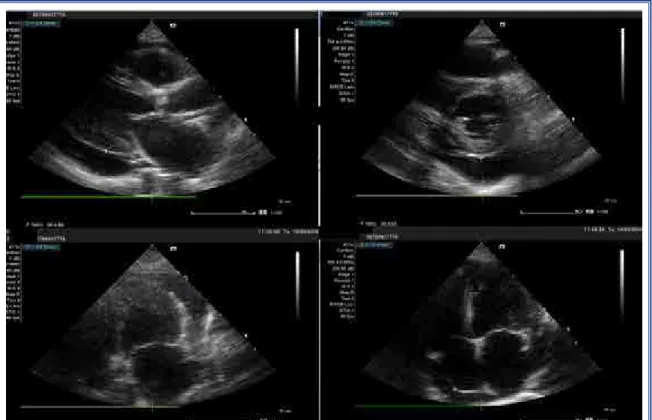
The physical examination upon the current pre-sentation yielded an arterial blood pressure of 120/46 mmHg and cardiac auscultation revealed muffled S1 and S2 sounds with an irregular heart rhythm. Auscul-tation of the lungs revealed decreased lung sounds in the left basal portion. There was no jugular venous dis-tention. The electrocardiogram (ECG) demonstrated atrial fibrillation with a heart rate of 47 bpm. A chest X-ray revealed left pleural effusion and an enlarged cardiac shadow. Echocardiography showed severe pericardial effusion with a diameter of 2.5 cm behind the left ventricle, 2.1 cm adjacent to the right atrium, and a 2.3 cm effusion adjacent to the right ventricular lateral wall, accompanied by signs of right ventricular collapse (Fig. 1) and respiratory variation of more than 25% in mitral and tricuspid inflow velocities. Moder-ate mitral regurgitation, as well as mild aortic and tri-cuspid regurgitation were observed. Cardiac chambers
and systolic pulmonary arterial pressure were normal. The ejection fraction was mildly decreased (50%).
A complete blood count indicated mild anemia, leukopenia, and thrombocytopenia consistent with MDS. His C-reactive protein (8.56 mg/L), troponin-T (30/91 ng/mL), and N-terminal-pro-brain natriuretic peptide (3748 pg/mL) levels were elevated. The crea-tinine value was 1.4 mg/dL. Liver function test results were within the normal range, but the total bilirubin level was slightly elevated (2.02 mg/dL).
Although the patient was clinically stable, due to the persistence of pericardial effusion and echocardio-graphic tamponade features, a pericardiocentesis was selected as the next management step, but the proce-dure was unsuccessful and the patient was referred to surgery for fluid drainage and sampling. A pericardial window was opened, and a left pleural catheter was inserted for thoracentesis. Postoperatively, the pa-tient developed acute respiratory failure and acidosis secondary to central hypopnea and acute renal fail-ure. The patient was extubated the following day and transferred to the coronary intensive care unit.
The pericardial fluid was classified as exudative according to the Light criteria[6] and E. rhusiopathiae
was isolated from the samples. The bacteria were sen-sitive to ceftriaxone and therapy was initiated. The isolates were identified as E. rhusiopathiae using the VITEK 2 system (BioMérieux, Marcy l’Etoile, France) (Fig. 2, 3).
Echocardiographic assessment was repeated ev-ery 2 days, since E. rhusiopathiae has been shown to cause endocarditis in human beings.[7] Due to
deterio-rating renal function, dialysis treatment was also
initi-ated. On the fourth day, the patient’s dialysis catheter became obstructed and dialysis was terminated earlier than expected. The patient experienced a bradycardic cardiopulmonary arrest, though the potassium level was normal. He was intubated, and after 10 minutes of cardiopulmonary resuscitation, his hemodynamic status was stabilized. The control echocardiography revealed mild pericardial effusion, normal left ven-tricular function, depressed right venven-tricular func-tion with right ventricular and atrial dilatafunc-tion, a pul-monary artery systolic pressure of 55–60 mmHg and suspected vegetation on the tricuspid leaflet. Trans-esophageal echocardiography (TEE) revealed fila-mentous vegetation on the atrial side of the tricuspid valve and a thrombus-like image attached to the tip of the dialysis catheter (Fig. 4). The dialysis catheter obstruction of the previous day and the thrombus seen in the catheter lumen suggested that the etiol-ogy of the cardiopulmonary arrest might have been a pulmonary embolism originating from the catheter. It was not possible to perform a pulmonary contrast angiography since the patient was in acute kidney failure. Thrombolysis for a possible acute pulmonary embolism was also not performed because though the patient was hemodynamically stable, severe anemia and thrombocytopenia were present. Dobutamine therapy was implemented to increase the contractility of the right ventricle.
Despite ceftriaxone therapy, the patient had a per-sistent fever of 38°C. Therefore, his antibiotic therapy was changed to meropenem and vancomycin empir-ically. Multidrug resistant Acinetobacter
bauman-nii (A. baumanbauman-nii) was detected in blood cultures of
pleural and pericardial fluid, and colistin was added
Figure 2. Colony morphology of Erysipelothrix rhusiopathiae.
veg-to the therapy. After the addition of colistin, he be-came afebrile and his C-reactive protein level grad-ually decreased. A follow-up echocardiography and control TEE revealed that the vegetation on the tri-cuspid valve had regressed. Once the infection was under control, his diuresis improved and dialysis was no longer necessary.
Antibiotic treatment was continued with only meropenem and colistin. A control blood culture was negative. Those two antibiotics were discontinued on the 14th day and ceftriaxone was re-initiated for E.
rhusiopathiae for the next 6 weeks. The patient had
no fever at the follow-up visit. A control echocardio-graphy revealed mild-moderate pericardial effusion of 15 mm behind the left ventricular posterior wall. No vegetation or thrombus was seen on the tricuspid valve. Due to chronic kidney disease, colchicine the-rapy was initiated instead of non-steroid anti-inflam-matory therapy.
DISCUSSION
To the best of our knowledge, this is the first case of E. rhusiopathiae pericarditis without signs of E.
rhusiopathiae septicemia. While it is important to
note that the lack of matrix assisted laser desorption ionization-time of flight mass spectrometry and 16S RNA sequencing is a limitation, the isolates were identified as E. rhusiopathiae using the VITEK 2 au-tomated microbiology system, and treatment was suc-cessfully managed according to this diagnosis.
Acine-tobacter bacteremia was confirmed in our patient, but Erysipelothrix bacteremia was not. E. rhusiopathiae
is an immobile, pleomorphic, non-sporulating, aero-bic or facultative anerobe, Gram-positive bacillus.[2]
E. rhusiopathiae is generally known as a cause of
infection in animals. It can be found in pigs, sheep, rabbits, chickens, turkeys, ducks, emus, pigeons, cows, guinea pigs, cats, dogs, and fish. Human infec-tion is typically acquired from animals through work-related exposure by those such as animal breeders, farmers and ranchers, veterinarians, furriers, butchers, fishermen and fishmongers, cooks, and grocers. Our patient had a history of contact with fowl. Three forms of human disease have been described: a localized cutaneous form, known as erysipeloid; a generalized cutaneous form; and a septicemic form that is often associated with endocarditis.[3] Rare manifestations
of E. rhusiopathiae infection have also been reported
in cases of persistent bacteremia, septic arthritis, os-teomyelitis, pneumonia intra-abdominal abscess, and meningitis.[8–10]
In immunocompromised patients, bacteremia without endocarditis is more common; there are no pericarditis cases in the literature thus far We could provide no histological evidence or immunohisto-chemical confirmation of myocardial involvement ac-cording to the state of knowledge position statements of the European Society of Cardiology Working Group on Myocardial and Pericardial Diseases. Car-diac magnetic resonance imagining was not available in order to provide other evidence of myocarditis.[11]
Though there are no data indicating myocardial in-volvement of Erysipelothrix infection, considering the past history of pericardial effusion, atrial fibrilla-tion with a fast ventricular rate, low ejecfibrilla-tion fracfibrilla-tion 8 months prior, and an elevated troponin level at the current presentation might indicate a myo-pericardi-tis recurrence. It may be that the prior low ejection fraction was a result of myocardial cell infection by
E. rhusiopathiae, or as a result of an inflammatory
re-sponse to the pathogen which temporarily depressed the myocardium via inflammatory cytokines.[12]
How-ever, we do not have enough evidence to prove that in our case. Although penicillin and cephalosporins are the first-line choice for the treatment of Erysipelothrix infection, we preferred to implement ceftriaxone ini-tially. Varied treatment periods have been applied for other rarely seen clinical entities.[10,13] However, the
patient had a healthcare-associated blood stream in-fection of carbapenem-resistant A. baumanni. These infections are a serious problem in healthcare facili-ties because of the limited options for antibiotic treat-ment. Carbapenem resistance among A. baumannii blood stream infections was reported at 94% in a re-cent multire-center study conducted in Turkey.[14]
We attributed the deterioration of the patient during follow-up in the intensive care unit to early postopera-tive acute respiratory failure and hospital-acquired A.
baumannii infection followed by renal failure.
Con-sidering the thrombus image on the dialysis catheter and motile hypoecogen mass on the tricuspid valve demonstrated with TEE in addition to right ventricu-lar enventricu-largement, an acute pulmonary embolism may have been responsible for the hemodynamic collapse in the follow-up period. Regression of the vegetation may have been a result of anticoagulant therapy or
antibiotherapy.
We report this patient as the first proven case report of pericarditis and possible pancarditis due to E.
rhu-siopathiae. The administration of positive inotropic
agents, the selection of appropriate antibiotics, and the timing of renal replacement therapy was essential in this complicated case.
Peer-review: Externally peer-reviewed. Conflict-of-interest: None.
Informed Consent: Written informed consent was ob-tained from the patient for the publication of the case report and the accompanying images.
Authorship contributions: Concept: F.Ö.; Design: F.Ö., S.P., H.A.; Supervision: S.P., H.A.; Materials: M.A., U.K.; Data collection: M.A.; Literature search: F.Ö.; Writing: S.P., H.A., U.K.
REFERENCES
1. Maisch B, Seferović PM, Ristić AD, Erbel R, Rienmüller R, Adler Y, et al; Task Force on the Diagnosis and Management of Pricardial Diseases of the European Society of Cardiology. Guidelines on the diagnosis and management of pericardial diseases executive summary; The Task force on the diagnosis and management of pericardial diseases of the European soci-ety of cardiology. Eur Heart J 2004;25:587−610. [CrossRef] 2. Vázquez L, De Los Santos C, Cichero M, Frantchez V, Batista
N, Palacio R, et al. Infective endocarditis caused by Erysip-elothrix rhusiopathie. Report of one case. [Article in Spanish] Rev Med Chil 2015;143:1598−600. [CrossRef]
3. Veraldi S, Girgenti V, Dassoni F, Gianotti R. Erysipeloid: a review. Clin Exp Dermatol 2009;34:859−62. [CrossRef] 4. Kaya S, Gençalioğlu E, Yildirim SS, Altun G, Yilmaz G,
Köksal I. Native valve endocarditis caused by Erysipelothrix rhusiopathiae in an immunocompetent individual. J Med
Mi-crobiol 2013;62:1911−3. [CrossRef]
5. Quabeck K, Müller J, Wendt F, Rosenthal E. Pericarditis in Erysipelothrix rhusipahiae septicemia. Infection 1986;14:301. 6. Light RW, Macgregor MI, Luchsinger PC, Ball WC Jr. Pleural
effusions: the diagnostic separation of transudates and exu-dates. Ann Intern Med 1972;77:507−13. [CrossRef]
7. Gorby GL, Peacock JE. Erysipelothrix rhusiopathiae endocar-ditis: Microbiologic, epidemiologic, and clinical features of oc-cupational disease. Rev Infect Dis 1988;10:317−25. [CrossRef] 8. Denes E, Camilleri Y, Fiorenza F, Martin C. First case of
osteomyelitis due to Erysipelothrix rhusiopathiae: pubic os-teomyelitis in a gored farmer. Int J Infect Dis 2015;30:133−4. 9. Meric M, Ozcan SK. Erysipelothrix rhusiopathiae pneu-monia in an immunocompetent patient. J Med Microbiol 2012;61:450−1. [CrossRef]
10. Feasi M, Bacigalupo L, Cappato S, Pontali E, Usiglio D, Rol-landi GA, et al. Erysipelothrix rhusiopathiae intra-abdominal abscess. Int J Infect 2010;14:81−3. [CrossRef]
11. Caforio ALP, Pankuweit S, Arbustini E, Basso C, Gime-no-Blanes J, Felix SB, et al. Current state of knowledge on aetiology, diagnosis, management, and therapy of myocardit: a position statement of the European Society of Cardiology Working Group on myocardial and pericardial Diseases. Eur J Cardiol 2013;34:2636−48. [CrossRef]
12. Imazio M, Cooper LT. Management of myopericarditis. Ex-pert Rev Cardiovasc Ther 2013;11:193−201. [CrossRef] 13. Kim SR, Kwon M J, Lee JH, Lee NY. Chronic meningitis
caused by Erysipelothrix rhusiopathiae. J Med Microbiol 2007;56:1405−6. [CrossRef]
14. Ergönül Ö, Aydin M, Azap A, Başaran S, Tekin S, Kaya Ş, et al. Healthcare-associated Gram-negative bloodstream infec-tions: antibiotic resistance and predictors of mortality. J Hosp Infect 2016;9:381−5. [CrossRef]
Keywords: Endocarditis; Erysipelothrix rhusiopathiae; myocarditis;
pancarditis; pericarditis
Anahtar sözcükler: Endokardit; Erysipelothrix rhusiopathiae;