Application of patch test in allergic (atopic) dogs and investigation of
dog allergy incidence in people
Aziz Arda SANCAK1, Mehmet SAHAL1, Sibel Yasa DURU1, Duygu CAKIROGLU2
1Department of Internal Medicine, Faculty of Veterinary Medicine, Ankara University, Turkey. 2Department of Internal Medicine,
Faculty of Veterinary Medicine, Ondokuz Mayis University, Turkey.
Summary: Incidence of canine allergic contact dermatitis (ACD) is difficult to determine, although it is regularly encountered in veterinary practice. Patch testing is a non-invasive method of determining contact allergens that may cause eczematous skin eruptions in dogs, although standardization of the procedure is yet to be completed. The aim of the present study was to determine the frequency of contact sensitizations in dogs with dermatitis, to interpret the results from standardized allergens, and to evaluate their clinical relevance. Sensitivity to pet animals is a frequent cause of allergic symptoms in atopic human patients and/or patients with asthma. Therefore, we also try to determine the possible allergic hypersensitivity of dog owners to their own dogs. European patch test standard serial was applied to 22 allergic dogs. Test results were positive in 9 (41%) dogs with allergic dermatitis. Nickel sulphate, potassium dichromate, bis-diethydithiocarbamato-zinc, p-t-butylph.-formaldehyde-resin, fragrance mix, benzocaine, ammoniated mercury, mercapto mix and Cetylstearylalcohol were the positive allergens determined. Specific IgE (dog epithelium and dandruff) floroenzymeimmunoassay was applied to blood samples of dog owners (n = 12), as well as to a control group of non-owners (n = 10). Although the dog owners were not demonstrating any signs of allergic symptoms, they all were positive for specific IgE. On the other hand, in the control group, only 4 people (40%) were positive with specific IgE. Our data suggest that complete avoidance of dog antigen may not be possible. Moreover, these findings support the potential involvement of contact allergens in dogs with atopic dermatitis.
Key words: Atopic dermatitis, contact allergy, dog, patch test.
Alerjik (atopik) köpeklerde yama testi uygulamaları ve insanlarda köpek alerjisi insidansının araştırılması
Özet: Köpeklerde alerjik kontak dermatitis (AKD) vakalarına pratikte rastlanmakla birlikte, gerçek insidansının belirlenmesi oldukça zordur. Yama testi, kontakt alerjenlere bağlı olarak gelişen ekzematöz tipte deri rahatsızlıklarının tanısına yönelik, fakat henüz köpeklerde standardizasyonu tamamlanmamış, invaziv olmayan bir testtir. Bu araştırmada, insan hekimliği için standardize edilmiş yama testi yöntemi kullanılarak dermatitisli köpeklerde görülen alerjenlerin çeşitleri ve bunların klinik tablo ile ilişkisi araştırılmıştır. Beslediği hayvana karşı duyarlılık, atopik veya astımlı insanlarda sık görülen alerji semptomlarıdır. Bu nedenle, çalışmanın ikinci aşamasında köpek sahiplerinde köpeklerine karşı bir alerjik duyarlılık gelişip gelişmediği de araştırılmıştır. Çalışmada 22 adet alerjik dermatitisli köpeğe, standart “Avrupa yama testi” uygulandı. Yama testi 9 (%41) köpekte pozitif sonuç verirken, pozitif alerjenler kapsamında nikel sülfat, potasyum dikromat, çinko-diethydithiokarbamat, p-t-butylph.-formaldesid, “fragrance“ karışımı, IPPD, benzokain, cıva (II)-amid-klorid, merkapto karışımı, setylstearylalkohol saptandı. Köpek sahiplerinin (n = 12) yanısıra kontrol olarak köpek sahibi olmayan kişilerin (n = 10), kan numunelerinde spesifik IgE (köpek epiteli ve kepeği) floroenzimimmunoassay çalışıldı. Köpek sahiplerinde herhangi bir alerji belirtisi gözlenmemiş olmasına rağmen, hepsi spesifik IgE’ye karşı pozitif bulundu. Diğer yanda kontrol grubunda, yalnızca 4 kişinin (40%) spesifik IgE açısından pozitif olduğu anlaşıldı. Bu çalışmanın verileri, köpek antijeninden tamamen ari olmanın mümkün olmayabileceğini göstermektedir. Ayrıca, bu bulgular atopik dermatitli köpeklerde, kontakt alerjenlerin potansiyel varlığını desteklemektedir.
Anahtar sözcükler: Atopik dermatit; kontact allerji; köpek, yama testi.
Introduction
Household or synthetic products that contain allergens are a major cause of disease in humans but they are rarely documented as causing allergic disease in dogs (10,15). Contact dermatitis is generally classified into two etiologic categories: irritant and allergic. Allergic contact dermatitis is immune mediated, while irritant
contact dermatitis is caused by nonimmunological factors of a physical or chemical nature (4,11).
Patch testing is a non-invasive method to identify contact allergens that may cause eczematous skin eruptions due to a delayed type of hypersensitivity (type IV) (4,11). Patch testing is extensively used to diagnose eczematous skin eruptions in people (5). However, the
Aziz Arda Sancak - Mehmet Sahal - Sibel Yasa Duru - Duygu Cakiroglu 114
incidence of naturally occurring cases of canine allergic contact dermatitis (ACD) are rarely reported in the veterinary literature and not well documented as humans (3,6,15). Although standardization of the procedure remains incomplete, recognition of canine ACD in dogs has been increased by the use of new standardized patch tests (12). Routine screening diagnostic tests and some definitive tests do not identify canine ACD. Many agents have been determined as causes of allergic contact dermatitis. It is important to remember that all contact reactions that are allergic in nature require repeated exposure to the allergen (6,8,11,18). Variations in environmental conditions strongly influence and determine the presence or absence of allergens (6,18). However, when contact dermatitis is suspected, an attempt should be made to identify the allergen or irritant.
Sensitivity to pet animals is a frequent cause of allergic symptoms in atopic human patients and/or patients with asthma (17). Dog ownership is common worldwide. Today, an increased number of pet animals are kept at homes in Turkey, therefore it is impossible for people to completely avoid exposure to dogs. Several studies have suggested the existence of a correlation between the presence of dog antigen in homes and the increased frequency of asthma (7,17). Therefore, in this study, we try to determine the possible allergic hypersensitivity of dog owners to their own dogs.
The current study describes the application of the patch test, determines the frequency of contact sensitizations in dogs with dermatitis, interprets the results from standardized allergens, and evaluates their clinical relevance.
Material and Methods
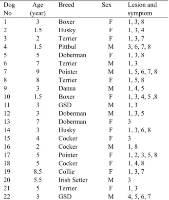
Twenty-two dogs exhibiting clinical signs of intense pruritus and erythematous dermatitis on the ventral abdomen were examined in the clinics of Veterinary Faculty of Ankara University. The dogs were at different ages and breeds and of both sexes. Demographic and clinical details of the cases are documented in Table 1. A diagnosis of contact dermatitis with secondary pyoderma was considered where there was marked restriction of lesions to the ventrum. Self-trauma secondary to pruritus is a common feature of atopy, which led the inclusion of such dogs in the differential diagnosis. Parasitological and microbiological examinations were performed for the differential diagnosis. All the dogs had received unsuccessful prior treatment with either anti fungal drugs, antibiotics or corticosteroids, and they had been referred to the faculty clinics after their condition had deteriorated. Steroids were avoided for 4 weeks prior to testing.
Table 1: Clinical findings in dogs Tablo 1: Köpeklerde klinik bulgular
Dog No
Age (year)
Breed Sex Lesion and
symptom 1 3 Boxer F 1, 3, 8 2 1.5 Husky F 1, 3, 4 3 2 Terrier F 1, 3, 7 4 1.5 Pittbul M 3, 6, 7, 8 5 5 Doberman F 1, 3, 8 6 7 Terrier M 1, 3 7 9 Pointer M 1, 5, 6, 7, 8 8 8 Terrier F 1, 5, 8 9 3 Danua M 1, 4, 5 10 1.5 Boxer F 1, 3, 4, 5 ,8 11 3 GSD M 1, 3 12 3 Doberman M 1, 3, 5 13 7 Doberman F 3 14 3 Husky F 1, 3, 6, 8 15 4 Cocker F 3 16 2 Cocker M 1, 8 17 5 Pointer F 1, 2, 3, 5, 8 18 5 Cocker F 1, 4, 8 19 8.5 Collie F 1, 3, 7 20 5.5 Irish Setter M 3 21 5 Terrier F 1, 3 22 3 GSD M 4, 5, 6, 7
M(ale), F(emale), GSD (German shepherd dog); 1 pruritus, 2 hyperpigmentation, 3 erythema, 4 lichenification, 5 serous
crusting, 6 macules, 7 pustules, 8 otitis
Since it was apparent that there was contact with some suspected substances in the dog’s environment, “European standard” (Brial, Allergen GmbH, D-Greven) patch test serial was applied to these 22 dogs to determine whether an allergic or irritant reaction was present.
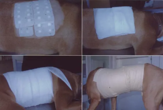
The hair on the right or left dorsolateral thorax was closely clipped (15 x 20 cm) with a No.40 blade, before the test was carried out. The test substances were applied to test chambers (Finn Chambers, Haye’s, Netherlands) and were placed directly on intact skin and a gauze pad (0.5 cm thick) was placed over the chambers. Before fixing to the skin with a hypoallergic tape, chambers, gauze pad and hypoallergic tape were secured under a body bandage. The bandaging was performed with gauze (10 x 10) and extended over both right and left thorax and abdomen. Strips of adhesive bandage at the edges fixed the wrapping to unshaven areas so that it could not move. In some dogs an Elizabethan collar was applied to prevent the dog from removing the bandage. Application of patch test is presented in Figure 1.
The first reading was performed after 48 hours. The test material was removed, and the condition of the underlying skin was examined 30 minutes later. The second reading was performed after a further 72 hours later. The interpretation method (13,18) recommended by the international contact dermatitis research group is:
(-) No reaction; (?) Doubtful reaction, faint macular erythema only; (+) Weak (nonvesicular) positive reaction (erythema, infiltration, possible papules); (++) Strong (vesicular) positive reaction (erythema, infiltration, papules, vesicular); (+++) Extreme positive reaction (bullous reaction, coalescing vesicles); (IR) Irritant reaction, discrete patchy erythema without infiltration.
Blood samples of control group of non-dog owners (n = 10) and dog owners (n = 12) were collected at the Department of Dermatology, Gazi University’s School of Medicine. Specific IgE against dog epithelium and dandruff were evaluated by using UnicapTM specific IgE fluoroenzyme immunoassay (FEIA) (Pharmacia & Upjohn diagnostics AB, Sweden) (7).
Results
The median age of the dogs tested for ACD was 4.3 years (range: 1.5 years to 9 years), of both sexes and various breeds (Table 1). In all dogs, abnormal physical findings were limited to the integument. Most lesions were present in the groin, ventral surface of the paw and ventral abdomen. Common patterns were erythematous papules, macules, lichenification and occasional pustules. Secondary to self-trauma, alopecia and edema were also detected
Otitis was found in 10 (50%) of the dogs. Information related to environment was obtained from dog owners. It was revealed that the indoor environment involved floors made of hard wood, concrete or ceramic. Waxes, rinses or cleaning agents were regularly applied
to the floor. All dogs were either sleeping on the floor or on a specific bed or bedding materials. The outdoor environment included thick vegetation. In their histories 2 pet owners reported increased pruritus associated with a particular environment such as one room and they reported improvement of clinical signs in association with travelling. One pet owner reported that only one dog in a multiple animal household was affected. Results of the culturing of samples and skin scrapings were negative in all dogs.
European patch test standard serial was applied to 22 allergic dogs. When the test chambers were removed after 48 hours, there was a markedly erythematous plaque composed of tiny papules at the location in which test material had been placed.
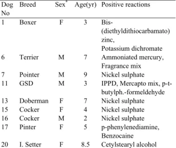
Positive patch test results were found in 9 dogs with allergic dermatitis (Table 2). The positive allergens were nickel sulphate (n = 4), potassium dichromate (n = 1), bis-diethydithiocarbamato-zinc (n = 1), p-t-butylph.-formaldehyde-resin (n=1), fragraence mix (n = 1), IPPD (n = 1), benzocaine (n = 1), ammoniated mercury (n = 1), mercapto mix (n = 1), cetylstearylalcohol (n = 1). In our study, 5 (55.5%) of the affected dogs involved in the study were monosensitive and 4 (44.5%) were sensitive to two or more allergens. Irritant reactions were not included and determined in only 3 dogs (dogs no. 3, 14 and 18). Two dogs (dogs no. 14 and 18) showed IR to potassium dichromate and one dog (dog no. 3) was reacted to p-t-butylph.-formeldehyde-resin.
Figure 1: Application of patch test in dogs Şekil 1. Köpeklerde yama testinin uygulanması
Aziz Arda Sancak - Mehmet Sahal - Sibel Yasa Duru - Duygu Cakiroglu 116
Table 2 : Results of Patch Test: Positive reactions were elicited by 11 different contact allergens.
Tablo 2: Yama testinin sonuçları: 11 farklı kontakt allergen ile pozitif sonuçlar elde edilmiştir.
Dog No
Breed Sex* Age(yr) Positive reactions
1 Boxer F 3
Bis-(diethyldithiocarbamato) zinc,
Potassium dichromate 6 Terrier M 7 Ammoniated mercury,
Fragrance mix 7 Pointer M 9 Nickel sulphate 11 GSD M 3 IPPD, Mercapto mix,
p-t-butylph.-formeldehyde 13 Doberman F 7 Nickel sulphate 15 Cocker F 4 Nickel sulphate 16 Cocker M 2 Nickel sulphate 17 Pinter F 5 p-phenylenediamine,
Benzocaine 20 I. Setter F 8.5 Cetylstearyl alcohol *: M(ale), F(emale), GSD (German shepherd dog)
A gauze pad placed over the test chambers was found to be useful for preventing the friction between layers; it also helped to prevent dermographism. Therefore the skin and the test chambers could move independently of the covering bandage when the dog moved around.
Patch test positive dogs were kept away from the relevant allergen. Only 4 dogs (dogs no. 13, 16, 17, 20) showed clinical improvement by avoiding allergens.
Specific IgE (dog epithelium and dander) FEIA assay were applied to blood samples of control group having no dogs (n=10) and dog owners (n=12). Although the dog owners did not show any signs of allergic symptoms, they were all positive for specific IgE. In the control group, only 4 (40%) people were positive with specific IgE.
Discussion and Conclusion
The patch test is a unique method for documenting contact hypersensitivity. In this report, characteristic cutaneous clinical signs and responses to patch testing confirmed canine allergic contact dermatitis.
The list of documented contact allergens for humans exceeds 800 (5), but naturally occurring contact hypersensitivity is only reported to account for about 1% to 20% of all canine dermatoses (6,12,18). In this study 41% of dogs have been found patch test positive. Moreover, 3 different dogs showed irritant reaction. It is difficult to differentiate the naturally occurring allergic contact dermatitis and irritant contact dermatitis (16). Irritant contact dermatitis, which may have a similar clinical presentation, is the result of direct, immediate chemical damage to the epidermis (9,11). In contrast to
allergic contact dermatitis, skin lesions appear within only a few hours after patch test challenge (18).
In spite of the frequent exposure of most dogs to allergens used in this patch test, they have rarely been recorded as contact allergens in the dog. It has been suggested that the relatively low incidence of contact allergies in dogs compared to humans is due to protection of the skin by the coat (6,9,11,18). Yet, there are no scientific data to substantiate this claim (17). Contact hypersensitivity in dogs was confirmed to the synthetic textiles and cement and the following substances were also documented: thiuram mix, cobalt chloride, nickel sulphate, quinoline mix, colophony, black rubber mix, ethylenediamine, primin and wood tar (15,16). Two other dogs were patch test positive to colophony, a pine oil resin, and this correlated with reaction to cleaning products (15).
This study reports that the most frequent allergen was nickel. It is very frequently utilised in most branches of industry. It can be present in dog toys, food containers and collars. Plastic dog toys contain zinc- diethyldithiocarbamato, IPPD and p-t-butylph.-formaldehyde. Ammoniated mercury and fragrance mix are used in the drug and cosmetic industry and cause canine CAD. It has been reported that carpet deodorizer was a contact allergen in a dog (3). Similar to our findings, in a Danish study, 63% of the affected dogs were monosensitive and 23% were sensitive to two allergens (16).
The age of onset varies since the development of an allergic response requires repeated exposure to the contactant and no specific breed susceptibility to allergic contact dermatitis has been verified (11). However, breed predilections have been reported in Terriers, Poodles, Labrador Retrievers and German Shepherd Dogs (10,15). In our study, the Terrier, GSD, Irish Setter, Boxer, Pointer and Doberman were the affected breeds.
It has been reported that contact hypersensitivity can cause otitis externa and atopic dogs have signs of otitis in 83% of the cases, with 24% having otic symptoms at the onset of their allergic disease (2,15). Similar to our study, it has been reported that at least 50% of dogs with atopy suffer from otitis externa (1).
It has been reported that canine ACD is generally characterized by erythema, macules in the acute phase, and scaling, hyperpigmentation and occasionally lichenification accompanied by alopecia (11). In our cases erythema and lichenifacation were the most common clinical manifestations. In this study lesions localized to the ventral area were suggestive of a reaction to floor cleaners and bedding.
As a result of this study, its not possible to say patch test negative dogs are not canine ACD, although a
standardized patch test kit has been recommended for dogs (14). Its difficult to say that such a high incidence of positive patch test results shows the real an etiology of atopic dogs because only 4 dogs recovered when we kept these dogs away from suspected material. However, specific identification of the allergen may lead to more rewarding case management. New lesions continued to occur when the complete avoidance of the relevant allergen was not possible in most of the cases (11). The length of time necessary for improvement once the animal has been removed from relevant substances varies considerably and depends on the chronicity of the disease and complications such as secondary pyoderma (2,8,11). It is likely that some of the cases reported with positive patch tests may reflect contact atopic disease and not true hapten-induced contact hypersensitivity (19).
The mechanical problems associated with application and fixing of the devices have resulted in limited usage of patch tests (11,15,19). In this study, difficulties on applying and securing patch test substances and patch test material have been improved by placing the gauze pad between the chambers and gauze dressing. In this way, the sliding of material as well as irritation from tape, and dermogrophism were avoided.
Although homes without dogs have decreased antigen levels compared to homes with dogs (14), many other studies suggest that complete avoidance of dog antigen may not be possible (17). In this study most people had dogs; the level of antigen was also high in people without dogs. The study revealed that dog antigens exist even in the case of people who have no contact with dogs and that increasing IgE levels to dog antigen occurs even in subjects who completely avoid contact with dogs. This finding suggests that avoidance of dogs may not prevent exposure to dog allergens and in non-atopic people finding positive dog antigen is of no clinical importance.
It can be concluded from the present study that, contact hypersensitivity may occur in dogs, and that these findings support the potential involvement of contact allergens in dogs with atopic dermatitis.
Acknowledgement
Funded by the Scientific and Technical Research Council of Turkey (TUBITAK, VHAG-1590ADP). We are grateful to our client Mr. Celasin EGEL and to the Department of Dermatology, University of Gazi School of Medicine, Turkey for their valuable support.
References
1. August JR (1988): Otitis externa: a disease of
multifactorial etiology. Vet Clin N Amer, 18, 731-742.
2. Baker EB (1990): Contact Allergy. 113-119. In: Small Animal Allergy: A Practical Guide. Lea & Febiger, London.
3. Comer KM (1988): Carpet deodorizer as a contact
allergen in a dog. JAVMA, 193, 1553-1554.
4. Diker KS (1998): İmmunoloji. 1st ed., Medisan, Ankara, Turkey.
5. Fisher AA (1973): Contact Dermatitis, 2nd ed, Lea & Febiger, Philadelphia.
6. Grant DI, Thoday KL (1980): Canine allergic contact
dermatitis: a clinical review. J Small Anim Pract, 21,17-27
7. Ingram JM, Sporik R, Rose G, Honsinger R, Chapman MD, Platts-Mills TA (1995): Quantitative assessment of
exposure to dog (Can f1) and cat (Fel d 1) allergens: Relation to sensitization and asthma among children in living in Los Alamos, New Mexico, J Allergy Clin Immun,
114, 449-456.
8. Kunkle GA, Gross TL (1983): Allergic contact dermatitis
to Tradescantia fluminensis (Wandering Jew) in a dog.
Comp Cont Educ 5, 925-930.
9. Kunkle GA (1988): Contact dermatitis. Vet Clin N Amer, 8, 1061-1068.
10. Muller GH, Kirk RW, Scott DW (1989). Ear diseases. 807-814. In: Small Animal Dermatology, 4th ed., W.B. Saunders, Philadelphia.
11. Nesbitt GH (1983): Canine and Feline Dermatology: A
systemic Approach. Lea & Febiger, Philadelphia.
12. Olivry T, Prelaud P, Heripret D, Atlee BA (1990):
Allergic contact dermatitis in the dog: principles and diagnosis. Vet Clin N Amer, 20, 1443-1456.
13. Özkaya-Bayazıt E, Özarmağan G (1997): Yama testi: review, Türkderm, 9, 57-66.
14. Schou C, Hansen GN, Lintner T, Lowenstein H (1999):
Assay for the major dog allergen. Can f 1: Investigation of house dust samples and commercial dog extracts. J
Allergy Clin Immunol, 88, 847-853.
15. Scott DW, Miller WH, Griffin CE (2001). Muller &
Kirk’s Small Animal Dermatology. 6th ed., W.B. Saunders,
Philadelphia.
16. Thomsen MK, Thomsen HK (1989). Histopathological
changes in canine allergic contact dermatitis patch test reactions: A study on spontaneously hypersensitive dogs.
Acta Vet Scand, 30, 379.
17. Tubiolo VC, Beall GN (1997): Dog allergy:
understanding our “best friend”, Review, Clin, Exp,
Allergy, 27, 354-357.
18. Walton GS. (1977): Allergic contact dermatitis. In: Kirk R.W.: Current Veterinary Therapy, 6th ed. W.B. Saunders Co, Philadelphia.
19. Walder EJ, Conroy JD (1994): Contact dermatitis in dogs
and cats: pathogenesis, histopathology, experimental induction and case reports. Vet Derm, 6, 149-162.
Geliş tarihi: 11.02.2008 / Kabul tarihi: 29.04.2008
Address for correspondance: A. Arda Sancak
Department of Internal Medicine Faculty of Veterinary Medicine Ankara University
Dışkapı-Ankara / Turkey.


