Journal of Clinical and Analytical Medicine |
O
h
r
c
i
r
g
a
in
e
a
s
l
R
e
1 Keziban Uçar Karabulut1, Hale Turan Özden2 ,Yıldız Uçar3 1Department of Emergency Medicine, Baskent University Faculty of Medicine, Ankara, 2Department of İnfection Dissease, Baskent University Faculty of Medicine, Ankara, 3Department of Pulmonary Dissease, Diyarbakır Memorial Hospital, Diyarbakır, TurkeyMPV and Acute Pancreatitis
The Significance of Mean Platelet Volume in Acute Pancreatitis
Akut Pankreatitte Ortalama Platelet Hacimlerinin Önemi
DOI: 10.4328/JCAM.4487 Received: 16.03.2016 Accepted: 05.04.2016 Printed: 01.02.2016 J Clin Anal Med 2016;1(suppl 1): 19-22 Corresponding Author: Keziban Ucar Karabulut, Baskent Universitesi, Konya Hastanesi Hocacihan Mah. Saray Cad. No:1, Selcuklu, Konya 42080 Turkey. T.: +90 3322570606 F.: +90 3322570637 E-Mail: [email protected]
Özet
Amaç: Akut pankreatit, önemli komplikasyonlar ve yüksek mortalite ile sey-redebilen bir hastalıktır. Pankreasta normalde inaktif halde bulunan sindirim enzimlerinin aktif hale geçerek pankreas dokularını sindirmesi ve buna bağlı ortaya çıkan inflamasyonla karakterize bir hastalıktır. Çalışmamızda; acil ser-vise karın ağrısı şikayeti ile başvurup Akut Pankreatit tanısı alan hastalarda Ortalama Trombosit Hacmi oranlarının seyrini izlemeyi amaçladık. Gereç ve Yöntem: Çalışmamız acil servise karın ağrısı şikayeti ile başvurup Akut Pank-reatit tanısı alan hastalarda üzerinde yapıldı. Çalışmaya toplam 104 hasta dahil edildi. Hastaların Akut Pankreatit tanısı konulduktan ve tam iyileşme sağlandıktan sonraki Ortalama Trombosit Hacmi, Lökosit ve Lipaz değerle-rine bakıldı. Bu değerler karşılaştırıldı. Ayrıca, hastaların akut pankreatit ta-nısı konulduktan ve tam iyileşme sağlandıktan sonraki Ortalama Trombosit Hacmi, Lökosit ve Lipaz değerleri kaydedildi. Bulgular: Çalışmaya alınan 104 akut pankreatitli hastanın hastalık esnasında ve hastalık tamamen düzeldik-ten sonraki Ortalama Trombosit Hacmi, Lökosit ve Lipaz değerleri istatistik-sel olarak karşılaştırıldı. Ortalama Trombosit Hacmi, Lökosit ve Lipazın baş-vuru esnasındaki değerleri hastalık sonrasına göre yüksek tespit edildi. Tar-tışma: Ortalama Trombosit Hacmi, Akut Pankreatite yükselen bir belirteçtir. Akut Pankreatit tanısının erken konulmasında ve hastalığın seyrinde fayda-lı olabileceğini düşünüyoruz.
Anahtar Kelimeler
Akut Pankreatit; Ortalama Trombosit Hacmi; Lökosit; Lipaz
Abstract
Aim: Acute pancreatitis is a high-mortality disease carrying significant risk of complications and characterized by intra-acinar cell activation of diges-tive enzymes, followed by a subsequent response via the release of proin-flammatory cytokines. Here, we aimed to investigate the development of mean platelet volume in patients diagnosed with acute pancreatitis. Material and Method: The study was performed in patients admitted to the emer-gency room with the complaint of abdominal pain and diagnosed with acute pancreatitis. A total of 116 patients were included in the study. Mean platelet volume, leucocytes or white blood cells and lipase values were investigated and compared after all patients had been diagnosed with acute pancreatitis and regained good health. Also, the values of mean platelet volume, white blood cells and lipase were recorded twice for these patients. Results: After diagnosis and complete treatment, the mean platelet volume, white blood cells and the lipase values of 116 acute pancreatitis patients were statisti-cally compared it was found that the values on admission were higher than after the treatment. Discussion: Mean platelet volume is an indicator that increases in acute pancreatitis so, we consider that mean platelet volume may be beneficial in the diagnosis and in monitoring the course of acute pancreatitis.
Keywords
Acute Pancreatitis; Mean Platelet Volume; Leucocyte; Lipase
| Journal of Clinical and Analytical Medicine MPV and Acute Pancreatitis
2
Introduction
Acute pancreatitis (AP) is a condition that may also have an effect on tissues outside of the pancreas (1) and is an inflam-matory process of the pancreas, advancing with an increase in enzymes such as amylase and lipase, and with abdominal pain. Because of diagnoses followingdeath or ignorance of the diag-nostic process, accurate incidence rates of AP remain unknown, but the incidence of AP is accepted as5–35/100,000 (2). An association was found between thrombocyte activation and many diseases developing with thrombosis and inflamma-tion(3). Mean platelet volume (MPV) is an indicator determined by an automated blood count device as part ofa complete blood count, but is generally neglected by medical professionals. MPV was found to be associated with mean thrombocyte volumeand the function and activation of thrombocytes (4,5). It has been suggested that as the number of thrombocytes is generally de-creased, MPV is inde-creased, and large thrombocytes are younger and more reactive. MPV is one of the most widely used surro-gate markers of platelet function and has been shown to reflect inflammatory burden and disease activity in several diseases including pre-eclampsia, acute appendicitis, unstable angina, myocardial infarction, and systemic inflammation such as ul-cerative colitis and Crohn’s disease (6,7). Studies related to the development of MPV in AP are present in the literature (8,9). In this studywe aimed to investigate the development of MPV val-ues in AP patients both during the disease and after complete healing. We also investigated the course of lipase and leucocyte values with MPV, and the correlation between these values. Material and Method
Admitted to the emergency room of Konya Training and Re-search Hospital, Baskent University and diagnosed with AP between March 2012 and April 2014, 116 patients were ret-rospectively investigated. Among the patients admitted to the emergency, those diagnosed with AP via clinical, laboratory, and radiological tests were included in the study. Those with a his-tory of oncologic diseases, iron deficiency anemia, conditions developing with thrombocytopenia, thrombocytosis, or thalas-semia, a history of metabolic diseases,orinflammatory bowel disorder were excluded from the study. Additionally, all patients were assessed according to severity classification using the Ranson’s criteria; a total of 55 patients with mortality or the features of a severity classification were excluded, while those with mild to moderate severe pancreatitis (edematous and in-terstitial types) were included in the study. Blood samples were drawn for MPV, leucocyte count or white blood cells (WBC), and lipase from 116 patients with AP. The patients were referred to the department of gastroenterology to commence treatment. According to the standard treatment modality, intravenous fluid resuscitation, electrolyte replacement, and analgesics were ad-ministered to all patients. For those with nausea and vomit-ing, nasogastric drainage was performed to prevent aspiration into the respiratory tract. Total parenteral nutrition (TPN) was started to support the patients nutritionally, and antibiothera-py was started to prevent septic complications. In addition to these treatment regimes, various therapeutic methods (such as endoscopic retrograde cholangiopancreatography (ERCP) were performed due to parenchymal injuries and other complications
in the pancreas to decrease mortality rate. The patients began oral feeding 5-10 days later and improvements as measured by clinical, radiological, and laboratory tests were monitored. After observing improvements, blood samples were obtained from 116 patients to evaluate the MPV, WBC, and lipase values. Laboratory measurements
For the measurements of MPV and WBC values, an electronic routineblood count device was used (Cell-Dyne 3700, Abbott, Abbott Park, IL, USA). Routine biochemistry kits were utilized for the measurement of lipase. As expected, the results of our laboratory tests for MPV values ranged from7.0 to 12.0 fl. Statistical analysis
Accumulated data were registered into previously prepared forms, and the statistical analyses were performed via SPSS 15.0 package. The data were analyzed as to appropriateness for normal distribution. For comparisons, the student’s t and paired t tests were performed in dependent groups. Numeric values were defined as mean±standard deviation, and p<0.05 was accepted to be significant. While performing our analysis, we evaluated the difference between the two groups. In other words, the correlation between MPV1 during the disease period and MPV2 after amelioration was assessed in our study. Results
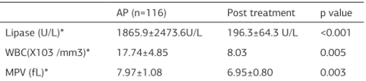
Admitted to the emergency room, a total of 116 patients hospi-talized in the emergency department of our hospital from March 2012 to April 2014 due to the diagnosis of AP were included into the study. Of these patients, 65 were men (56.03%) and 51 were women (43.97%). The mean age rate was detected as 61.2±3.6. In their etiology, 94 patients (81.03%) were seen to have bili-ary causes of AP. Twenty-two patients (18.96%) were aged 70 or over. While the mean lipase value was 1865.9±2473.6 U/L in the acute phase, it was found to be196.3±64.3 U/L in the post-disease phase. When compared, this rate was evaluated to be statistically significant (p<0.05). In terms of mean WBC values, while the rate was found to be17.74±4.85/mm3 in the acute phase, it was determined to be 8.03±1.72/mm3 after the disease period; these rates were also accepted asstatistically significant (p<0.05). Another statistically significant rate was related to mean MPV values (p<0.05), found to be7.97±1.08 fL in the acute phase and detected as 6.95±0.80 fL after amelio-ration (Table 1).
In the patient group, the correlations between MPV, WBC, and lipase were evaluated, and a moderate correlation was found between MPV and lipase and MPV and WBC, although no corre-lation was detected between MPV1 and MPV2 values (Table 2).
Table 1. Labaratory value of patients with AP
AP (n=116) Post treatment p value Lipase (U/L)* 1865.9±2473.6U/L 196.3±64.3 U/L <0.001
WBC(X103 /mm3)* 17.74±4.85 8.03 0.005
MPV (fL)* 7.97±1.08 6.95±0.80 0.003
*Mean±standard deviation values, WBC: white blood count, MPV: mean platelet volume
I Journal of Clinical and Analytical Medicine
20
| Journal of Clinical and Analytical Medicine MPV and Acute Pancreatitis
3
Discussion
AP is an important gastroenterological emergencywith high-rates of morbidity and mortality, requiring long hospital stays and with the possibility of several local and systemic com-plications (10). Early diagnosis and classification are of vital importance, both before and after the development of severe AP (11). However, a reliable laboratory test that diagnoses AP accurately and that can inform boththe early diagnosis and determination of the condition’s etiology has yet to be found (12). Available studies are mostly related to the determination of secondary inflammation. Levels of blood and urine amylase, serum lipase, serum elastase 1, serum trypsin, serum phospho-lypase A2, C-reactive protein, interleukin 6-8, and procalcitonin may be observed as increased in AP cases (13,14). In this study, we aimed to monitor the course of MPV, as well as WBC and
lipase, during and after diagnosis of AP.
Various factors are present in the etiology of AP.Gallbladder is-sues and alcohol consumption are amongthe responsible fac-torsin 90% of cases. Among those other factors leading to AP, an increase in cholesterol level, pancreatic tumors, abdominal trauma, drugs, hypothermia, infections, ERCP, or surgical inter-ventions are reported. In 10% of patients, however, no reasons that might causeAP are determined(15,16). As consistent with findings in the literature, biliary reasons leading to AP were wit-nessed in 81.03% of the patientsin our study.
In a study where the literature was reviewed by DiMagno et al.(17), AP was reported to be most frequently seen in the pa-tients’ 60’s. Consistent with this finding, the mean age in our study was 61.2±3.6.
Because the clinical presentation of AP exhibits changeable features and there are factors restricting diagnostic proce-dures, sometimes difficulties may occur in the diagnosis of AP. Different parameters are used in the diagnosis and follow-up of AP. Levels of blood and urine amylase, serum lypase, serum elastase 1, serum trypsin, serum phospholypase A2, C-reactive protein, interleukin 6-8, and procalcitonin may be increased in cases of AP. The increase seen in serum lipase is more specific, compared with that in amylase. Because the increase in serum lipase continues longer than that of serum amylase, serum li-pase is a more beneficial parameter in patients clinically diag-nosed late. Additionally, serum lipase may also increase in such diseases as acute cholecystitis, peptic ulcer perforation, and mesentric embolism (13). In the diagnosis of lipase-suspected AP cases, amylasin is known as the first step treatment option (18). Also in our study, lipase values were developed at higher levels than normal during AP.
AP is a disease that developswith inflammation of the pancreas as a result of the activation of enzymes of acute pancreatitis, and levels of WBC are seen as increased in such an inflamma-tory condition. Further, the rate of WBC>16.000/ml isa com-ponent of the Ranson’s criteria and is an important marker in the determination of prognosis and the severity of AP (19). As consistent with the literature, in our study, WBC progression-also increased during AP.
MPV is an easily measured value and a useful marker for throm-bocyte function and activity. High levels of MPV usually indicate thrombocyte activity and the existence of large young throm-bocytes, which are metabolically more active (4,20). It is ac-cepted that there is an association between inflammation and thrombocytes. Active platelets play a key role in both inflam-mation and thrombosis. Hematology analyzers measure this activity with different indicators, and MPV is also one of these indicators (21,22). MPV is a cost-effective and cheap parameter showing platelet function and activation, which may be demon-strated in whole blood count and affected by inflammation (23). For example, the values of MPV are affected by inflammation and acute cholecystitis (24).
In the literature, a few studies follow the course of MPV in pa-tients with AP. In one of these studies, MPV values were de-tected to progress at a higher rate in AP patients compared to controls. The same study also emphasized that MPV has a posi-tive correlation with other pancreatic enzymes (8). In another study, MPV values were detected to be higher in severe AP, and
Figure 1. Comparison of lypase values during AP treatment (Lipase 1) and after AP treatment (Lipase 2)
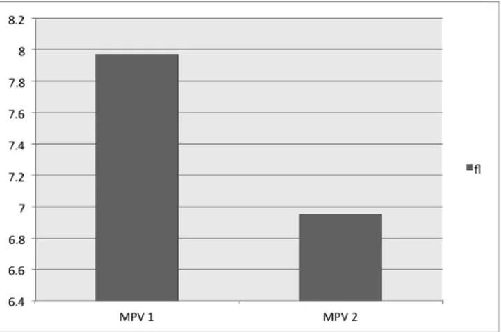
Figure 2. Comparison of MPV values during AP treatment (MPV 1) and after AP treatment (MPV 2)
AP: acute pacreatitis
AP: acute pacreatitis, MPV: mean platelet volume
Table 2. Correlation of MPV values between lipase and WBC in AP patients Parameters Correlation value (r) p value
MPV 1-WBC 1 0.34 0.001
MPV 1-Lipase 1 0.56 <0.001
MPV 1-MPV 2 0.003 0.974
WBC: white blood count, AP: acute pacreatitis, MPV: mean platelet volume
Journal of Clinical and Analytical Medicine I 21
| Journal of Clinical and Analytical Medicine MPV and Acute Pancreatitis
4
it was considered that MPV may be a valuable marker for the prognosis of AP. Another finding was that MPV values also de-creased after treatment (9).
However, in our study, MPV values displayed a higher progres-sion in AP patients, as is consistent with literature. After the patients regained good health, MPV values were observed at normal levels, even more decreased in the patients A positive correlation was also detected between MPV values and lipase, a trypsinogen ezyme, and WBC, an indicator of inflammation. Conclusion
In conclusion, MPV may be a beneficial marker in conjuction with others in the early diagnosis of AP in emergency rooms. Further studies with larger sample sizes are needed to support our findings.
Conflict of interest and source of funding statement: The au-thors declare that they have no competing interests and no funding statement.
All authors had access to the data and approved the current version of the manuscript. Keziban Ucar Karabulut was instru-mental in data collection and assessment, prepared the first version of the manuscript. Dr. Hale Turan Ozden helped with clinical data collection, statistical analysis and preparation of the first version of the manuscript. Yildiz Ucar was instrumental in protocol generation, statistical analysis, data analysis and manuscript preparation revising it critically for important intel-lectual content.
Competing interests
The authors declare that they have no competing interests. References
1. Banks PA, Bollen TL, Dervenis C, et al. Classification of acute pancreati-tis--2012: Revision of the Atlanta classification and definitions by international consensus. Gut 2013;62(1):102-11.
2. Vege SS, Yadav D, Chari ST. Pancreatitis. In: GI Epidemiology, 1st ed, Talley NJ, Locke GR, Saito YA (Eds), Blackwell Publishing, Malden, MA 2007
3. Gasparyan AY, Ayvazyan L, Mikhailidis DP, Kitas GD. Mean platelet volume: a link between thrombosis and inflammation? Curr Pharm Des 2011;17:47-58. 4. Park Y, Schoene N, Harris W. Mean platelet volume as an indicator of platelet activation: methodological issues. Platelets 2002;13:301-6.
5. Vagdatli E, Gounari E, Lazaridou E, Katsibourlia E, Tsikopoulou F, Labrianou I. Platelet distribution width: a simple, practical and specific marker of activation of coagulation. Hippokratia 2010;14:28-32
6. Freitas LG, Alpoim PN, Komatsuzaki F, Carvalho Md, Dusse LM. Preeclamp-sia: are platelet count and indices useful for its prognostic? Hematology 2013;18(6):360-4.
7. Narci H, Turk E, Karagulle E, Togan T, Karabulut K. The role of mean platelet vol-ume in the diagnosis of acute appendicitis: a retrospective case-controlled study. Iran Red Crescent Med J 2013;15(12):11934.
8. Erdem A, Selim D, Erdem K, Seyfettin K, Ömer B, Yaşar T. Alterations of platelet function and coagulation parameters during acute pancreatitis. Blood Coagula-tion and Fibrinolysis 2013;24.3.
9. Yavuz B, Abdurrahim S, Serkan T, Burak S, Yusuf Y, Tuğrul P, Erkin Ö, Mevlüt K, Murat K, Mehmet İ. Mean Platelet volume as an indicator of disease severity in patients with acute pancreatitis. Clinics and Research in Hepatology and Gastro-enterelogy 2012;36:162-8.
10. Vishal Sharma, Surinder S. Rana, Ravi K. Sharma, Rajesh Gupta and Deepak K. Bhasin. Clinical outcomes and prognostic significance of early vs. late computed tomography in acute pancreatitis. Gastroenterol Rep (Oxf). 2015 May;3(2):144-7. 11. Thomas L Bollen, Vikesh K Singh, Rie Maurer, Kathryn Repas, Hendrik W van Es, Peter A Banks and Koenraad J Mortele. A Comparative Evaluation of Radio-logic and Clinical Scoring Systems in the Early Prediction of Severity in Acute Pancreatitis. The American Journal of Gastroenterology 2012;107:612-9. 12. Brunicardi FC, Andersen DK, Billiar TR. Schwartz’s principles of surgery. Eighth edition 2005;1222-96.
13. Carroll JK, Herrick B, Gipson T, Lee SP. Acute pancreatitis: diagnosis, prognosis, and treatment. Am Fam Physician 2007;75:1513-20.
14. Koizumi M, Takada T, Kawarada Y, et al. JPN Guidelines for the management of acute pancreatitis: diagnostic criteria for acute pancreatitis. J Hepatobiliary
Pancreat Surg 2006;13:25-32.
15. Sargent S. Pathophsiology, diagnosis and management of acute pancreatitis. Br J Nurs 2006;15:999-1005.
16. Mitchell RM, Byrne MF, Baillie J. Pancreatitis. Lancet 2003;361:1447-55. 17. DiMagno MJ, DiMagno EP. New advances in acute Panreatitis. Curr Opin Gas-troenterol 2007;23:494-501.
18. Hofmeyr S, Meyer C, Warren BL. Serum lipase should be the laboratory test of choice for suspected acute pancreatitis. S Afr J Surg 2014;52(3):72-5.
19. Wu BU, Johannes RS, Sun X, Tabak Y, Conwell DL, Banks PA. The early pre-diction of mortality in acute pancreatitis: a large population-based study. Gut 2008;57:1698-703.
20. Lance MD, Sloep M, Henskens YMC, Marcus MAE. Mean platelet volume as a diagnostic marker for cardiovascular disease: Drawbacks of preanalytical condi-tions and measuring techniques. Clin and Appl Thromb Hemost 2012;18:561-8. 21. Li B, Liu X, Cao ZG, Li Y, Liu TM, Wang RT. Elevated mean platelet volume is associated with silent cerebral infarction. Intern Med J 2014;44(7):653-7. 22. Nkambule BB, Davison GM, Ipp H The evaluation of platelet indices and mark-ers of inflammation, coagulation and disease progression in treatment-naïve, as-ymptomatic HIV-infected individuals. Int J Lab Hematol 2015;37(4):450-8. 23. Dastjerdi MS, Emami T, Najafian A, Amini M. Mean platelet volume measure-ment EDTA or citrate? Hematology 2006;11:317-9.
24. Sayit AT, Gunbey PH, Terzi Y. Is the Mean Platelet Volume in Patients with Acute Cholecystitis an Inflammatory Marker? J Clin Diagn Res 2015;9(6):05-7.
How to cite this article:
Karabulut KU, Özden HT, Uçar Y. The Significance of Mean Platelet Volume in Acute Pancreatitis. J Clin Anal Med 2016;1(suppl 1): 19-22.
I Journal of Clinical and Analytical Medicine
22