The Role of Selvester Score on 12-Lead ECG
in Determination of Left Ventricular Systolic
Dysfunction Among Patients Receiving
Trastuzumab Therapy
Orçun Çiftci
1, Kerem Can Yılmaz
1, Emir Karaçağlar
1, Arzu Neslihan Akgün
1,
Mustafa Yılmaz
1, Arzu Oğuz
2, İbrahim Haldun Müderrisoğlu
11Department of Cardiology, Başkent Üniversitesi Faculty of Medicine, Ankara, Turkey
2Department of Internal Medicine, Division of Medical Oncology, Başkent Üniversitesi Faculty of Medicine, Ankara, Turkey
ABSTRACT
Objective: Breast cancer is the most common cancer in women. Trastuzumab is an effective breast cancer agent. The most
signifi-cant side effect of trastuzumab is left ventricular systolic dysfunction. Selvester score calculated from 12-lead electrocardiography (ECG) has a proven accuracy in predicting left ventricular infarct area and scar volume. We aimed to determine its role in detection of left ventricular systolic dysfunction among trastuzumab-treated breast cancer patients.
Methods: A total of 60 trastuzumab-treated patients were retrospectively included. The patients were grouped into two groups
with trastuzumab-induced left ventricular systolic dysfunction (left ventricular ejection fraction (LVEF) <55%) (Group 1) and with-out (Group 2). The left ventricular systolic dysfunction group was divided into two subgroups: LVEF <50% and (Group 1a) and LVEF 50–54% (Group 1b). The Selvester score was compared between Group 1 and Group 2, and between Group 1a, Group 1b, and Group 2. The predictive role of Selvester score in trastuzumab-induced left ventricular systolic dysfunction was determined with univariate and multivariate analysis.
Results: The mean age of the patients was 56.7±13.7 years. Twenty (21.1%) patients had trastuzumab-induced left ventricular
systolic dysfunction. The Selvester score was similar between Group 1 and Group 2. Group 1a had a significantly greater Selvester score compared to Group 1b and Group 2 (p<0.05); however, Group 1b and Group 2 had similar Selvester scores (p>0.05). The Selvester score was significantly correlated with left ventricular systolic dysfunction in univariate analysis (r=0.189, p<0.05) but not in multivariate analysis.
Conclusion: Selvester score may be useful especially for detecting severe trastuzumab-induced left ventricular systolic dysfunction. Keywords: Breast cancer, heart, selvester score, systolic dysfunction, trastuzumab
ORCID IDs of the authors: O.Ç. 0001-8926-9142; K.C.Y. 0003-3320-9508; E.K. 0002-2538-1642; A.N.A. 0000-0002-1752-4877; M.Y. 0000-0002-2557-9579; A.O. 0000-0001-7974-3074; İ.H.M. 0000-0002-9635-6313.
Corresponding Author: Orçun Çiftci E-mail: [email protected] Received: 12.11.2018 • Accepted: 12.02.2019
Original Research
69
INTRODUCTION
Breast cancer is the most common cancer in women, and it is associated with the highest number of cancer-related deaths in the same gender (1). Although survival rates have been recently improved by surgery and chemotherapy regimens, the need for novel therapies remains. Trastuzumab is a monoclonal antibody against HE-2 protein and has a confirmed efficacy against met-astatic and early breast cancers overexpressing HER-2 as mono-therapy or in combination with other agents (2-7). Cardiotoxic-ity, particularly left ventricular systolic dysfunction, is the most important side effect of trastuzumab (8). Trastuzumab-induced cardiotoxicity has been described with monotherapy and/or combination therapy (9), with the risk particularly being partic-ularly increased by a history of anthracycline use (10, 11).
Trastu-zumab cardiotoxicity appears to be mediated by the inhibition of the human epidermal growth factor receptor 2, causing ATP depletion and loss of contractile properties and strength (12); however, other mechanisms involving immune destruction of cardiomyocytes have also been described (12, 13).
Transthoracic echocardiography plays a central role in determin-ing trastuzumab-induced left ventricular systolic dysfunction. However, it is operator dependent, costly, and time-consuming. Moreover, it is not always available, particularly in the emergency setting. Therefore, simpler tools are needed to determine trastu-zumab cardiotoxicity. Twelve-lead electrocardiography (ECG) is a quick, relatively easy-to-interpret, cheap, and widely available non-invasive tool. It is less operator dependent, and has a high
Content of this journal is licensed under a Creative
reproducibility. The Selvester score is a 12-lead ECG tool to in-dicate left ventricular infarct size and extent, particularly in the case of myocardial infarction (14). The original Selvester score is composed of 54 criteria scored with a total of 32 points. A mod-ified, simplified form of the Selvester score was developed later, consisting of 50 criteria with a total score of 31 points (15, 16). ECG criteria in Selvester score include wave durations (Q or R), wave amplitude (R or S), and amplitude ratios (R/Q or R/S). The original score’s each point roughly corresponds to 3% of left ventricular mass/volume, and the score provides the closest ECG approximation to infarct and scar volume of the left ventricle (16, 17). To date, the role of the Selvester score has been shown for a number of disorders and conditions characterized by ventric-ular scarring, such as ischemic and non-ischemic cardiomyopa-thy (18, 19), Chagas disease (20), and acute myocardial infarction (21). Nevertheless, to the best of our knowledge, the Selvester score has not been studied previously among breast cancer pa-tients who were administered trastuzumab and developed left ventricular systolic dysfunction. Herein, we aimed to assess the Selvester score in 12-lead ECG in detecting left ventricular sys-tolic dysfunction among breast cancer patients who received trastuzumab.
METHODS
Başkent University Institutional Review Board and Ethics Com-mittee approved this study and Başkent University Research Fund supported the study (Project No: KA16/57). No informed consent was obtained from the patients since this was a retro-spective study performed with screening of medical records. The patients who received trastuzumab between 01.01.2011 and 16.02.2016 were determined from among 726 patients who presented to our hospital’s division of medical oncology and diagnosed with breast cancer. A total of 130 patients that received trastuzumab were identified. All the patients under-went serial clinical and echocardiographic cardiac examination after trastuzumab initiation. All the patients were examined by echocardiography after each trastuzumab course by expert echocardiographer physicians using a General Electric Vivid E9 (Horten, Norway) Echocardiography device. Left ventricular sys-tolic dysfunction was considered as a left ventricular ejection fraction (LVEF) drop of at least 5% with a final LVEF dropping be-low 55% in symptomatic patients or a LVEF drop of at least 10% with a final LVEF dropping below 55% in asymptomatic patients (8). The patients were deemed to have trastuzumab-induced left ventricular systolic dysfunction anytime when they were detect-ed to have a drop in LVEF compardetect-ed to baseline, as describdetect-ed above. Patients with previous left ventricular systolic dysfunction (LVEF<55%) and those with left ventricular systolic dysfunction attributable to other causes (acute myocardial infarction, cardiac ischemia, myocarditis, toxins, etc.) were excluded. Additionally, patients with left or right bundle branch block, left anterior or posterior fascicular block, left or right ventricular hypertrophy, preexcitation, low voltage, or ventricular pacing on 12-lead ECG were excluded. The Selvester score was calculated using stan-dard 12-lead ECGs taken with the patient in supine position. The Selvester score was composed of 38 ECG criteria rated by 22 points, excluding the posterior V1 and V2 derivations included in the original score (Table 1) (22).
Table 1. The 38-criteria, 22-point, modified Selvester
QRS-scor-ing system excludQRS-scor-ing the posterior V1 and V2 leads. If ≥2 criteria within the same gray-shaded box were met, only the criterion generating the highest number of points was considered. In each lead, the maximum lead score is seen within parenthesis
Lead Criterion Points
I (1) Q≥30 ms 1 R/Q≤1 1 R≤0.2 mV 1 II (2) Q≥40 ms 2 Q≥30 ms 1 aVL (2) Q≥30 ms 1 R/Q<1 1 aVF (5) Q≥50 ms 3 Q≥40 ms 2 Q≥30 ms 1 R/Q≤1 2 R/Q≤2 1 V1 (1) Any Q 1 V2 (1) Any Q 1 R<RV1 1 R≤10 ms 1 R≥0.1 mV 1 V3 (1) Any Q 1 R≥20 ms 1 R≥0.2 mV 1 V4 (3) Q≥20 ms 1 R/Q≤0.5 2 R/S≤0.5 2 R/Q≤1 1 R/S≤1 1 R≤0.7 mV 1 V5 (3) Q≥30 ms 1 R/Q≤1 2 R/S≤1 2 R/Q≤2 1 R/S≤2 1 R≤0.7 mV 1 V6 (3) Q≥30 ms 1 R/Q≤1 2 R/S≤1 2 R/Q≤3 1 R/S≤3 1 R≤0.6 mV 1
V: Chest electrodes (V1-V6); Q: Q wave in QRS complex; R: R wave in QRS complex; S: S wave in QRS complex; aVL: stands for augmented unipolar limb lead in which the positive electrode is on the left arm. aVF: stands for augmented unipolar limb lead in which the positive electrode is on the right leg.
The patients were divided into two main groups on the basis of left ventricular systolic dysfunction (Group 1, left ventricular systolic dysfunction present; and Group 2, left ventricular sys-tolic dysfunction absent). Patients with left ventricular syssys-tolic dysfunction were also divided further into subgroups of more severe left ventricular systolic dysfunction (LVEF <50%) and less severe left ventricular systolic dysfunction (LVEF 50–54%). Group 1 and Group 2 were compared regarding the Selvester score on 12-lead ECG; this was followed by the comparison of Group 1a, Group 1b, and Group 2 for the same variable. The Selvester score was also assessed in univariate and multivariate analyses to seek its correlation with left ventricular systolic dysfunction.
Statistical Analysis
IBM Statistical Package for the Social Sciences Statistics Version 20.0 (SPSS IBM Corp.; Armonk, NY, USA) software package was used to perform the statistical analyses. Descriptive statistics included mean and standard deviation for normally distribut-ed quantitative variables; mdistribut-edian (minimum-maximum) for non-normally distributed continuous variables; and number (frequency) for categorical variables. The Kolmogorov-Smirnov test was used to test the distribution of quantitative variables. The normally distributed quantitative variables were compared between Group 1 and Group 2 using Student’s t test and the non-normally distributed continuous variables were compared with the Mann Whitney-U test. Three-group comparisons be-tween Group 1a, Group 1b, and Group 2 were performed with the Kruskal Wallis test, and when the comparison was statistical-ly significant, the Mann Whitney-U test was used to determine the significantly different pair(s). Categorical variables were compared with the Chi-square test. Correlation analyses were performed with Pearson correlation analysis or Spearman cor-relation analysis, depending on data distribution. Multivariate analysis of left ventricular systolic dysfunction was performed with Binary Logistic Regression Analysis to determine the inde-pendent predictors of left ventricular systolic dysfunction.
RESULTS
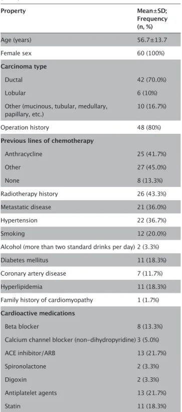
Among 726 patients who presented to Başkent University Faculty of Medicine, Department of Internal Medicine Division of Medi-cal Oncology between 01.01.2011 and 16.02.2016, a total of 130 patients received trastuzumab therapy. Among these, 70 patients were excluded due to missing ECG data or meeting the exclusion criteria. Of the remainder 60 patients, all (100%) patients were female. The mean age was 56.7±13.7 years. The breast cancer subtypes were ductal carcinoma in 42 (70.0%) patients, lobular carcinoma in 6 (10.0%) patients, and other types (mucinous, tu-bular, medullary, or papillary) in 10 (16.7%) patients. Twenty-one (36.0%) patients had metastatic disease; 48 (50.0%) patients were operated; and 26 (43.3%) received radiotherapy. While 8 (13.3%) patients did not receive any previous line of chemotherapy, 25 (41.7%) had been administered anthracyclines and 27 (45.0%) other chemotherapy agents. Table 2 presents the demographic and clinical parameters of the general study population.
A total of 20 (15.4% of the original trastuzumab population and 33.3% of the final study population) patients developed trastu-zumab-induced left ventricular systolic dysfunction. Of these,
14 (70%) were asymptomatic and 6 (30%) were symptomatic. Among 20 patients developing trastuzumab-induced left ven-tricular systolic dysfunction, 9 (45%) had a LVEF of <50% (Group 1a) and 11 (55%) had an LVEF of 50–54% (Group 1b). Those in Group1, 12 (60%) patients had persistent LVEF drop and 8 (40%) patients had reversible LVEF drop. All the patients in Group 1a
Table 2. Demographic properties of the whole study population
(n=60) Property Mean±SD; Frequency (n, %) Age (years) 56.7±13.7 Female sex 60 (100%) Carcinoma type Ductal 42 (70.0%) Lobular 6 (10%)
Other (mucinous, tubular, medullary,
papillary, etc.) 10 (16.7%)
Operation history 48 (80%)
Previous lines of chemotherapy
Anthracycline 25 (41.7%) Other 27 (45.0%) None 8 (13.3%) Radiotherapy history 26 (43.3%) Metastatic disease 21 (36.0%) Hypertension 22 (36.7%) Smoking 12 (20.0%)
Alcohol (more than two standard drinks per day) 2 (3.3%)
Diabetes mellitus 11 (18.3%)
Coronary artery disease 7 (11.7%)
Hyperlipidemia 11 (18.3%)
Family history of cardiomyopathy 1 (1.7%)
Cardioactive medications
Beta blocker 8 (13.3%)
Calcium channel blocker (non-dihydropyridine) 3 (5.0%)
ACE inhibitor/ARB 13 (21.7%)
Spironolactone 2 (3.3%)
Digoxin 2 (3.3%)
Antiplatelet agents 13 (21.7%)
Statin 11 (18.3%)
ACE: angiotensin converting enzyme; ARB: angiotensin receptor blocker
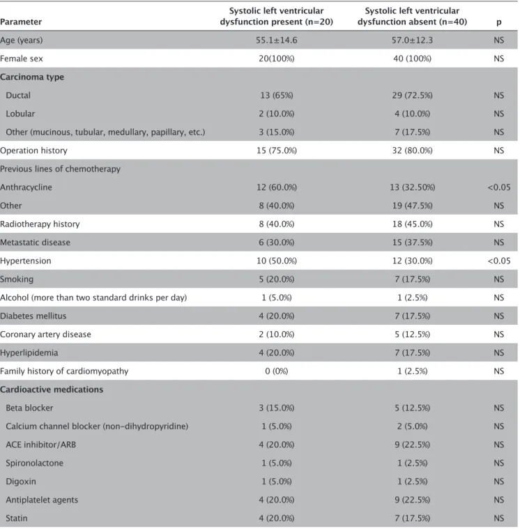
(n=9) had persistent LV dysfunction while three (27.3%) of pa-tients in the mild LV dysfunction had persistent LV dysfunction while 8 (72.7%) of them had reversible LV dysfunction, that is all patients with reversible LV dysfunction were in the mild LV dysfunction group. A comparison of Group 1 and Group 2 re-vealed that a history of anthracycline chemotherapy and hy-pertension were significantly more common in patients with trastuzumab-induced left ventricular systolic dysfunction than those without (for both, p<0.05) (Table 3); the other
demo-graphic and clinical parameters were similar between the two groups. A comparison of the Selvester score between Group 1 and Group 2 revealed no significant difference regarding trastu-zumab-induced left ventricular systolic dysfunction (5.83±1.11 vs 4.89±1.19 p>0.05). A comparison of Group 1a, Group 1b, and Group 2, on the other hand, revealed that Group 1a showed a significantly greater Selvester score than Group 1b and Group 2 did (7.29±1.61 vs 4.63±1.47; p<0.05 and 7.29±1.81 vs 4.89±1.29, respectively; p<0.05). However, there was no significant
differ-Table 3. Comparison of demographic, treatment and clinical parameters between the study groups with vs without systolic left
ven-tricular dysfunction
Parameter dysfunction present (n=20)Systolic left ventricular dysfunction absent (n=40)Systolic left ventricular p
Age (years) 55.1±14.6 57.0±12.3 NS
Female sex 20(100%) 40 (100%) NS
Carcinoma type
Ductal 13 (65%) 29 (72.5%) NS
Lobular 2 (10.0%) 4 (10.0%) NS
Other (mucinous, tubular, medullary, papillary, etc.) 3 (15.0%) 7 (17.5%) NS
Operation history 15 (75.0%) 32 (80.0%) NS
Previous lines of chemotherapy
Anthracycline 12 (60.0%) 13 (32.50%) <0.05 Other 8 (40.0%) 19 (47.5%) NS Radiotherapy history 8 (40.0%) 18 (45.0%) NS Metastatic disease 6 (30.0%) 15 (37.5%) NS Hypertension 10 (50.0%) 12 (30.0%) <0.05 Smoking 5 (20.0%) 7 (17.5%) NS
Alcohol (more than two standard drinks per day) 1 (5.0%) 1 (2.5%) NS
Diabetes mellitus 4 (20.0%) 7 (17.5%) NS
Coronary artery disease 2 (10.0%) 5 (12.5%) NS
Hyperlipidemia 4 (20.0%) 7 (17.5%) NS
Family history of cardiomyopathy 0 (0%) 1 (2.5%) NS
Cardioactive medications
Beta blocker 3 (15.0%) 5 (12.5%) NS
Calcium channel blocker (non-dihydropyridine) 1 (5.0%) 2 (5.0%) NS
ACE inhibitor/ARB 4 (20.0%) 9 (22.5%) NS
Spironolactone 1 (5.0%) 1 (2.5%) NS
Digoxin 1 (5.0%) 1 (2.5%) NS
Antiplatelet agents 4 (20.0%) 9 (22.5%) NS
Statin 4 (20.0%) 7 (17.5%) NS
ACE: angiotensin converting enzyme; ARB: angiotensin receptor blocker
ence between Group 1b and Group 2 regarding the Selvester score (p>0.05). In the comparison of the Selvester score within the reversible LV dysfunction group (n=8), no significant differ-ence was noted between the Selvester scores at the time of LV dysfunction and after recovery of LV dysfunction (5.13±1.29 vs 4.99±1.78, p>0.05) The Selvester score was significantly, albeit only moderately strongly, correlated to trastuzumab-induced left ventricular systolic dysfunction (r=0.189, p<0.05). A multi-variate analysis with binary logistic regression showed that the Selvester score was not significantly and independently correlat-ed to trastuzumab-induccorrelat-ed left ventricular systolic dysfunction (χ2=1.14; %95 GA 0.92–1.26, p>0.05).
DISCUSSION
Breast cancer is the most common cancer type that is respon-sible for the most cancer-related deaths among women world-wide (1). It has been reported that HER2-positive (HER2+) sub-types constitute 20% of all breast cancer sub-types and are related to poor prognosis. However, with the advent of anti-HER2 targeted therapies, this has started to change. Trastuzumab is a mono-clonal antibody that binds to the extracellular domain of HER2 and acts via activation of antibody-dependent cytotoxicity, inhi-bition of signal conduction, inhiinhi-bition of neoangiogenesis, and inhibition of repair of treatment-induced DNA injury (23). Trastu-zumab is used alone or in combination as neoadjuvant, adjuvant therapies, and in metastatic disease (24). Cardiotoxicity is the most important side effect of trastuzumab, which is most com-monly seen as asymptomatic LVEF depression or, less comcom-monly as overt congestive heart failure (8). Available data about trastu-zumab cardiotoxicity is limited, and various mechanisms, includ-ing angiotensin II upregulation and resultinclud-ing cell death due to oxidative stress (24), antiapoptotic protein BCL-XL’s downregu-lation and the proapoptotic protein BCL-XS’s upregudownregu-lation (25) and inability of maintaining sarcomeres’ structure and function and dysregulated scavenging mechanism for the proapoptotic oxidative subproducts (26, 27) have been postulated.
Trastuzumab’s cardiotoxicity is shown by a variety of echocar-diographic methods, of which biplane disks method (modified Simpson’s rule) is the most commonly used one, as in our study. Additionally, M-Mode echocardiography, three dimensional echo-cardiography, speckle tracking echoecho-cardiography, and deforma-tion imaging (strain and strain rate), as well as cardiac magnetic resonance imaging, are also utilized (28-30). However, most of the above-mentioned techniques are technologically demanding and time-consuming, and associated with increased cost and limited availability. Moreover, most echocardiographic techniques are operator dependent and flawed by a significant inter-observer variability in LVEF measurement. Therefore, simpler, cheap, readily available techniques are needed to assess left ventricular systol-ic function in trastuzumab-treated patients. Twelve-lead ECG is a simple, rapidly and widely available test relatively independent of operator interpretation. Therefore, it may offer promise in detect-ing trastuzumab-induced left ventricular systolic dysfunction. The Selvester score is a 12-lead ECG score that indicates left ventricular infarct size and extent (14). The original Selvester score is composed of 54 ECG criteria with a total of 32 points.
The subsequently developed modified Selvester score consists of 50 criteria with a total of 31 points (15, 16). The Selvester score represents the best electrocardiographic approximation of in-farct volume (17). Its role has been shown in a number of disor-ders and clinical scenarios characterized by myocardial scarring, including ischemic and non-ischemic cardiomyopathy (18, 19), Chagas disease (20), and acute myocardial infarction (21). How-ever, as far as we know, the Selvester score has not been studied in breast cancer patients with trastuzumab-induced left ventric-ular systolic dysfunction.
In our study, the modified Selvester score did not show any sta-tistically significant difference between the study groups with and without trastuzumab-induced left ventricular systolic dys-function (Group 1 and Group 2, respectively). However, the pa-tient subgroup with more severely depressed LVEF (LVEF<50%) (Group 1a) had a significantly greater Selvester score compared to both less severely affected patients (LVEF 50–54%) (Group 1b) and the controls (Group 2). These results suggest that the Sel-vester score was significantly increased among patients with more severe left ventricular systolic dysfunction, who possibly had myocardial scarring. Unfortunately, we had no MRI data to show myocardial scarring in such patients. However, 12 (60%) of those who had LVEF drop had a persistent LVEF drop, and these patients encompassed all the patients in Group 1a (more severe trastuzumab left ventricular systolic dysfunction). Furthermore, all the patients with reversible LV dysfunction had mild LV dys-function. These findings suggests that as more severe left ventric-ular systolic function occurs, more scarring or persistent myocar-dial fibrosis occurs, increasing the Selvester score. This finding is in line with the previous observations where the Selvester score was closely related to myocardial scar after myocardial infarction (14). In fact, the original definition of trastuzumab-induced left ventricular systolic dysfunction, that is a 10% LVEF drop to be-low 55% in asymptomatic patients or a 5 percent LVEF drop to below 55% in symptomatic patients, may not necessarily show severe left ventricular injury in every patient. Some patients may temporarily develop such dysfunction, and some due to contrac-tile dysfunction rather than scarring. Additionally, echocardiog-raphy, in its all forms, is heavily operator dependent and, thus, may show LVEF variability. In relation to this information, pa-tients with mild LVEF drop may in fact be included as depressed LVEF, which approximated the Selvester scores in both groups, precluding any significant difference between the patient and control groups regarding the Selvester score. However, as sub-group analyses showed a significantly greater Selvester score in patients with more severely depressed LVEF (LVEF<50%), those patients may have true LVEF depression. One may speculate that ECG changes may not occur without, or with small amount of, scarring or fibrosis in the left ventricular myocardium. The Sel-vester score, as a constellation of ECG parameters, may actually be expected to better delineate any left ventricular systolic dys-function than single ECG parameters like isolated Q waves, loss of R wave height, or persistent ST elevation. The lack of statistical significance between patients with and without left ventricular systolic dysfunction may have resulted from a lower number of patients with more severely depressed LVEF, thus, limiting our statistical power.
73
A reduction of the Selvester score in patients with reversible tras-tuzumab-induced LV dysfunction after normalization of LV func-tion would be a plausible finding. However, when we re-calcu-lated the Selvester score after normalization of LV function and compared it with the initial Selvester score among patients with LV dysfunction, we identified no significant difference between the two scores. This was interpreted in three ways: First, the low number of subjects in reversible LV dysfunction subgroup masked a statistically significant change in the Selvester score; second, the Selvester scores may be similar in patients with and without subtle degrees of LV dysfunction. This hypothesis was in-deed corroborated by the lack of statistical significance between the initial Selvester scores of control subjects and those with a minor LV dysfunction; third, some minor degree of LV dysfunc-tion (LVEF 50–54%) may have actually been a measurement dif-ference between independent echocardiographers performing the echocardiographic studies, and, thus, there may have not been true LV dysfunction and/or normalization at all in these pa-tients. Therefore, we could not ascertain the exact role of the Sel-vester score in reversible trastuzumab-induced LV dysfunction. This issue should be addressed in a study prospective random-ized, controlled study.
Our study has some limitations. First, this was a retrospective study with a relatively small study sample. Second, we could not assess posterior ECGs and, therefore, excluded posterior ECG criteria of the ECG leads V1 and V2, which limited the sensitivity of the study. Third, we did not exclude coronary artery disease as the cause of left ventricular systolic dysfunction. However, we determined trastuzumab-induced left ventricular systolic dysfunction on the basis of temporal and clinical features, with coronary artery disease being unlikely in our patients consider-ing the lack of ischemic symptoms, gender (all female), and age (predominantly of middle age) of the study population.
CONCLUSION
The Selvester score assessed from 12-lead ECG may be indica-tive of severe left ventricular systolic dysfunction evidenced by subnormal LVEF (<50%) among patients receiving trastuzumab therapy. However, patients with less severe left ventricular sys-tolic impairment may not be detected by the Selvester score as they may have no scarring in the left ventricular myocardium but only reversible systolic dysfunction. Further large-scale studies are needed to answer these questions.
Ethics Committee Approval: Ethics committee approval was received
for this study from the ethics committee of Başkent University (Approval Date: 09/02/2016; Approval No: KA16/57).
Informed Consent: As the present study had a retrospective design, no
written/verbal consent was obtained from the patients.
Peer-review: Externally peer-reviewed.
Author Contributions: Concept – O.Ç., A.N.A.; Design – O.Ç., K.C.Y.;
Su-pervision – İ.H.M., A.O.; Resources – İ.H.M.; Materials – E.K., A.O.; Data Col-lection and/or Processing – O.Ç., K.C.Y., M.Y.; Analysis and/or Interpreta-tion – O.Ç., M.Y., K.C.Y.; Literature Search – O.Ç., A.N.A.; Writing Manuscript – O.Ç.; Critical Review – A.N.A., A.O.; Other – İ.H.M.
Conflict of Interest: The authors have no conflicts of interest to declare. Financial Disclosure: This study was supported by Başkent University
Research Fund.
REFERENCES
1. Torre LA, Bray F, Siegel RL, Ferlay J, Lortet-Tieulent J, Jemal A. Global cancer statistics, 2012. CA Cancer J Clin 2015; 65: 87-108. [CrossRef] 2. Untch M, Ditsch N, Hermelink K. Immunotherapy: New options in
breast cancer treatment. Expert Rev Anticancer Ther 2003; 3: 403-8. [CrossRef]
3. Sengupta PP, Northfelt DW, Gentile F, Zamorano JL, Khandheria BK. Trastuzumab-induced cardiotoxicity: Heart failure at the crossroads. Mayo Clin Proc 2008; 83: 197-203. [CrossRef]
4. Marty M, Cognetti F, Maraninchi D, Snyder R, Mauriac L, Tubiana-Hu-lin M, et al. Randomized phase II trial of the efficacy and safety of trastuzumab combined with docetaxel in patients with human epi-dermal growth factor receptor 2-positive metastatic breast cancer administered as first-line treatment: the M77001 Study Group. J Clin Oncol 2005; 23: 4265274. [CrossRef]
5. Piccart-Gebhart MJ, Procter M, Leyland-Jones B, Goldhirsch A, Untch M, Smith I, et al. Trastuzumab after adjuvant chemotherapy in HER-2 positive breast cancer. N Engl J Med 2005; 353: 1659-72. [CrossRef] 6. Romond EH, Perez EA, Bryant J, Suman VJ, Geyer CE Jr, Davidson
NE, et al. Trastuzumab plus adjuvant chemotherapy for operable HER2-positive breast cancer. N Engl J Med 2005; 353: 1673-84. [CrossRef]
7. Vogel CL, Cobleigh MA, Tripathy D, Gutheil JC, Harris LN, Fehren-bacher L et al. Efficacy and safety of trastuzumab as a single agent in first-line treatment of HER2-overexpressing metastatic breast cancer. J Clin Oncol 2002; 20: 719-26. [CrossRef]
8. Seidman A, Hudis C, Pierri MK, Shak S, Paton V, Ashby M, et al. Car-diac dysfunction in the trastuzumab clinical trials experience. J Clin Oncol 2002; 20: 1215-21. [CrossRef]
9. Keefe DL. Trastuzumab-associated cardiotoxicity. Cancer 2002; 95: 1592-600. [CrossRef]
10. Romond EH, Jeong JH, Rastogi P, Swain SM, Geyer CE Jr, Ewer MS, et al. Seven-year follow-up assessment of cardiac function in NSABP B-31, a randomized trial comparing doxorubicin and cyclophosphamide followed by paclitaxel (ACP) with ACP plus trastuzumab as adjuvant therapy for patients with node-positive, human epidermal growth factor receptor 2-positive breast cancer. J Clin Oncol 2012; 30: 3792-9. [CrossRef]
11. Slamon DJ, Leyland-Jones B, Shak S, Fuchs H, Paton V, Bajamonde A, et al. Use of chemotherapy plus a monoclonal antibody against HER2 for metastatic breast cancer that overexpresses HER2. N Engl J Med 2001; 344: 783-92. [CrossRef]
12. Yeh ET, Bickford CL. Cardiovascular complications of cancer thera-py: incidence, pathogenesis, diagnosis, and management. J Am Coll Cardiol 2009; 53: 2231-47. [CrossRef]
13. Vasu S, Hundley WG. Understanding cardiovascular injury after treatment for cancer: an overview of current uses and future di-rections of cardiovascular magnetic resonance. J Cardiovasc Magn Reson 2013; 15: 66. [CrossRef]
14. Selvester RH, Wagner GS, Hindman NB. The Selvester QRS scoring system for estimating myocardial infarct size. The development and application of the system. Arch Intern Med 1985; 145: 1877-81. [CrossRef]
15. Wagner GS, Freye CJ, Palmeri ST, Roark SF, Stack NC, Ideker RE, et al. Evaluation of a QRS scoring system for estimating myocardial infarct size. I. Specificity and observer agreement. Circulation 1982; 65: 342-7. [CrossRef]
16. Hindman NB, Schocken DD, Widmann M, Anderson WD, White RD, Leggett S, et al. Evaluation of a QRS scoring system for estimating
myocardial infarct size. V. Specificity and method of application of the complete system. Am J Cardiol 1985; 55: 1485-90. [CrossRef] 17. Carey MG, Luisi AJ, Baldwa S, Al-Zaiti S, Veneziano MJ, deKemp RA,
et al. The Selvester QRS Score is more accurate than Q waves and fragmented QRS complexes using the Mason-Likar configuration in estimating infarct volume in patients with ischemic cardiomyopa-thy. J Electrocardiol 2010; 43: 318-25. [CrossRef]
18. Strauss DG, Selvester RH, Lima JA, Arheden H, Miller JM, Gersten-blith G, et al. ECG quantification of myocardial scar in cardiomyop-athy patients with or without conduction defects: correlation with cardiac magnetic resonance and arrhythmogenesis. Circ Arrhythm Electrophysiol 2008; 1: 327-36. [CrossRef]
19. Strauss DG, Selvester RH. The QRS complex--a biomarker that “im-ages” the heart: QRS scores to quantify myocardial scar in the pres-ence of normal and abnormal ventricular conduction. J Electrocar-diol 2009; 42: 85-96. [CrossRef]
20. Strauss DG, Cardoso S, Lima JA, Rochitte CE, Wu KC. ECG scar quan-tification correlates with cardiac magnetic resonance scar size and prognostic factors in Chagas’ disease. Heart 2011; 97: 357-61. [CrossRef]
21. Tjandrawidjaja MC, Fu Y, Westerhout CM, Wagner GS, Granger CB, Armstrong PW. Usefulness of the QRS score as a strong prognostic marker in patients discharged after undergoing primary percuta-neous coronary intervention for ST-segment elevation myocardial infarction. Am J Cardiol 2010; 106: 630-4. [CrossRef]
22. Engblom H, Wagner GS, Setser RM, Selvester RH, Billgren T, Kasper JM, et al. Quantitative clinical assessment of chronic anterior myo-cardial infarction with delayedenhancement magnetic resonance imaging and QRS scoring. Am Heart J 2003; 146: 359-66. [CrossRef] 23. Spector NL, Blackwell K. Understanding the mechanisms behind
trastuzumab therapy for human epidermal growth factor receptor 2-positive breast cancer. J Clin Oncol 2009; 27: 5838-47. [CrossRef] 24. Pondé NF, Lambertini M, de Azambuja E. Twenty years of anti-HER2
therapy-associated cardiotoxicity. ESMO Open 2016; 1: e000073. [CrossRef]
25. Grazette LP, Boecker W, Matsui T, Semigran M, Force TL, Hajjar RJ, et al. Inhibition of ErbB2 causes mitochondrial dysfunction in car-diomyocytes: implications for herceptin-induced cardiomyopathy. J Am Coll Cardiol 2004; 44: 2231-8. [CrossRef]
26. Kuramochi Y, Guo X, Sawyer DB. Neuregulin activates erbB2-depen-dent src/FAK signaling and cytoskeletal remodeling in isolated adult rat cardiac myocytes. J Mol Cell Cardiol 2006; 41: 228-35. [CrossRef] 27. ElZarrad MK, Mukhopadhyay P, Mohan N, Hao E, Dokmanovic M,
Hirsch DS, et al. Trastuzumab alters the expression of genes essen-tial for cardiac function and induces ultrastructural changes of car-diomyocytes in mice. PLoS One 2013; 8: e79543. [CrossRef] 28. Walker JR, Sharma A, Lytwyn M, Bohonis S, Thliveris J, Singal PK, et
al. The cardioprotective role of probucol against anthracycline and trastuzumab-mediated cardiotoxicity. J Am Soc Echocardiogr 2011; 24: 699-705. [CrossRef]
29. Walker J, Bhullar N, Fallah-Rad N, Lytwyn M, Golian M, Fang T, et al. Role of three-dimensional echocardiography in breast cancer: com-parison with two-dimensional echocardiography, multiple-gated acquisition scans, and cardiac magnetic resonance imaging. J Clin Oncol 2010 20; 28: 3429-36. [CrossRef]
30. Fallah-Rad N, Walker JR, Wassef A, Lytwyn M, Bohonis S, Fang T, et al. The utility of cardiac biomarkers, tissue velocity and strain imaging, and cardiac magnetic resonance imaging in predicting early left ventricular dysfunction in patients with human epidermal growth factor receptor II-positive breast cancer treated with adjuvant tras-tuzumab therapy. J Am Coll Cardiol 2011; 57: 2263-70. [CrossRef]
How to cite:
Çiftci O, Yılmaz KC, Karaçağlar E, Akgün AN, Yılmaz M, Oğuz A, et al. The Role of Selvester Score on 12-Lead ECG in Determi-nation of Left Ventricular Systolic Dysfunction Among Patients Receiving Trastuzumab Therapy. Eur J Ther 2019; 25(1): 69-75.