Department of Pathology, Pt. BDS, PGIMS, University of Health Sciences, Rohtak, India
Yazışma Adresi /Correspondence: Monika Gupta,
17/8 FM, Medical enclave PGIMS Rohtak, Haryana, India Email: [email protected]
Geliş Tarihi / Received: 20.07.2015, Kabul Tarihi / Accepted: 27.01.2016 ORIGINAL ARTICLE / ÖZGÜN ARAŞTIRMA
Clinicohematological Profile of Pancytopenia: A Study from a Tertiary Care
Hospital
Pansitopeninin Klinik ve Hematolojik Profili: Bir Üçüncü Basamak Hastane Çalışması
Monika Gupta, Abha Chandna, Sanjay Kumar, Sant Prakash Kataria, Sonia Hasija, Gajender Singh, Rajeev Sen
ÖZET
Amaç: Pansitopeni geniş bir ayırıcı tanı yelpazesi olan
klinik ve hematolojik bir problemdir. Hastaların detaylı bir incelemesi gerekmektedir çünkü etiyolojide geri dönü-şümlü bir etiyoloji olabileceği gibi erken patolojinin hayat kurtaracağı ciddi bir patolojide sorumlu olabilir. Pansito-peni tanısında kemik iliğinin hücresel ve morfolojik ince-lenmesi için kemik iliği aspirasyonu gerekmektedir. Bu çalışma, kemik iliği bulguları temelinde pansitopeninin nedenini bulmak amacıyla yapılmıştır.
Yöntemler: Bu çalışma bir yıl süre içinde patoloji
bölü-münde gerçekleştirilmiştir. Pansitopeni kriterlerini karşı-layan 169 hasta çalışmaya dahil edildi. Tüm olgularda, tam kan sayımı, periferik yayma incelemesi ve kemik iliği aspirasyonu takiben ayrıntılı klinik öykü ve fizik muayene yapıldı.
Bulgular: Erkek kadın oranında erkek lehine hafif bir
bas-kınlık vardı: 1/1.2. Hastaların büyük bir çoğunluğu, 10 lu ve 20 li yaşlarındaydılar. Pansitopenin ana nedeni mega-loblastik anemi ve ardından beslenme yetersizliği anemisi ve diğer nedenler olarak görüldü.
Sonuç: Bu çalışma göstermiştir ki Hindistan gibi
geliş-mekte olan ülkelerde pansitopeni etiyolojisi çoğunlukla geri dönüşümlü nedenlere bağlıdır ve bu ülkelerde hasta-lara kan yapıcı vererek yakın hematolojik takipleri önem-lidir.
Anahtar kelimeler: Pansitopeni, kemik iliği,
megaloblas-tik anemi, aplasmegaloblas-tik anemi
ABSTRACT
Objective: Pancytopenia is a clinicohematological
prob-lem with wide spread discriminated diagnosis. A through evaluation of patient is necessary to identify the cause as large number of patients has reversible etiology and early diagnosis may be lifesaving. Diagnosis of pancyto-penia requires microscopic examination of bone marrow aspirates to assess the overall cellularity and morphol-ogy. This study was conducted with the aim to find out the cause of pancytopenia on the basis of bone marrow findings.
Methods: The present study was conducted in the
de-partment of pathology over a period of one year. A total of 169 patients were included in the study that fulfilled the criteria of pancytopenia. A detailed clinical history and physical examination followed by complete blood count, peripheral smear examination and bone marrow aspira-tion was done in all cases.
Results: There was slight male predominance with male
to female ratio of 1.2:1. The majority of patients were in second and third decade. The main cause of pancytope-nia was megaloblastic anemia followed by mixed nutri-tional deficiency anemia and others.
Conclusion: This study emphasized that in developing
countries like India majority of the patients had reversible etiology and patients can be put on a trial of hematinics and close haematological follow up.
Key words: Pancytopenia, bone marrow, megaloblastic
INTRODUCTION
Pancytopenia is an important clinicohematological entity encountered in our day-to-day clinical prac-tice. A multitude of disorders primarily or second-arily affecting the bone marrow manifests with vari-ous haematological derangements which is com-monly presented as pancytopenia. In pancytopenia there is reduction of all the three formed elements of blood below the normal reference range [1].
The presenting symptoms such as pallor, dys-pnea, bleeding and bruising are usually due to ane-mia and thrombocytopenia. Increased propensity to infections due to leucopenia is an uncommon cause of initial presentation of the patient [2]. Pancytope-nia in a patient with associated organomegaly and lymphadenopathy usually suggest the possibility of malignancies or bone marrow failure syndrome [3].
Incidence of various disorders causing pancyto-penia varies according to geographical distribution and genetic differences [4]. Underlying pathology determines the management and prognosis of the patients [5]. Identifying the correct etiopathology in a given case is crucial and helps in implementing timely and appropriate treatment [6]. Bone marrow examination plays a significant role in investigation of pancytopenia. This study was conducted with the aim to find out the cause of pancytopenia on the ba-sis of bone marrow findings.
METHODS
The present study was conducted in the department of pathology from January 2012 to December 2012. A total of 169 patients were included in the study that fulfilled the criteria of pancytopenia. The inclusion criteria for pancytopenia were haemoglobin (Hb) <10g/dl, total leukocyte count (TLC) <4.0×10⁹/µl and platelets<100×109/µl [7]. A detailed relevant
clinical history for fever, pain, weight loss, weak-ness, treatment history, history of drug intake, radia-tion exposure or any other significant history was taken. Physical examination was done for pallor, jaundice, hepatomegaly, splenomegaly and lymph-adenopathy. Complete blood count was performed using an automated five- part hematology analyzer (Mindray BC 5800). Peripheral smears were ex-amined after staining with Leishman’s stain. Bone marrow aspiration was performed in all cases using
Salah needle from posterior superior iliac spine un-der aseptic condition. All the bone marrow aspirate smears were stained with May Grunewald Giemsa stain. Special staining for Sudan Black B, Period-ic acid Schiff and Perl’s stain on the smears done wherever indicated. Bone marrow biopsy was done for evaluation of bone marrow in cases with insuf-ficient cells, dry tap or hypoplastic bone marrow. Work up for Paroxysmal Nocturnal Hemoglobin-uria (PNH) was done for CD55 and CD59 by flow cytometry wherever required.
RESULTS
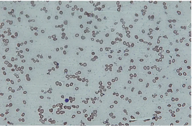
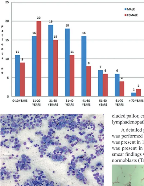
A total of 822 bone marrow aspirations were re-ceived from January 2012 to December 2012 for various haematological studies. Out of them, 169 cases (20.6%) showing evidence of pancytope-nia on peripheral smear examination were includ-ed in the present study (Figure 1). There were 94 (55.6%) males and 75 (44.4%) females. There was slight male predominance with male to female ra-tio of 1.2:1.The patients had age ranging from 1 to 75 years, maximum number of patients 36 (21.3%) were in age group of 11-20 years (Figure 2).
Figure 1. Peripheral smear showing macrocytic picture
with occasional lymphocyte (Leishman stain × 200X)
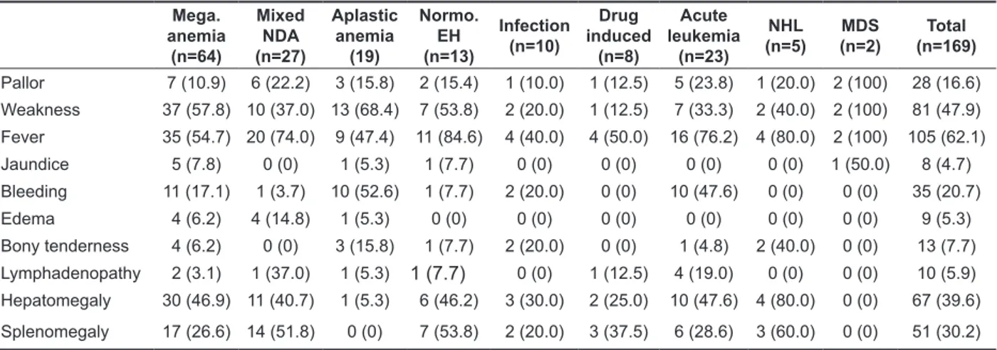
Bone marrow examination was helpful in iden-tifying the etiology of pancytopenia. Among non-malignant causes, megaloblastic anemia was the predominant cause 64 (37.87%) (Figure 3) followed by mixed nutritional deficiency anemia 27(15.98%) and aplastic anemia 19 (11.24%) (Figure 4).Other non-malignant causes were normoblastic erythroid hyperplasia, infections (hepatitis, HIV, Parvovirus) and drug induced bone marrow suppression. Drugs induced bone suppression was present in eight
pa-tients. Out of eight, three patients were taking non-steroidal anti-inflammatory drugs for joint pains while three patients gave history of intake of anti-biotic (chloramphenicol). Two patients were on di-uretics for hypertension. Among malignant causes,
acute leukemia forms the major part 21 (12.4%) (Figure 5) followed by non-Hodgkin’s lymphoma (NHL) and myelodysplastic syndrome (MDS) (Fig-ure 6).
Figure 2. Age distribution of
pancy-topenic patients
Figure 3. Bone marrow Aspirate showed megaloblastic
erythroid hyperplasia (Leishman stain × 200X)
The results of clinical manifestation according to cause of pancytopenia in all patients reflected major complaint of fever 105(62%), followed by weakness 81(47.9%). Hepatomegaly was the most common sign 67 (39.6%) followed by splenomeg-aly (30.2%) and bleeding manifestations (20.7%). Seven patients with normoblastic erythroid hyper-plasia had splenomegaly. Other manifestation
in-cluded pallor, edema, jaundice, bony tenderness and lymphadenopathy (Table 1).
A detailed peripheral blood smear examination was performed on all patients. Anisopoikilocytosis was present in 119(70.4%) cases and lymphocytosis was present in 97(57.4%) cases. Other peripheral smear findings were polychromasia and presence of normoblasts (Table 2).
Figure 4. Bone marrow biopsy showed hypoplastic
Figure 5. Bone marrow Aspirate showed blasts in
pancy-topenic patient (Leishman stain × 200X)
Haematological parameters revealed mean Hb 5.6 g/dl (lowest in megaloblastic anemia 4.2 g/dl), mean TLC 2.14 ×10⁹/L (lowest in MDS
1.09×10⁹/L),mean platelet count 34.1 × 10⁹/L (
lowest in aplastic anemia 19 ×109/L),mean MCV
91.76 fl, mean MCH 29.34 pg and mean MCHC 34.2+2.7%. Except drug induced pancytopenia and
acute leukemia cases, there was no significant dif-ference in red cell indices (Table 3). PNH work up in all cases with normoblastic erythroid hyperplasia by flow cytometry for CD55 and CD59 were nega-tive.
Figure 6. Etiological profile of pancytopenic patients (%) Table 1. Clinical profile according to cause of pancytopenia, n (%)
Mega. anemia (n=64) Mixed NDA (n=27) Aplastic anemia (19) Normo. EH (n=13) Infection (n=10) Drug induced (n=8) Acute leukemia (n=23) NHL (n=5) (n=2)MDS (n=169)Total Pallor 7 (10.9) 6 (22.2) 3 (15.8) 2 (15.4) 1 (10.0) 1 (12.5) 5 (23.8) 1 (20.0) 2 (100) 28 (16.6) Weakness 37 (57.8) 10 (37.0) 13 (68.4) 7 (53.8) 2 (20.0) 1 (12.5) 7 (33.3) 2 (40.0) 2 (100) 81 (47.9) Fever 35 (54.7) 20 (74.0) 9 (47.4) 11 (84.6) 4 (40.0) 4 (50.0) 16 (76.2) 4 (80.0) 2 (100) 105 (62.1) Jaundice 5 (7.8) 0 (0) 1 (5.3) 1 (7.7) 0 (0) 0 (0) 0 (0) 0 (0) 1 (50.0) 8 (4.7) Bleeding 11 (17.1) 1 (3.7) 10 (52.6) 1 (7.7) 2 (20.0) 0 (0) 10 (47.6) 0 (0) 0 (0) 35 (20.7) Edema 4 (6.2) 4 (14.8) 1 (5.3) 0 (0) 0 (0) 0 (0) 0 (0) 0 (0) 0 (0) 9 (5.3) Bony tenderness 4 (6.2) 0 (0) 3 (15.8) 1 (7.7) 2 (20.0) 0 (0) 1 (4.8) 2 (40.0) 0 (0) 13 (7.7) Lymphadenopathy 2 (3.1) 1 (37.0) 1 (5.3) 1 (7.7) 0 (0) 1 (12.5) 4 (19.0) 0 (0) 0 (0) 10 (5.9) Hepatomegaly 30 (46.9) 11 (40.7) 1 (5.3) 6 (46.2) 3 (30.0) 2 (25.0) 10 (47.6) 4 (80.0) 0 (0) 67 (39.6) Splenomegaly 17 (26.6) 14 (51.8) 0 (0) 7 (53.8) 2 (20.0) 3 (37.5) 6 (28.6) 3 (60.0) 0 (0) 51 (30.2)
Mega.: Megaloblastic, NDA: Nutritional deficiency anemia, Normo. EH: Normoblastic Erythroid hyperplasia, NHL: Non-Hodgkin lymphoma, MDS: Myelodysplastic syndrome
Table 2. Peripheral blood findings
Mega. anemia (n=64) Mixed NDA (n=27) Aplastic anemia (n=19) Normoblastic EH (n=13) Infection (n=10) Drug induced (n=8) Acute leukemia (n=23) NHL (n=5) (n=2)MDS (n=169)Total Anisopoikilo-cytosis 62 (96.9) 22 (81.5) 7 (36.8) 10 (76.9) 3 (30) 5 (62.5) 4 (19.0) 4 (80) 2 (100) 119 (70.4) nRBCs/ 100 WBCs 4 (6.2) 2 (7.4) 0 (0) 1 (7.7) 2 (20) 1 (1.2) 7 (33.3) 1 (20) 0 (0) 18 (4.1) Polychromasia 7 (10.9) 2 (7.4) 0 (0) 1 (7.7) 1 (10) 0 (0) 2 (9.5) 0 (0) 0 (0) 13 (5.9) Lymphocytosis 38 (59.4) 10 (37.0) 19 (100) 9 (69.3) 6 (60) 4 (50) 5 (23.8) 5 (100) 1 (50) 97 (57.4)
Mega.: Megaloblastic, NDA: Nutritional deficiency anemia, Normo. EH: Normoblastic Erythroid hyperplasia, NHL: Non-Hodgkin lymphoma, MDS: Myelodysplastic syndrome
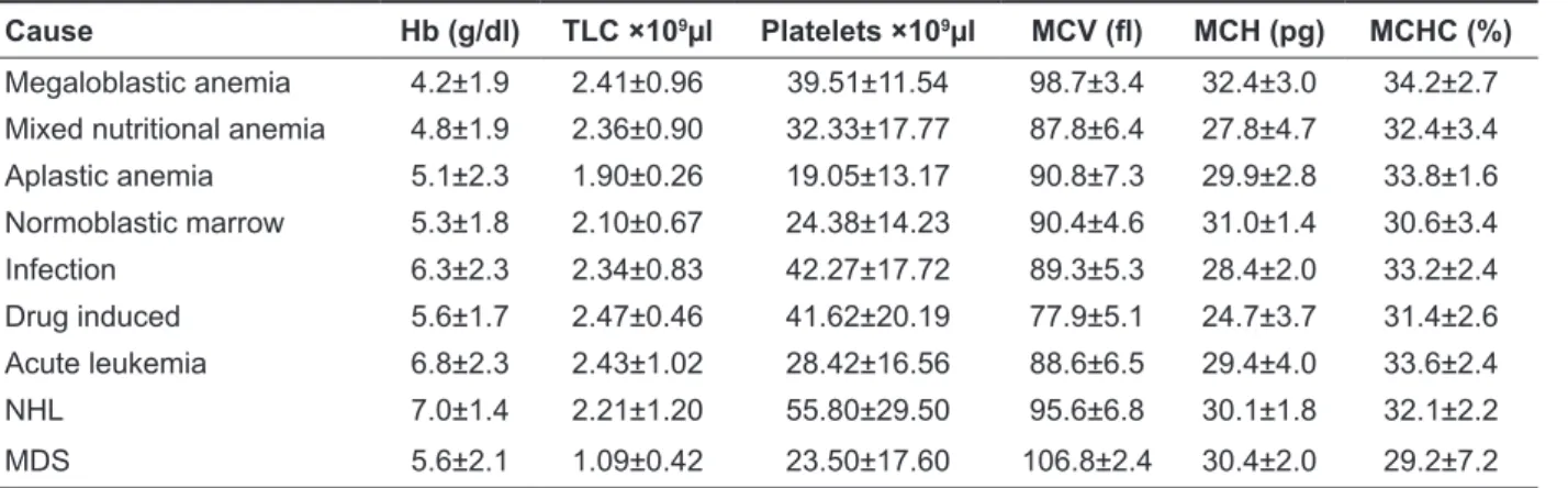
Table 3. Mean peripheral blood indices
Cause Hb (g/dl) TLC ×109µl Platelets ×109µl MCV (fl) MCH (pg) MCHC (%)
Megaloblastic anemia 4.2±1.9 2.41±0.96 39.51±11.54 98.7±3.4 32.4±3.0 34.2±2.7
Mixed nutritional anemia 4.8±1.9 2.36±0.90 32.33±17.77 87.8±6.4 27.8±4.7 32.4±3.4
Aplastic anemia 5.1±2.3 1.90±0.26 19.05±13.17 90.8±7.3 29.9±2.8 33.8±1.6 Normoblastic marrow 5.3±1.8 2.10±0.67 24.38±14.23 90.4±4.6 31.0±1.4 30.6±3.4 Infection 6.3±2.3 2.34±0.83 42.27±17.72 89.3±5.3 28.4±2.0 33.2±2.4 Drug induced 5.6±1.7 2.47±0.46 41.62±20.19 77.9±5.1 24.7±3.7 31.4±2.6 Acute leukemia 6.8±2.3 2.43±1.02 28.42±16.56 88.6±6.5 29.4±4.0 33.6±2.4 NHL 7.0±1.4 2.21±1.20 55.80±29.50 95.6±6.8 30.1±1.8 32.1±2.2 MDS 5.6±2.1 1.09±0.42 23.50±17.60 106.8±2.4 30.4±2.0 29.2±7.2
NHL: Non-Hodgkin lymphoma, MDS: Myelodysplastic syndrome
Table 4. Comparison with different studies
Author Year Place No of cases First commonCause Second common cause
Imbert, et al. 1989 France 213 Malignant myeloid andlymphoid disorders Aplastic anemia
Varma, et al. 1992 India 202 Aplastic anemia Megaloblastic anemia
Tilak, et al. 1999 India 77 Megaloblastic anemia Aplastic anemia
Kumar, et al. 2001 India 166 Aplastic anemia Megaloblastic anemia
Khodke 2001 India 50 Megaloblastic anemia Aplastic anemia
Khunger, et al. 2002 India 200 Megaloblastic anemia Aplastic anemia
Jha, et al. 2008 Nepal 148 Hypoplastic bone marrowin children, megaloblastic anemia in adults
Erythroid hyperplasia in children and hypoplastic marrow in adults
Memon, et al. 2008 Pakistan 230 Aplastic anemia Megaloblastic anemia
Jain&Naniwadekar 2013 India 250 Hypersplenism Infections
Present study India 169 Megaloblastic anemia Mixed nutritionaldeficiency anemia DISCUSSION
Pancytopenia is not an uncommon haematological problem encountered in our day to day clinical prac-tice and should be suspected on clinical grounds when patient presents with unexplained anemia, fever and bleeding tendencies. The normal adult bone marrow produces about 1.7x10¹¹ RBC,1x1011
neutrophils and 2x10¹¹ platelets each day and thus
have tremendous capacity to increase output of these cells when necessary with help of growth factors and other cytokines [8].The mechanisms contributing to pancytopenia include decrease in hematopoietic cell production ,marrow replacement by abnormal cells, suppression of marrow growth and differentiation, ineffective hematopoiesis with cell death, defective cell formation, destruction of
cells in hypertrophied and overactive reticuloendo-thelial system [9].Till date there are limited number of studies from Indian subcontinent on the clinico-hematological correlation and frequency of various causes of pancytopenia.
The commonest cause in the present study was megaloblastic anemia (37.87%). In other similar studies the incidence of megaloblastic anemia var-ies from 37 to 68% [8-10]. Bone marrow was hy-per cellular in majority of cases with megaloblastic erythropoiesis, giant band forms, metamyelocytes and increased iron stores. Megaloblastic anemia due to vitamin B12 or folic acid deficiency is now a well-recognized and established cause of cytope-nias [11-13].It can either present as bicytopenia or pancytopenia or rarely with thrombocytopenia only
[14]. The possible explanation of folate and vita-min B12 deficiency in our study could be various chronic inflammatory disorders of gut like parasitic infestations, chronic diarrhea and malabsorption states along with poor nutrition mostly vegetarian.
Mixed nutritional deficiency anemia (15.98%) was the second commonest cause in the present study. This cause is not reported from Indian stud-ies but two studstud-ies from Pakistan Menon et al [15] (8.69%) and Khan et al [16] (5.3%) reported as cause of pancytopenia in children. Both the above causes reflect higher prevalence of nutritional ane-mias in Indian subjects as well as in other devel-oping countries. This is in sharp contrast to studies done in west where aplastic anemia was the com-monest cause, incidence varies from 10 to 52.7% [4,17,18].
In present study incidence of aplastic anemia was 11.24%. Similar findings were observed by several researchers [12,19] from India while Varma et al [5] found higher incidence of aplastic anemia (40.6%). Relative lymphocytosis was seen in all cases of aplastic anemia in peripheral smear exami-nation. Bone marrow aspiration yielded dry tap in most of these cases and bone marrow biopsy was performed subsequently to confirm the diagnosis. The pathophysiology of aplastic anaemia is now be-lieved to be immune mediated [19]. The immune re-sponse may be triggered by environmental exposure of chemicals or drugs. In our cases it could be due to exposure of pesticides as most of our patients were from agricultural background and use of pesticide is very common.
Normoblastic erythroid hyperplasia was pres-ent in 7.69% cases in prespres-ent study with normal iron stores. Half of the patients had splenomegaly and pancytopenia could be due to hypersplenism. In the remaining cases peripheral pancytopenia may represent a refractory anemia or evolution of hypo-plasia/aplasia [11]. Differentiation of these groups remains unsatisfactory and these patients should be kept under regular haematological follow up.
In the present study 5.9% cases showed evi-dence of infective etiology which included case of malaria, HIV, hepatitis, parvovirus infection. Ma-laria related cytopenia was also noted in other stud-ies [20,21]. The incidence of malaria was noted in
low income group with poor sanitation facilities. Pancytopenia occurs late in course of HIV infec-tion. Parvovirus B19 is a cause of chronic anemia in individuals with AIDS. All the patients with infec-tious related pancytopenia recovered after appropri-ate treatment.
Drug induced pancytopenia was seen in 4.73% cases. Majority of cases it was due to prolonged use of causing drugs .The role of chloramphenicol as the causative agent for aplastic anemia has been in-vestigated extensively however; restriction of drug will help in reducing the cases of this preventable cause of pancytopenia [22].
In the present study 26 cases showed malig-nant etiology of which acute leukemia constituted 12.43% cases of pancytopenia. The incidence of acute leukemia in similar studies varies from 1.8-19.59% [7,9,10] while Varma et al [5] reported acute leukemia as third most common cause. In the present study these patients presented with normo-cytic normochromic anemia, reduced leucocyte and platelet count and presence of circulating immature cells in peripheral smear. Bone marrow was hyper-cellular in all cases with reduced erythropoiesis and megakaryopoiesis.
In present study, lymphoproliferative disorders constituted 2.96% of cases. This group included multiple myeloma, chronic lymphoproliferative disorder and splenic lymphoma cases. Other simi-lar studies [12,19] also reported NHL as uncommon cause of pancytopenia. Pancytopenia in CLL can be explained by the immunologic mechanism, bone marrow infiltration or hypersplenism. Pancytopenia with MDS as diagnosis was noted in 1.18% of our cases. Hypercellular marrow with presence of dys-plastic erythroblasts, myeloid cells and megakaryo-cytes confirmed the diagnosis (Table 4).
The variation in the frequency of various diag-nostic entities causing pancytopenia has been attrib-uted to differences in methodology and stringency of diagnostic criteria, geographic area, and period of observation, genetic differences and varying ex-posure to myelotoxic agents etc [4].
Routine haematological parameters were non-specific and showed a significant overlap among major causes of cytopenias. However, the periph-eral blood films were valuable in pointing towards
cause in patients with megaloblastic anemia and leukemia. Considerable degrees of anisopoikilocy-tosis with macro-ovalocytes and hypersegmented neutrophils were main features in majority of cases of megaloblastic anemia. The importance of bone marrow examination in pancytopenic patients has been well emphasised in earlier studies [8,17,18]. Bone marrow aspirate was found to be sufficient for the diagnosis in most cases. However, biopsy was mandatory for the diagnosis of aplastic anemia. Common clinical presentations were fever, weak-ness, hepatosplenomegaly, bleeding manifestations and pallor.
In conclusion, pancytopenia is a common en-tity. However, it has received inadequate attention in the Indian subcontinent. A study of pancytope-nia using easily available diagnostic techniques is therefore important for early diagnosis and timely management of patients. Megaloblastic anemia was the commonest cause of pancytopenia in the present study followed by mixed nutritional deficiency ane-mia and aplastic aneane-mia among the non-malignant disorders. Acute leukemia constitutes most common malignant haematological disorder causing pancy-topenia. A comprehensive clinical, haematological workup and bone marrow study of patients usually help in evaluating the aetiology of pancytopenia. In addition attempt should be made for an early rec-ognition of underlying aetiology so that treatable causes are identified without delay and prognosis can be improved.
Declaration of Conflicting Interests: The
au-thors declare that they have no conflict of interest.
Financial Disclosure: No financial support
was received.
REFERENCES
1. Raz I, Shinar E, Polliack A. Pancytopenia with hypercel-lular bone marrow a possible paraneoplastic syndrome in carcinoma of lungs: a report of three cases. Am J Hematol 1984;16:403-408.
2. De Gruchy GC: Pancytopenia; Aplastic anemia. In Firkin F, Chestermann C, Penington D (eds). De Gruchy’s Clinical Haematology in Medical Practice.5th edition. Oxford Uni-versity Press. 1991:p: 119-136.
3. Chhabra A, Chander V, Patel A, et al. Clinico-aetiological profile of pancytopenia in paediatric practice. J Indian Acad Clin Med 2012;13:282-285.
4. International Agranulocytosis and Aplastic Anaemia Study group: Incidence of aplastic anaemia: the relevance of di-agnostic criteria. Blood 1987;70:1718-1721.
5. Verma N, Dash S. Reappraisal of underlying pathology in adult patients presenting with pancytopenia. Trop Geog Med 1992;44:322-327.
6. Jain and Naniwadekar. An etiological reappraisal of pancy-topenia- largest series reported to date from single tertiary care teaching hospital. BMC Hematology 2013;13:10. Doi:10.1186/2052-1839-13-10
7. Naseem S, Varma N, Das R, et al. Paediatric patients with bi-cytopenia/pancytopenia: review of aetiologies and clinico-haematological profile at a tertiary centre. Indian J Pathol Microbiol 2011;54:75-80.
8. Bunch C. Bone marrow failure. Med Int 1995;10:495-499. 9. Jha A, Sayami G, Adhikari RC, et al. Bone marrow
ex-amination in cases of pancytopenia. J Nepal Med Assoc. 2008;47:12-17.
10. Kumar R, Kalra SP, Kumar H, et al. Pancytopenia-a six year study. J Assoc Physicians India 2001;49:1078-1081. 11. Khodke K, Marwah S, Buxi G, et al. Bone marrow
exami-nation in cases of pancytopenia. J Indian Acad Clin Med 2001:2:55-59.
12. Tilak V, Jain R. Pancytopenia-a clinicohematological analy-sis of 77 cases. Indian J Pathol Microbiol 1999;42:399-404. 13. Chandra J, Jain V, Narayan S, et al. Folate and cobalamin
deficiency in megaloblastic anemia in children. Indian Pe-diatr 2002;39:453-457.
14. Jan MA.Thrombocytopenia in children. J Postgrad Med Inst 2004;18:353-358.
15. Menon S, Shaikh S, Nizamani MA. Etiological spectrum of pancytopenia based on bone marrow examination in chil-dren. J Coll Physicians Surg Pak 2008;18:163-167. 16. Khan FS, Hasan RF. Bone marrow examination of
pancyto-penic children. J Pak Med Assoc 2012;62:660-663. 17. Imbert M, Scoazec JY, Mary JY, et al. Adult patients
pre-senting with pancytopenia: A reappraisal of underlying pa-thology and diagnostic procedures in 213 cases. Hematol Pathol 1989;3:159-167.
18. Keisu M, Ost A. Diagnosis in patients with severe pancyto-penia suspected of having aplastic anemia. Euro J Haema-tol 1990;45:11-14.
19. Khunger JM, Arun selvi S, Sharma U, et al. Pancytopenia-a clinicohematological analysis of 200 cases. Indian J Pathol Microbiol 2002;45:375-379.
20. Latger Cannard V, Bibes B, Dao A, et al. Malaria related cytopenia. Ann Biol Clin 2002;60:213-216.
21. Aouba A, Noguera ME, Clawel JP, et al. Haemophagocytic syndrome associated with plasmodium vivax infection. Br J Haematol 2000;108:832-833.
22. Modan B, Segal S, Shani M, et al. Aplastic anemia in Israel: evaluation of the etiological role of chloramphenicol on a community wide basis. Am J Med Sci 1975;270:441-445.