Biliary Atresia Splenic Malformation Syndrome: A Single Center
Experience
A B S T R A C TObjective: Biliary atresia splenic malformation (BASM) syndrome which is a subgroup of BA is associated with situs inversus, intestinal malrotation, polysplenia, preduodenal portal vein, interrupted vena cava, congenital por-tocaval shunts and cardiac anomalies. We aimed to report our experiences in BASM management and association of CMV infection.
Materials and Methods: The data were collected retrospectively from med-ical records of patients treated in Cukurova University between 2005-2017. Sex, age, liver function tests, serological test results, BA types, surgical find-ings, and mortality were noted.
Results: Fifty-nine BA patients were diagnosed in the study period. Seven of them were classified as BASM. The median age was 60 days (45-90 days) with a female/male ratio of 3/4. The main complaint of all patients was jaun-dice. The jaundice of 6 patients began since birth and one began at 20 days-age. Median total/direct blood bilirubin levels were 9.6/5.4 mg/dL. Median values of liver function tests; ALT, AST, and GGT were 77 IU/L, 201 IU/L and 607 IU/L respectively. Five of the patients showed positive results for anti-CMV Ig M. All patients had positive anti-CMV Ig G. One patient had type 2 BA and all others had type 3 BA. Associated anomalies were polys-plenia (n=4), aspolys-plenia (n=1), preduodenal portal vein (n=5), midgut malrota-tion (n=7), inferior vena cava interrupmalrota-tion (n=1) and hepatic artery originat-ing from superior mesenteric artery (SMA) (n=1). Patients had Ladd proce-dure (n=7), duodenoduodenostomy (n=5) along with Kasai portoenterosto-my. The median follow-up time was 4 years (1-5 years). All patients are alive and one had liver transplantation.
Conclusion: Patients with BASM represent a distinct subgroup of BA which may have additional gastrointestinal anomalies such as midgut malrota-tion and preduodenal portal vein. Thus addimalrota-tional procedures such as du-odenoduodenostomy and Ladd procedure may be added to Kasai portoen-terostomy. Further research is recommended for CMV infections role in BASM pathogenesis.
ORIGINAL ARTICLE
acta medica
Onder Ozden1 ,[MD]
ORCID: 0000-0001-5683-204X Seref Selcuk Kilic1,[MD]
ORCID: 0000-0002-1427-0285 Murat Alkan1 ,[MD] ORCID: 0000-0001-5558-9404 Gokhan Tumgor2 ,[MD] ORCID: 0000-0002-3919-002X Recep Tuncer1 ,[MD] ORCID: 0000-0003-4670-8461
* Corresponding Author : Dr. Önder Özden
Cukurova University, Department of Pediatric Surgery Address: Çukurova Üniversitesi Balcalı Hastanesi Çocuk Cerrahisi Anabilim Dalı 01330 Sarıçam/Adana, Turkey Email: [email protected]
Tel: +905373104769
INTRODUCTION
Biliary atresia (BA) is a rare, progressive, inflamma-tory disease of the bile ducts. It is a destructive chol-angiopathy that eventually leads to liver cirrhosis. The etiology of BA is still unknown and its incidence varies between 1/9000 and 1/15000, depending on the country [1]. Biliary atresia splenic malforma-tion (BASM) syndrome which is a subgroup of BA
is associated with congenital abnormalities, includ-ing situs inversus, intestinal malrotation, polysplen-ia, preduodenal portal vein, interrupted vena cava, congenital portocaval shunts, and cardiac anoma-lies. The incidence of BASM also varies according to the country. The incidence of BASM cases among all cases of BA is 5% in China, 10% in England, 13% in Received: 29 April 2019, Accepted: 21 November 2019,
Published online: 31 December 2019
1,*Cukurova University Department of Pediatric
Surgery, Adana, Turkey
2 Cukurova University Department of Pediatric Gastroenterology,
Canada and 5% in Japan [2-5].
The Japanese Association of Pediatric Surgeons classifies BA into 3 types. There is common bile duct atresia with patent proximal bile ducts in type 1 BA. In type 2 BA, the main hepatic, cystic, and common bile ducts are atretic with patent right and left he-patic ducts. Finally, in type 3 BA, the intrahepat-ic bile ducts are also atretintrahepat-ic. Another classifintrahepat-ication scheme according to Davenport, divides BA into 4 clinical groups. These are syndromic BA, cystic BA, CMV-associated BA, and isolated BA. In this classifi-cation system, BASM is a subgroup of syndromic BA [6].
There is a well-established association between BA and cytomegalovirus (CMV) infection, and such cas-es are termed as CMV associated BA.
In this study, we aimed to present our data with spe-cial emphasis on additional interventions such as duodenoduodenostomy in cases with preduodenal portal vein and Ladd procedure in midgut malrota-tion besides associamalrota-tion of BASM and CMV infecmalrota-tion.
MATERIALS and METHODS
Medical records of patients who were diagnosed as BA between 2005 and 2017 were evaluated. Patients with BA were included in the study if they had one of the following anomalies: asplenia, polysplenia, heterotaxy syndrome, inferior vena cava portal vein anomalies, intestinal rotational anomalies, and car-diac anomalies. Patients with an association of BA and other common congenital abnormalities (unde-scended testis, hypospadias) were excluded. The pa-tient’s sex, age, liver function tests, serological tests of Toxoplasma infection, syphilis, varicella-zoster, parvovirus B19, Rubella, CMV and Herpes (TORCH) infections in the blood, BA type, surgical findings, and mortality were retrospectively analyzed.
RESULTS
Fifty-nine patients whose diagnose were confirmed by operative cholangiography had a surgical inter-vention for BA in our institution. Seven of these pa-tients (3 girls and 4 boys with a median age of 60 days, (45–90 days) were classified as BASM. The ma-jor complaint of all patients was jaundice, with six pa-tients exhibiting the symptoms since birth and the five with the disease onset at 20-days of age. The
me-aspartate aminotransferase and gamma-glutamyl transferase levels were 77 (53–147) IU/L, 201 (121– 503) IU/L, and 607 (247–780) IU/L, respectively. The levels of TORCH antibodies in blood were routine-ly evaluated by performing immunohistochemical analysis in all jaundiced patients. Five of the patients showed positive results for anti-CMV immunoglob-ulin M. All patients showed positive results for an-ti-CMV IgG and anti-toxoplasma IgG. All other sero-logical tests revealed normal results.
One patient had type 2 BA and all others had type 3 BA.
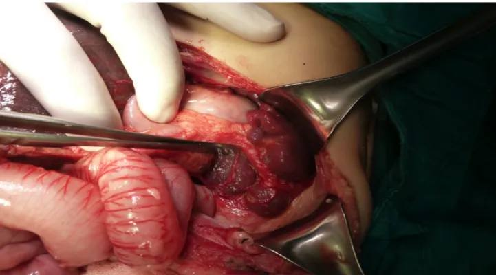
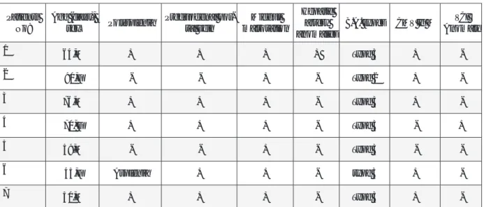
Surgical findings were used for the diagnosis of BASM. Further analysis of these findings revealed that four of the seven patients had polysplenia, one had asplenia, (Figure 1). Five patients had preduo-denal portal vein (Figure 2) and underwent duode-noduodenostomy. All patients had midgut malrota-tion and they had Ladd procedure. One patient had inferior vena cava interruption and one had a he-patic artery originating from the superior mesenter-ic artery. All patient data has been summarized in Table 1. The median time of follow-up was 4 years (1-5 years). All patients survived and 1 had a liver transplantation.
DISCUSSION
Twenty-five percent of BA cases are of the BASM subtype, which involves splenic malformations. Other anomalies are situs inversus, intestinal mal-rotation, preduodenal portal vein, interrupted vena cava, congenital portocaval shunts, cardiac anom-alies, and central nervous system anomalies [14]. A major component of BASM is polysplenia. Not all patients have polysplenia or splenic malformation and the term “biliary atresia congenital structur-al anomstructur-alies” has been used instead but not wide-ly accepted [7]. In this study, we found a BASM inci-dence rate of 11%. Two of the patients in our study did not have any splenic malformation.
The cause of BA is not understood as well as BASM. BA is destructive biliary fibrosis and the etiology is thought to be multifactorial (genetic, infection, tox-in, immunological). The extrahepatic bile duct orig-inates from the primordial bud of the intestine at 5 weeks gestation, followed by normal canaliza-tion at 6 weeks. BASM has some component which is definitely congenital such as midgut
malrota-Figure 1. Polysplenia of patient 4.
Figure 2. Preduodenal portal vein (black arrow), annular pancreas (white arrow) and atretic biliary ducts (red arrow) of patient 4.
Patients
No: Age (days), sex Polysplenia Preduodenal por-tal vein malrotationMidgut
Hepatic artery anomalies
B.A. types CMV Ig M AnomalyVCI
1 64,M + + + + Type 3 + -2 90,F - - + - Type 2 + -3 76,M + + + - Type 3 + -4 70, F + + + - Type 3 - + 5 59,M - - + - Type 3 - -6 45,F Asplenia + + - type 3 + -7 50,M + + + - Type 3 +
-genetic, infectious or immunological factors in the early fetal developmental period [8,9].
In their case reports, Makin et al have discussed the etiology of BASM. Three patients had surgical inter-vention in early life because of other conditions (je-junal atresia and duodenal atresia). They observed BASM, but only took liver biopsies since enteral feeding was a higher priority and liver morphology was normal. Liver biopsies showed some non-diag-nostic liver injury. Later on, they diagnosed and op-erated those patients due to BASM. This study sug-gests that bile duct injuries begin at birth in cases of BASM[10]
Lorent at all showed an association of cholangio-cyte toxin called biliatresone with biliary atresia [11]. Biliatresone is first discovered in Australian livestock when investigating the cause of biliary atresia out-break [12].
Regarding viral etiologic factors, the first report is published by Landing suggested in 1974. He sug-gested viral factors may play the role of the patho-genesis of neonatal hepatitis, biliary atresia and choledochal cyst [13]. Reovirus, rotavirus, and CMV have been showed to play a role in the etiology of BA[14,15]. CMV is the most common viral cause of BA. Serological evidence of CMV infection has been observed in 20–40% of infants with BA in studies from Sweden, China and England[16-19]. CMV DNA was identified in 60% BA patients [20]. Therefore, as-sociation of CMV infection and BA is absolute but association of CMV infection and BASM is not well documented. When we searched the PubMed
da-studies, even though there are several reports on BA and CMV infection. One of the important prospec-tive studies concerning CMV IgM and BA was per-formed at King’s College Hospital. The researchers evaluated and compared 20 CMV IgM-positive and 111 CMV IgM-negative patients. One of the 20 CMV IgM-positive and 17 of the 111 CMV IgM-negative patients studied, had BASM. We recalculated the in-cidence of CMV infection among BAPS patients and found it to be 5% [16]. Despite there were region-al differences in the incidence of BA and BASM, the BASM incidence in the current study was similar to King’s College Hospital. However, we found a higher rate of CMV infection with BASM at 71% compared with the study from the King’s College Hospital. CMV infection may also play an important role in BASM. However, our sample size was small. Further research concerning CMV infection in patients with BASM is recommended.
Preduodenal Portal Vein (PDPV):
The portal vein is usually formed by the conflu-ence of the superior mesenteric and splenic veins and also receives blood from the inferior mesenter-ic, gastrmesenter-ic, and cystic veins. The preduodenal por-tal vein passes the duodenum anteriorly and some-times causes intestinal obstruction without BA [21]. The preduodenal portal vein is very rare and was first described by Knight İn 1921[22]. The exact incidence is unknown since it may be asymptomatic. But PDPV is found 3 in 1000 biliary operations[23]. PDPV can be accompanied by other anomalies like situs inver-sus, biliary atresia, duodenal atresia or web, annular
obstruction, partial duodenal obstruction and as-ymptomatic”. Choice of treatment is duodenodu-odenostomy when a complete or partial duode-nal obstruction is relevant. If it is asymptomatic, no operation may be performed and duodenoduode-nostomy may be applied later when asymptomatic PDPV leads complete or partial duodenal obstruc-tion. Some people with PDPV may survive with-out any symptoms related to PDPV [25]. However, we did prefer to perform duodenoduodenostomy even the patients did not have any symptom relat-ed to PDPV because it is known that asymptomat-ic PDPV may later progress into a symptomatasymptomat-ic duo-denal obstruction. Secondary surgeries to treat du-odenal obstruction caused by PDPV in biliary atresia are dangerous by two reasons. One is surgical adhe-sion [26]. Second and our main reason to prefer du-odenoduodenostomy is the secondary surgical area is very close to portoenterestomy site and second-ary surgery may cause damage to this surgical site. Therefore, we prefer and recommend duodenodu-odenostomy initially in order to avoid secondary
surgery and the possibility of duodenal obstruction. Several reports claim that BASM has a worse progno-sis than BA with a mortality rate of 74.1%. However, the prognosis for BASM is no worse according to other reports [8,9,27,28]. Recent studies show that there is no difference in survival rates in BASM and BA [5]. The seven patients in our study have been followed for 5 years (1-5 years) uneventfully. One pa-tient required liver transplantation.
CONCLUSIONS
The association of BASM with other gastrointestinal anomalies and necessity of additional surgical pro-cedures, such as the Ladd procedure and duodeno-duodenostomy should be considered by the sur-geon. Duodenoduodenostomy may be performed when there is a preduodenal portal vein even if the anomaly is asymptomatic.
Further research concerning CMV infection and BASM association is recommended.
R E F E R E N C E S [1] Nio M, Ohi R, Miyano T, et al. Five- and 10-year
surviv-al rates after surgery for biliary atresia: a report from the Japanese Biliary Atresia Registry. Journal of pediatric sur-gery 2003; 38(7): 997-00.
[2] Nio M, Wada M, Sasaki H, et al. Long-term outcomes of bil-iary atresia with splenic malformation. J Pediatr Surg 2015; 50(12): 2124-21.
[3] Zhan J, Feng J, Chen Y, et al. Incidence of biliary atresia as-sociated congenital malformations: A retrospective multi-center study in China. Asian J Surg 2016.
[4] Davenport M, Tizzard SA, Underhill J, et al. The biliary atre-sia splenic malformation syndrome: a 28-year single-cen-ter retrospective study. J Pediatr 2006; 149(3): 393-00. [5] Guttman OR, Roberts EA, Schreiber RA, et al. Biliary atresia
with associated structural malformations in Canadian in-fants. Liver Int 2011; 31(10): 1485-93.
[6] Davenport M. Biliary atresia: clinical aspects. Semin Pediatr Surg 2012; 21(3): 175-84.
[7] Tanano H, Hasegawa T, Kawahara H, et al. Biliary atresia associated with congenital structural anomalies. J Pediatr Surg 1999; 34(11): 1687-90.
[8] Davenport M, Savage M, Mowat AP, et al. Biliary atresia splenic malformation syndrome: an etiologic and prog-nostic subgroup. Surgery 1993; 113(6): 662-68.
[9] Silveira TR, Salzano FM, Howard ER, et al. Congenital struc-tural abnormalities in biliary atresia: evidence for etio-pathogenic heterogeneity and therapeutic implications.
Acta Paediatr Scand 1991; 80(12): 1192-99.
[10] Makin E, Quaglia A, Kvist N, et al. Congenital biliary atre-sia: liver injury begins at birth. J Pediatr Surg 2009; 44(3): 630-33.
[11] Lorent K, Gong W, Koo KA, et al. Identification of a plant isoflavonoid that causes biliary atresia. Science transla-tional medicine 2015; 7(286) :286ra267.
[12] Waisbourd-Zinman O, Koh H, Tsai S, et al. The toxin bili-atresone causes mouse extrahepatic cholangiocyte dam-age and fibrosis through decreased glutathione and SOX17. Hepatology 2016; 64(3): 880-93.
[13] Landing BH. Considerations of the pathogenesis of neo-natal hepatitis, biliary atresia and choledochal cyst--the concept of infantile obstructive cholangiopathy. Prog Pediatr Surg 1974; 6:113-39.
[14] Tyler KL, Sokol RJ, Oberhaus SM, et al. Detection of reovi-rus RNA in hepatobiliary tissues from patients with extra-hepatic biliary atresia and choledochal cysts. Hepatology 1998; 27(6): 1475-82.
[15] Riepenhoff-Talty M, Gouvea V, Evans MJ, et al. Detection of group C rotavirus in infants with extrahepatic biliary atre-sia. J Infect Dis 1996; 174(1): 8-15.
[16] Zani A, Quaglia A, Hadzic N, et al. Cytomegalovirus-associated biliary atresia: An aetiological and prognostic subgroup. J Pediatr Surg 2015; 50(10): 1739-45.
[17] Fischler B, Ehrnst A, Forsgren M, et al. The viral associa-tion of neonatal cholestasis in Sweden: a possible link
between cytomegalovirus infection and extrahepatic bil-iary atresia. J Pediatr Gastroenterol Nutr 1998; 27(1): 57-64. [18] Fischler B, Svensson JF, Nemeth A. Early cytomegalovi-rus infection and the long-term outcome of biliary atresia. Acta Paediatr 2009; 98(10): 1600-02.
[19] Shen C, Zheng S, Wang W, et al. Relationship between prognosis of biliary atresia and infection of cytomegalo-virus. World J Pediatr 2008; 4(2): 123-26.
[20] Xu Y, Yu J, Zhang R, et al. The perinatal infection of cyto-megalovirus is an important etiology for biliary atresia in China. Clinical pediatrics 2012; 51(2): 109-13.
[21] Masumoto K, Teshiba R, Esumi G, et al. Duodenal steno-sis resulting from a preduodenal portal vein and an op-eration for scoliosis. World J Gastroenterol 2009; 15(31): 3950-53.
[22] Knight HO. An Anomalous Portal Vein with Its Surgical Dangers. Annals of surgery 1921; 74(6): 697-99.
[23] Stevens JC, Morton D, McElwee R, et al. Preduodenal por-tal vein: Two cases with differing presentation. Archives of
surgery 1978; 113(3): 311-13.
[24] Esscher T. Preduodenal portal vein--a cause of intesti-nal obstruction? Jourintesti-nal of pediatric surgery 1980; 15(5): 609-12.
[25] Yi SQ, Tanaka S, Tanaka A, et al. An extremely rare inver-sion of the preduodenal portal vein and common bile duct associated with multiple malformations. Report of an adult cadaver case with a brief review of the literature. Anatomy and embryology 2004; 208(2): 87-96.
[26] Miyake H, Fukumoto K, Yamoto M, et al. Surgical Management of Hiatal Hernia in Children with Asplenia Syndrome. European journal of pediatric surgery : official journal of Austrian Association of Pediatric Surgery [et al] = Zeitschrift fur Kinderchirurgie 2017; 27(3): 274-79. [27] Vazquez J, Lopez Gutierrez JC, Gamez M, et al. Biliary
atre-sia and the polysplenia syndrome: its impact on final out-come. J Pediatr Surg 1995; 30(3): 485-87.
[28] Karrer FM, Hall RJ, Lilly JR. Biliary atresia and the polysple-nia syndrome. J Pediatr Surg 1991; 26(5): 524-27.