492
Ceramic
Processing Research
A simple chemical method for conversion of Turritella terebra sea snail into
nanobioceramics
Yesim Muge Sahina,b,*, Zeynep Ormanc and Sevil Yucelc
aArelPOTKAM (Polymer Technologies and Composite Application and Research Center), Istanbul Arel University 34537, Turkey bDepartment of Biomedical Engineering, Istanbul Arel University 34537, Turkey
cDepartment of Bioengineering, Yildiz Technical University 34220, Turkey
In this study, a sea shell was converted into bioceramic phases at three different sintering temperatures (450oC, 850oC, 1000oC). Among the obtained bioceramic phases, a valuable β-TCP was produced via mechanochemical conversion method from sea snail Turritella terebra at 1000oC sintering temperature. For this reason, only the bioceramic sintered at 1000oC was concentrated on and FT-IR, SEM/EDX, BET, XRD, ICP-OES analyses were carried out for the complete characterization of β-TCP phase. Biodegradation test in Tris-buffer solution, bioactivity tests in simulated body fluid (SBF) and cell studies were conducted. Bioactivity test results were promising and high rate of cell viability was observed in MTT assay after 24 hours and 7 days incubation. Results demonstrated that the produced β-TCP bioceramic is qualified for further consideration and experimentation with its features of pore size and ability to support bone tissue growth and cell proliferation. This study suggests an easy, economic method of nanobioceramic production.
Key words: Bioactivity, Bioceramic, Bone regeneration, TCP, Mechanochemical conversion.
Introduction
Calcium-phophate-based ceramics have been used successfully as an augment hard tissue. Among these bioceramics the most well-known ones are hydroxyapatite [HA, Ca10(PO4)6(OH)2] and β-tricalcium phosphate [β-TCP, Ca3(PO4)2] phases. HA, is a natural component of bones and teeth; thus hydroxyapatite was employed in bone defects and voids as coatings on implant materials. β-TCP is on the other hand a biocompatible, bioresorbable calcium-phosphate apatite and has been utilized for orthopedic and dental studies. β-TCP concentration plays an important role in bone regereration by accelerating resorption speed and apatite formation. Main advantage of β-TCP is, its biodegradability which can be adjusted by modifying the composition. In the last decades, biological resources that contain calcium such as sea shells [1], bovine bone [2], corals [3] etc. have been used to produce calcium phophate apatites for bone grafts. It is reported that sonochemical method providing a fast and easy convertion technique gives out these promising bioceramic materials [4]. Rocha et al. [5] produced HA from cuttlefish via hydrothermal method. Kang et. al. [6] produced β-TCP from the shell of Haliotis sp. (abalone shell) via chemical reation between CaHPO4 and CaO from 950oC to 1100oC for
3 hours. Also marine origins such as land snails(helix pomatia) [7], razor shells (ensis ensis) [8], sea snail Cerithium vulgatum [9] were employed to produce calcium phosphate apatites via mechanochemical method. The final method to produce bioceramics, mechanochemical method, is advantages over other hydrothermal, and microwave production methods since it does not need expensive and complex instruments and challenging conditions such as high pressure, temperature etc. Because of this reason, we suggest an easy, economic method for nanobioceramic production in the proposed study.
Materials and Methods
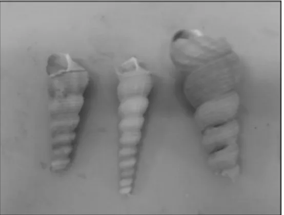
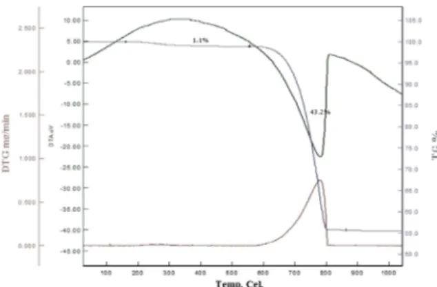
Turritella terebra sea snail shells, was shown in Fig. 1, were purchased in Istanbul. Shells were cleaned and dried in the vacuum furnace for a day at 100 °C to cleanse them from dirt and organic remains. They were crushed in an agate mortar by hand grinding and processed in a high energy ball mill (Retsch Planetary Ball Mill PM 400) for an hour and sieved to obtain standard powder size under 100 μm. The seashell powders were characterized by differential thermal analysis combined with thermogravimetry (DTA/TG SII6000 Exstar TG/DTA 6300, heating rate 5 K/min, in air) to obtain the exact CaCO3 amount in the raw shell, as illustrated in Fig. 2. In order to set the exact stoichiometry of TCP, the required ortho-phosphoric acid (H3PO42-) was calculated and Ca/P ratio was
*Corresponding author: Tel : +90-537-975-75-07 Fax: +90-212-860-04-81 E-mail: ymugesahin@ arel.edu.tr
adjusted as 1.50 [10]. The reaction was conducted in 50 ml of distilled water on hotplate stirrer for chemical precipitation. The temperature was set to 80oC and an equivalent amount of H3PO4 was added dropwise to the mixture and the reaction was conducted for 2 hours under continuous stirring.
Powders were filtered and dried in a vacuum furnace at 100oC overnight. Finally, the synthesized powder were sintered in an oven increasing 5oC per minute and kept at a sintering temperature of 450oC, 850oC and 1000oC for 4 hours. Sample were sintered at 450oC, 850oC and 1000oC will be indicated as TT450, TT850 and TT1000 respectively.
FT-IR (Shimadzu) with an ATR unit in the range of 4000 cm−1-650 cm−1 was used to evaluate chemical functional groups of bioceramics before and after being soaked into SBF. XRD patterns were recorded with PANalytical Xpert Pro diffractometer and the analyses were performed in a range of 10o-80o on a 2θ step of 0.02o/s. Copper Kα radiation (λ = 1.5406 nm) at operating power of 30 kV and 25 mA intensity of the current were characterization parameters
Phases were identified by comparing the experimental XRD patterns depending on the Joint Committee of Powder Diffraction Standards (JCPDS) using the cards 17-0499 for Ca2P2O7 and 9-0169 for β-TCP. SEM analysis was employed (Zeiss EVO® LS 10) to analyse morphological properties of the bioceramic powders before and after being soaked in the SBF. Samples were coated with gold prior of the SEM analysis by using a Sputter Coater device (Emitech K 550X) in Ar atmosphere due to amplify the secondary electrons signal. Energy Dispersive X-ray Spectroscopy (EDX) was conducted to identify the elemental composition of the samples. Specific surface area of the samples was measured with BET analysis with Tristar II 3020. The samples were degassed at 100oC for 2 h under nitrogen purging prior of BET measurements. Pore size and pore volume were calculated using BJH adsorption method.
In vitro bioctivity of the obtained bioceramic powder was tested in SBF medium. Method of Kokubo [11]
was used for SBF which consists of inorganic ionic composition equal to human blood plasma and buffered with TRIS at a physiological pH. Each sample was pressed under 100 bar pressure for 60 seconds to achieve pellet form. Eq. 1 was used to obtain the pellet size:
(Eq. 1) where Vs is the volume of SBF (ml) and Sa is the apparent surface area of specimen (mm2). Pellets were sintered at 1000oC increasing temperature 10oC per minute for 2 hours. Then samples were immersed into SBF containing falcon tubes at 37oC and were stored
in an oven at of 37oC. SBF was renewed on
7th,14th,21th days. Inductively Coupled Plasma/Optical Emission Spectrometry (ICPE9000) was used to determine the level of calcium and phosphorus ion release during certain periods in SBF solution. Pellets were immersed into tris-buffer solution having a pH value of 8 and at 37oC to analyse their degradation behaviour. pH was measured each day at an exact time, during 7 days and data was recorded. SBF and Tris analyses were conducted in dublicate and their averages were calculated.
Cell viability study was conducted by preparing concentration of 20 mg/mL (powder / DMEM-F12); 0.1 g of material was soaked in a 5 ml DMEM-F12 solution and left at 37oC for 24 hours. The mixture was centrifuged subsequently, the supernatant passed through a 0.2 μm filter. Cell viability and cytotoxicity of the obtained bioceramic were investigated by culturing of Saos-2 osteoblast-like cells in DMEM-F12 supplemented with 10% FBS (fetal bovine serum) at 37oC in 5% CO2. The culture medium was replaced every two days. MTT assay was carried out according to the manufacturer's protocol (Sigma USA). Cells were seeded in a 96-well culture plate to obtain 1x10³ cells for per well. Incubation took for 24 hours, then the sample extracts were prepared with DMEM-F12 at different concentrations. After 7 days, 20 μl MTT solution (5 mg/ml) was put to each well. Following additional incubation for 4 hours, 100 μl of DMSO was added. Absorbance was measured at 540 nm with a microplate reader (Multiskan Ascent 96/384 Plate Reader).
Results and Discussion
TG/DTA Analysis
CaO content of bioceramic powder was determined by differential and gravimetric thermal analysis (TG/DGA) which indicate mass loss% of CaCO3 corresponding to heat induced processes data (Fig. 2). The decomposition of raw powders starts at ~600oC and finalized at ~800oC. A 56.8% mass loss denoted at 800oC which accompanied by decomposition of CaCO3 and CO2
Vs Sa⁄ =10
removal. The decomposition of CaCO3 into CaO was shown in the following chemical reaction:
CaCO3→ CaO + CO2
Concentration of H3PO4 solution was calculated according to the ratio of Ca:P = 1.5.
X-Ray Diffraction analysis
As the produced samples were heated from 450oC to 1000oC, the peak height of the samples increases substantially, with an associated narrowing of the peak corresponding to increase of crystallinity. Characteristic peaks of calcium pyrophosphate (Ca2O7P2) was observed when the samples were sintered at 450oC and 850oC (Fig. 3). β-TCP obtained after the heating temperature had reached to 850oC. Peaks of β-TCP (JCPDS 09-0169) were registered at 21.9o, 28o, 30.9o, 32.1o, 34.4o for sample TT1000 (Fig. 4). It can be suggested that as the sintering temperature increases more intense and narrower peaks were obtained. The increase of the peak intensity can be attributed to the crystallinity increment.
Fourier transform infrared spectroscopy (FT-IR) analysis
The FTIR spectra for Ca2P2O7 revealed various bands from the respective phosphate groups of Ca2P2O7 as displayed in Fig 5. The bands around 1028.06 cm−1 for TT450 and 1026.13 cm−1 for TT850 was determined
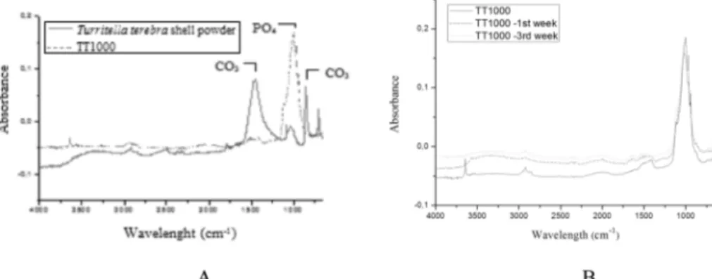
to triply degenerate antisymmetric P-O stretching mode. P-O symmetric stretching mode was observed at 923.90 cm−1 for TT450 and 939.33 for TT850 [12]. FT-IR spectra of before and after heat treatment of the Turritella terebra raw powder at 1000oC was shown in Fig. 6A. CO3 bands at1465.9, 858.32, 711.73 cm−1 were disappeared and PO4 bands were appeared at 1116.78, 1078.21, 1001.06, 970.19, 943.19 for TT1000. Samples were immersed into SBF and changes were observed regularly every week. Fig. 6B displays FT-IR spectra of samples before and after being soaked into SBF. After being soaked into SBF, phosphate group intensity increased for TT1000 and there was no evidence of CO32− band. HA contains internal hydrogen bonds between oxygen of adjacent orthophosphate groups, resulting in the presence of HPO24. This band was referred as the absorption band in some biological Fig. 2. DTA/TGA analysis graphics of raw powders of Turritella
terebra.
Fig. 3. XRD patterns of A) TT450 B) TT850.
Fig. 4. XRD patterns of TT1000.
apatites. It is suggested that typical FT-IR spectrum after being soaked into SBF, gave evidence that HA containing HPO2−4 appeared [13]. These peaks can confirm that HCA (hydroxyl carbonate apatite layer) formed on the obtained bioceramic. Also broad hydroxyl peak at around 3400 cm−1 was observed giving clue for the HCA layer formation on the biomaterial.
BET analysis
Porous structure analysis was carried out by adsorption-desorption gas analysis. BET analysis revealed that specific surface area and pore volume decreases with increasing sintering temperature. Sintering is a heat treatment results to strength increase, reduction in surface area and generally condensation of a particle that has a high surface area. In sintering process, the formation of the bonds between the particles, the closure of the pore channels, the condensation or shrinking of the pores and the pore fusion together occurs.
Average particle diameter was calculated by following Eq. 2:
(Eq. 2) Sintering effect was evaluated for the obtained bioceramics in Table 1. It can be revealed that, as the sintering temperature increased specific surface area decresed. Average pore diameter decreased as the sintering temperature increased from 450oC to 850oC
and it did not change as the sintering temperature increased from 850oC to 1000oC [14].
ICP-OES analysis of simulation body fluid
Bioactivity studies of the synthesized β-TCP were investigated by SBF analyses. For this purpose HA accumulation on the produced bioceramic surfaces were analysed by ICP-OES measurements. Apatite formation occurs in SBF needs enough saturation of Ca and P ion concentration. It is a process of heterogeneous nucleation on calcium phosphate ceramic surface following the spontaneous growth of crystals. Apatite nuclei consumes Ca+2 and HPO42− ions from SBF solution and it takes spherulitic shape. Positive ions in the SBF are adsorbed to surface of the calcium phosphate ceramic which leads to attraction of negative ions. Thus Ca2+ ions are attracted following by HPO42− on the solid-liquid surface. Calcium and phosphate ion change was illustrated in Fig. 7. Ca level of TT1000 decreased from 100 to 79.6 ppm in the 1th week and decreased slightly until 3rd week. P concentration of TT1000 decreased drastically from 28.8
d 6
BET sur face Area inm
2 g ---⎝ ⎠ ⎛ ⎞ Density in g cm3 ---⎝ ⎠ ⎛ ⎞ × ---=
Fig. 6. A) FT-IR spectra of before and after heat treatment of the Turritella terebra shell powder B) TT1000: before and after being soaked in SBF.
Table 1. Sintering temperature effect on specific surface area, pore volume of samples.
Origin Sintering temperature (oC) Specific surface area (m²/g) Pore volume (cm3/g) Average pore diameter Turritella terebra 450 11.72 0.01 6.30 850 2.36 0.0027 4.60 1000 1.09 0.0012 4.60
Fig. 7. A) Ca ion content of TT1000 in SBF B) P ion content of samples in SBF.
to 10.3 ppm. Decrease of Ca and P level in the SBF can be attributed to accumulation of the ions on the
sample surface. This is known as bone-like apatite formation and occurs when Ca and P level reach to a certain value in the solution.
pH changes give information about dissolution of the samples. Fig. 8 reveals pH change of the solution in SBF. Dissolution of β-TCP can be explained in the following chemical reaction:
3Ca3(PO4)2 (s) + H2O 9Ca2+ (aq) + HPO42-(aq) + 5PO43 (aq) + OH− (aq)
Fig. 9. SEM images of Turritella terebra shell before sieving and milling (5000x).
Fig. 10. SEM images of TT1000 produced from Turritella terebra sea snail before and after being soaked into SBF A) sea snail B) after sintering at 1000oC C) after 1st week in SBF D) after 2nd week in SBF E) after 3rd week in SBF (5000x).
Table 2. Ca/P ratio of the samples before and after after being soaked into SBF.
Sample
Before being soaked in
SBF
Day 7th Day 14 th Day 21 th
TT1000 1.88 1.95 1.35 1.52
H+ and OH- ion concentrations changes as a result of dynamic nature of this dissolution process. pH increased for 7 days. After 7 days, a pH decrease was observed up to the 21th day to a final value of 7.01. The increament of pH can be attributed to HPO42− formation in SBF.
SEM/EDX analysis
The typical microstructure at a fracture surface of the shell is exhibited in SEM image in Fig.9. A plate-like structure can be attributed largely to aragonite crystals [9].
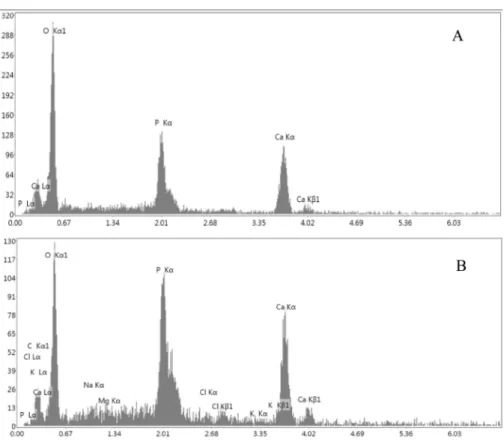
Before and after being soaked in SBF, morphologies were monitored by SEM. SBF results revealed accumulation of HA after immersing into SBF. The surface of the material was observed to be covered with HA at the end of each week (Fig. 10). EDX analysis demonstrated that there are measurable
quantities of Cl−, Na+, Mg2+ in addition to Ca+2, P-, O−2 ions. Ca/P ratio was calculated according to EDX data (Fig. 11). It was determined that the Cl−, Na+, Mg2+ ions present in SBF and bonded to the bioceramic sample as a trace element.
Since β-TCP is a valuable bioceramic for tissue engineering purposes, bioactivity studies were performed for only TT1000 bioceramic sample. Ca/P ratio of the sample before and after being soaked in SBF was shown in Table 2. Ca/P ratio of TT1000 was changed from 1.35 to 1.52 between 14th and 21th days. Ca/P composition of the surface is lower than stoichiometric HA (1.67), indicates a calcium-deficient HA.
Being consistent with the ICP-OES analysis SEM results were also show the HA formation on the synthesized β-TCP bioceramics.
Before being soaked into SBF, Ca/P ratio were obtained very similar to the data for the 7 days treated sample. This result can be atrributed to the formation of a thin layer of HA [15]. Within 5-10 days a subsequent degredation of TCP phase and an accumulation of HA layer on the surface of the material took place simulteniously. A slight decrease in Ca/P ratio can be attributed to this fact, where the degredation of β-TCP is much more faster than the HA accumulation. Ca/P molar ratio is 1.52 after 21 days. Ca/P ratio decrease indicates that a newly formed Ca-deficient hydroxyapatite layer accumulated on the surface of the sample [13].
pH Changes in tris-buffer solution
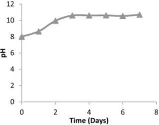
In vitro biodegradation behaviours were investigated in Tris solution. pH changes of samples give information of their biodegradation because Ca+2 and P ions release in SBF. Tris-buffer solution, [(CH2OH)3CNH2], contains three active hydroxyl groups and can be stabilized by Fig. 12. pH changes of TT1000 in a Tris-buffer solution over 7
days.
HCl. Therefore, it can react with calcium ions released from sample. This environment enables more dissolution to occur until it reaches to equilibrium stage comparing to deionized water. Due to the dissolution reactions in the Tris-buffer solution, H+ and OH- ions that released so pH of the solution changes. Fig. 12 indicates that pH of TT1000 increased excessively to 10.6. whereas, after 2 days pH remained almost the same. This can be attributed to fast dissolution of the material for the first two days.
Cell viability test
In- vitro cell viability test was conducted by seeding to SAOS-2 osteoblast-like cells at concentrations of 20 μg/ml, 50 μg/ml, 100 μg/ml and 200 μg/ml for 24 hours and 7 days. Fig. 13 indicates cell viability of the samples at different concentrations. ISO10993-5 standard represents that if the viability of treated human cell line culture is more than 70% means there is no toxic effect of the material [16]. According to ISO10993-5 standard, samples not only have non-toxic effect on the cells but also have the ability of cell adhesion, spreading, and resuts show increased viability over time. Highest cell viability was noted for the concentration of 200 µm.
Conclusions
β-TCP was produced from sea snail Turritella terebra via mechanochemical method. Bioactivity tests and SEM images indicate that the produced β-TCP was covered with HCA after immersing into SBF. Tris-buffer solution test results suggested that synthesized β-TCP is highly biodegredable.
Additionally, SBF studies showed a high bioactivity in the first week. MTT assay proposed non-toxic effect on SAOS-2 osteoblast-like cells. Along with their biological-natural origin, the costless Turritella terebra shells are attractive source for bioceramic production. On the other hand, the proposed mechanochemical conversion method is a simple and economic method in comparison to its analogues and guaranties nanoparticle
bioceramic production.
References
1. F. N. Oktar, H. Gokce, O. Gunduz, Y.M. Sahin, D. Agaogullari, I.G. Turner, L.S. Ozyegin, B. Ben-Nissan, Key Eng. Mater. 631 (2014) 137-142.
2. O. Gündüz, C. Gode, Z. Ahmad, H. Gökçe, M. Yetmez, C. Kalkandelen, F. N. Oktar, Journal Of The Mechanical Behavior Of Biomedical Materials 35 (2014) 70-76. 3. I. Karaca, O. Gunduz, L.S. Ozyegin, H. Gökçe, B.
Ben-Nissan, S. Akyol, F. N. Oktar, Journal of the Australian Ceramic Society 54 (2017) 317-329.
4. Y. M. Şahin, O. Gunduz, B. Bulut, L.S. Ozyeğin, H. Gökçe, D. Alaoğulları, F. N. Oktar, Acta Physica Polonica A. 127[4] (2015) 1055-1058.
5. J. H. G. Rocha, A.F.Lemosa, S.Agathopoulos, P.Valério, S.Kannan, F.N.Oktar, J.M.F.Ferreira, Bone 37[6] (2005) 850-857.
6. K.-R. Kang, Z.-G. Piao, J.-S., In-A Cho, M.-J. Yim, B.-H. this chemical Kim, J.-S. Oh, J. S. Son, C. S. Kim, D. K. Kim, S.-Y. Lee, S.-G. Kim Implant Dent. 26[3] (2017) 378-387.
7. D. Kel, H. Gökçe, D. Bilgiç, D. Aðaoðullarý, I. Duman, M.L. Öveçoðlu, E. S. Kayali, I. A. Kiyici, S. Agathopoulos, F.N. Oktar, Key Eng. Mater. 493-494 (2011) 287-292. 8. S. Agathopoulos, L.S. Ozyegin, Z. Ahmad, O. Gunduz,
E.S. Kayali, O. Meydanoglu, F.N. Oktar, Key Eng. Mater. 493-494 (2011) 775-780.
9. O. Gunduz, Y. M. Sahin, S. Agathopoulos, B. Ben-Nissan, and F. N. Oktar, J. Nanomater. 2014 (2014) 1-6.
10. O. Gunduz, Y.M. Sahin, S. Agathopoulos, D. Aðaoðullarý, H. Gökçe, E.S. Kayali, C. Aktas, B. Ben-Nissan, F.N. Oktar, Key Eng. Mater. 587 (2013) 80-85.
11. T. Kokubo, H. Takadama, Biomaterials 27[15] (2006) 2907-2915.
12. B. Mehdikhani, G. Borhani, J. Ceram. Process. Res. 16[3] (2015) 308-312.
13. A. Sobczak-Kupiec, Z. Wzorek, R. Kijkowska, Z. Kowalski, Bull. Mater. Sci. 36[4] (2013) 755-764. 14. A. Ibrahim, W. Wei, D. Zhang, H. Wang, J. Li, Materials
Letter 110(2013)195-197.
15. Bui, X. V., & Thang, T. D., ASEAN Journal on Science and Technology for Development. 33[2] (2016)38-68. 16. P. Sobierajska, A. Dorotkiewicz Jach, K. Zawisza, J. Okal,
T. Olszak, Z. Drulis-Kawa, R. J.Wiglusz J. Alloys Compd. 748 (2018) 179-187.