Abstract
Objectives: This study evaluated the microtensile bond strength (µTBS) of a resin composite bonded to bleached enamel as a function of bleaching conditions.
Materials and Methods: The whiteness hydrogen peroxide (HP) bleaching agent containing 35% HP was applied to the central incisors’ facial enamel surface and irradiated as follows: No treatment (G1; control); no light (G2); light‑emitting diode, the 40s (G3); diode laser, the 20s (G4); and neodymium:yttrium aluminum garnet laser, 20s (G5). A Variolink II resin composite structure was then built up incrementally on the surface. The teeth were sectioned into three 1.2 mm × 1.2 mm wide “I”‑shaped sections. The specimens were then subjected to microtensile testing at a crosshead speed of 1 mm/min. Data were analyzed using one‑way ANOVA (α =0.05) followed by the Tukey Honestly Significant Difference post‑hoc test. The fractured surfaces were observed with a stereomicroscope at × 100 magnification. Results: One‑way ANOVA revealed no statistical differences among the groups (P > 0.05). No differences appeared between the groups bonded 14 days after bleaching (P > 0.05). Mean µTBS values (MPa) were as follows: 22.05 ± 5.01 (G1); 19.6 ± 5.6 (G2); 19.3 ± 5.4 (G3); 20.08 ± 2.08 (G4); and 18.1 ± 4.8 (G5). Many adhesive failures occurred at the bleached and irradiated enamel surfaces.
Conclusion: The various irradiation treatments following the application of the whiteness HP bleaching agent to enamel did not significantly reduce the µTBS within a 14‑day period.
Key words: Bleaching agents, lasers, lasers neodymium: yttrium aluminum garnet, resin bonding, tooth bleaching Date of Acceptance: 02‑Sep‑2015
Bond strength of resin composite to light activated
bleached enamel
T Yavuz, OY Ozyilmaz1, AN Ozturk2, F Aykent2 Department of Prosthodontics, Faculty of Dentistry, Abant Izzet Baysal University, Bolu, 1Department of
Prosthodontics, Faculty of Dentistry, Medipol University, Istanbul, 2Department of Prosthodontics, Faculty of Dentistry,
Selcuk University, Konya, Turkey
Address for correspondence:
Dr. T Yavuz,
Department of Prosthodontics, Faculty of Dentistry, Abant Izzet Baysal University, Golkoy, Bolu, Turkey. E‑mail: drtevfikyavuz@gmail.com
Introduction
Dental bleaching is one of the most rapidly developing areas of dentistry. Demand is increasing for tooth bleaching to improve the whiteness and perceived the esthetic appearance of tooth tissue.[1,2] Contemporary tooth
bleaching methods provide real‑time, successful, and affordable tooth whitening.[3,4]
Several types of irradiation sources are currently used to accelerate the in‑office bleaching procedure.[5,6] These
techniques, using coherent[4,7] or incoherent[8] light sources,
are quick and convenient.[5]
The bleaching procedure is an oxidation reaction that releases free radicals. Hydrogen peroxide (HP; HOOH) Access this article online
Quick Response Code:
Website: www.njcponline.com DOI: 10.4103/1119-3077.178909
PMID: *******
How to cite this article: Yavuz T, Ozyilmaz OY, Ozturk AN, Aykent F. Bond
strength of resin composite to light activated bleached enamel. Niger J Clin Pract 2016;19:766‑71.
This is an open access article distributed under the terms of the Creative Commons Attribution‑NonCommercial‑ShareAlike 3.0 License, which allows others to remix, tweak, and build upon the work non‑commercially, as long as the author is credited and the new creations are licensed under the identical terms.
degradation can lead to various species having differing reactivities.[9] These include hydroxyl ions (OH−);
perhydroxyl ions (HOO−), which are considered stronger
free radicals; hydrogen ions (H+); water molecules (H 2O);
and oxygen ions (O−2). Water (H
2O) and oxygen
molecules (O2) can form in the presence of salivary peroxidase enzymes.[4]
Decomposition of HP is a unidirectional reaction that ultimately leads to the formation of water. In the first step, HP dissociates into free radicals such as HOO− and
O−2, which oxidize the organic dark stains on teeth to
a much lighter shade.[1] The saturation point is reached
when the bleaching intensity has stabilized.[9] The O−2
ion is stabilized by removing an electron from surrounding molecules, such as the color pigments present in the enamel surface. This degradation of the pigments can make them lighter.[10]
The in‑office bleaching technique involves using high concentrations of carbamide or HP for faster and more effective treatment.[9,11] The effectiveness of these chemical
agents can be accelerated by light or heat.[4] The purpose
of the light source is not to directly bleach the teeth, but to accelerate the activation of the bleaching product by absorbing light from a photosensitizer (a dye). The energy absorbed from the light accelerates the oxidation–reduction reaction.[12]
Bleaching treatment is frequently recommended before porcelain restorations or adhesive esthetic restorations are performed.[13] However, reductions in bond strength
to enamel and dentin have been reported when these restorations are made immediately after the bleaching treatment.[13,14] Approximately 7–14 days are required to
achieve the normal bond strength.[13] Various treatments
with antioxidants have been used to reverse this effect, such as 10% sodium ascorbate and the catalase enzyme. However, these are typically not routine clinical procedures.[13]
The development of new technologies and methods, such as laser appliances, has greatly benefitted modern esthetic dentistry.[13] Erbium:yttrium aluminum garnet
and neodymium:yttrium aluminum garnet (Nd:YAG) lasers have been widely used in dentistry, for example in soft‑tissue surgery, canal disinfectants in endodontic procedures, and pit and fissure sealing. Laser irradiation increases fluoride uptake in enamel and promotes dentin melting, which creates a natural seal on exposed dentin. Such sealing reduces sensitivity on the cervical areas of teeth by minimizing marginal microleakage and improves bond strength.[13,15,16] Use of Nd:YAG
lasers has been reported to cause morphological and chemical changes in the dental structure, that is,
melting of dental hard tissues (dentin and enamel), as well as increased distribution of calcium, phosphorus, magnesium, and oxygen in the enamel, thereby affecting acid resistance.[15,17]
Use of a Nd:YAG laser promotes mineralized tissue recrystallization. The laser appears to influence post bleaching bonding by providing substrate heating and causes alterations in enamel and dentin morphologies.[13] This laser
treatment eliminates the formation of residual free radicals and neutralizes the immediate effects of bleaching agents on bond strength; it also allows restorations to be replaced immediately.[13]
The previous study performed diode lasers and indicated that the laser has been demonstrated to being the most valuable energy source for power bleaching, with simple, and short application in the dental practice.[18,19] The researcher
used infrared laser light and reported that irradiation with infrared laser light can produce some beneficial effects on sensitivity.[4,5] The inflammatory response of the pulpar
tissue could be increased, and the pulp damage and the pain could be reduced after the whitening process with the near‑infrared laser.[10,20]
Tensile and shear tests are commonly used to evaluate bond strengths.[21,22] However, traditional tensile and shear
tests have been criticized for using relatively large bonded surfaces, over which the stress distribution is likely to be uneven relative to the density of intrinsic faults, possibly acting as stress raisers.[21] The microtensile technique is
considered a more reliable test. It more closely reflects the interfacial bond strength because it provides a more uniform stress distribution.[21,23]
Thus, the aim of this study was to evaluate the microtensile bond strength (µTBS) of a resin composite to bleached enamel as a function of different Nd:YAG, light‑emitting diode (LED), and diode laser light‑activating applications. The null hypothesis was that no statistically significant differences would appear in the bleaching photoactivation, and the µTBS would be unchanged.
Materials and Methods
Twenty sound, recently extracted, human maxillary central incisors were cleaned and polished. Those with cracks and carious lesions were excluded. The teeth were stored in distilled water at room temperature immediately after extraction until the bleaching process was conducted. Before bleaching, the teeth were randomly divided into five groups of four teeth each for subsequent exposure to the different light sources. Then the whiteness HP bleaching agent (FGM Produtos Odontológicos Ltda, Joinville, SC,
Brazil) containing 35% HP was applied to the facial enamel surface according to the groups.
The red activator was mixed into the colorless bleaching gel at the moment of use according to the manufacturer’s instructions. The mixture was brushed on the buccal surface of a tooth to produce a uniform layer approximately 1 mm thick.
• G1 (control): No treatment
• G2 (no photoactivation): Bleaching gel was applied, but no photoactivation device was used
• G3 (blue‑emitting diode): Bleaching gel was applied and photoactivated using a blue LED (light intensity 470 mW/cm2) for 40 s
• G4 (diode): Bleaching gel was applied and photoactivated using a diode laser (Lasersmile, Biolase, San Clemente, CA, USA) (continuous mode, output power 4.0 W, 1 mm distance, energy density 17.7 J/cm2, wavelength 980 nm, focal spot area 4,25
cm2) for 20 s
• G5 (Nd:YAG): Bleaching gel was applied and photoactivated using an Nd:YAG laser (Fotona, At Fidelis, Ljubljana, Slovenia) (noncontact bleaching handpiece, output power 4.0 W, pulse repetition rate 60 Hz, 1 mm distance, pulse length 320 µs, energy density 0.23 J/cm2, wavelength 1064 nm, focal spot
area 0,08 cm2) for 20 s.
Adhesives and resin cements were applied according to the manufacturers’ instructions 14 days after the bleaching process. Total Etch etching gel (37% phosphoric acid) was applied only on the enamel for 15 s, then rinsed and dried in air. Syntac primer was then applied for 15 s, and air‑dried. Syntac adhesive was applied for 10 s, and dried in air. Heliobond light‑curing bonding agent was finally applied. Total Etch, Syntac, and Heliobond were all obtained from Ivoclar Vivadent AG, Schaan, Liechtenstein.
After setting of the cement, a composite resin (Variolink II) block was incrementally built‑up on the cement surface to a height of 5 mm. Each increment of 2 mm was light‑cured for 40 s. All curing steps were performed using the same halogen light‑curing unit (Bluephase). All specimens were stored in distilled water at 37°C for 24 h. Variolink II and Bluephase were obtained from Ivoclar Vivadent AG, Schaan, Liechtenstein.
The specimens (resin block/tooth) were sectioned perpendicularly to the cement/tooth interface using a cutting machine (Buehler IsoMet 1000 Low Speed Saw, Buehler Ltd., Lake Bluff, IL, USA). A diamond saw blade was used at low speed with water cooling and irrigation. The teeth were sectioned into three “I”‑shaped sections with dimensions of 1.2 mm × 1.2 mm [Figure 1]. The specimens were prepared from the middle third of teeth for
specimens’ standardization. Approximately 12 rectangular specimens (8 mm in length) were obtained from each tooth for microtensile testing.
Microtensile bond strength test
Cyanoacrylate adhesive (Zap‑It, Dental Ventures of America, Inc., Corona, CA, USA) was used to attach a specimen to the opposing arms of a µTBS testing device (Harvard Apparatus Co. Inc., Holliston, MA, USA). The bonded area was kept perpendicular to the long axis [Figure 2] to avoid torsion and shear forces. The mounting adhesive was applied sparingly to the edges of each specimen. The specimen was fractured under tension at a crosshead speed of 1 mm/min. The crosshead movement was stopped automatically upon fracture. µTBSs were recorded and tabulated for statistical analysis. The two halves of a fractured sample were retained to assess the fracture mode.
The bond strength data were subjected to the Shapiro– Wilk normality test. Normal distributions were observed, so a parametric test (one‑way ANOVA, α = 0.05) was used to determine the significance between the groups, followed by the Tukey Honestly Significant Difference
post‑hoc test. The fracture surfaces were observed using a
stereomicroscope at ×100 magnification to identify the fracture mode.
Results
Table 1 lists the mean µTBS data for the five groups with their standard deviations. Table 2 lists the ANOVA results.
One‑way ANOVA revealed no statistical differences between the groups (P > 0.05). No differences appeared between the groups bonded 14 days after bleaching
Table 1: MBS of testing groups (in MPa) Group n Mean±SD 1 12 22.05±5.01 2 12 19.6±5.6 3 12 19.3±5.4 4 12 20.08±2.08 5 12 18.1±4.8
No treatment: G1=Control; G2=No light; G3=An light‑emitting diode, 40 s; G4=A diode laser, 20 s and G5=Nd: YAG laser, 20 s. SD=Standard deviation; MBS=Microtensile bond strengths; Nd: YAG=Neodymium: yttrium aluminum garnet
Table 2: Results of one‑way ANOVA
Sum of squares df Mean square F Significant
Between groups 107.329 4 26.832 1.13 0.352
Within groups 1305.782 55 23.741
Total 1413.111 59
Figure 4: Scanning electron microscope images of the enamel surface (a) no treatment (b) nonphotoactivated (c) photoactivated with light‑emitting diode (d) diode laser and (e) neodymium: yttrium aluminum garnet laser
d
c b
a
e
(P > 0.05), although the untreated control group (G1,
22.05 ± 5.01 MPa) had the highest mean µTBS [Figure 3]. A stereomicroscope was used to analyze the type of fracture. Many adhesive failures occurred at the bleached and irradiated enamel surfaces. Adhesive failures occurred in 67% of the G1, 75% of the G2, 83% of the G3, 83% of the G4, and 92% of the G5 specimens. In the control group (G1), 33% of the specimens failed within the resin cement.
Figure 4 presents scanning electron microscope images of the bleached enamel surfaces. Specimens from groups bleached with the photoactivation unit had similar topographies. However, scratches were observed in bleached enamel surfaces that had been treated with the diode and the Nd:YAG lasers (i.e. G4 and G5). No morphological changes were observed in enamel surfaces bleached with the Nd:YAG laser or those not photoactivated.
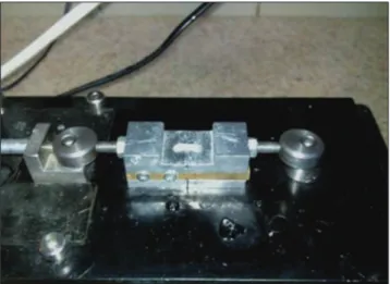
Figure 2: Microtensile test device
Discussion
This study evaluated the effects of four different bleaching techniques on the µTBS of resin bonded to a bleached enamel surface. Photoactivation and chemically‑activated HP negatively influenced the µTBS when the bonding procedure was performed 14 days after bleaching. G1 (control) had the maximum mean µTBS value, followed by G4 (the group bleached with diode laser activation), G2 (no light), G3 (LED), and G5 (Nd:YAG laser). However, no statistically significant differences appeared between the five groups. The null hypothesis of this study was accepted for the µTBS of all groups treated with or without photoactivated bleaching.
HP is one of the primary agents used as a preservative treatment for bleaching pigmented or darkened teeth.[23,24]
HP has a low molecular size and readily releases oxygen free radicals (OH and OOH) and the perhydroxyl anion (–OOH). These species can penetrate the enamel
surface and migrate along the dental structure to reach the dentin.[8,24,25] Commercial HP solutions have a
concentration of 3–35%.[25,26] The concentration does
not appear to affect the whitening achievable with HP or carbamide peroxide.[27‑29] Highly concentrated bleaching gels
can be used to bleach much more quickly.[24,30]
Most approaches to dental whitening have focused on accelerating peroxide bleaching of the anterior teeth with light sources. These light sources vary widely in their wavelengths and powers, e.g. plasma arc lamps, lasers and LEDs, and halogen curing lights.[5,25] The Nd:YAG laser, in
particular, has great potential for whitening teeth because it produces the heat necessary for activating the HP and the analgesia produced in the pulp through biostimulation.[15]
Previous study showed that there was no difference between LED and diode laser groups.[19] Although these results
were consistent statistically with our results they obtained higher shear bond strength for the LED. However, the values suggested by Can‑Karabulut and Karabulut[19] are
based on shear bond strength, whereas, in this study, µTBS values were evaluated. This study showed that there was no statistical difference diode laser and control groups, additionally higher µTBS values were obtained for the control group. The lower µTBS values of enamel that were found for the diode laser may be due to higher power density of the diode laser[5,19,31,32] and also the specification
of the applied gel might have played a role in this result.[19]
The required delay between the bleaching and restoration steps is an issue.[15] Cavalli et al. and others reported an
eduction in the µTBS of bleached teeth.[33] However,
those restoration treatments (porcelain or composite) were applied just 24 h after the bleaching procedure.[15] Nour
El‑din et al. reported that bleached teeth produced smaller
and thinner resin tags than unbleached teeth,[34] but the
restorative treatments were applied immediately after the bleaching protocols.[15] Sung et al. found that the bond
strength of bleached teeth did not differ statistically from those stored in a physiological solution for 5 days before adhesive procedures.[35] Giannini performed a carbamide
peroxide treatment on specimens that had been stored in artificial saliva for longer than 2 weeks.[36,37] In that study,
the peroxide absorbed in the enamel was released during the bleaching treatment; remineralization occurred to reestablish the surface morphology.[15]
This study showed that no statistical differences between the control and treated groups on the µTBS values. Residual oxygen could either inhibit free radical polymerization of the resins[38] or interfere with resin infiltration into enamel and
dentin.[39,40] The time delay after bleaching could account
for these findings.[15] The time delay period is supposed to
be sufficient for any residual oxygen to leave the dental hard tissues.[41] Sung et al. and Giannini reported similar µTBS
results for teeth stored in artificial saliva for 2 weeks after bleaching.[35,42]
Various factors contribute to the reduced bond strength of an adhesive applied to bleached enamel and dentin.[41]
HP bleaching or agents released from the decomposition of HP may significantly reduce the amount of calcium and phosphate in the enamel and alter the chemical structure of the crystallites in the superficial enamel.[41‑43]
Additionally, the bond strength reduction in enamel and dentin treated with HP could be caused by residues in the enamel and dentin pores after completion of the bleaching treatment.[41] Residual oxygen could either interfere
with resin infiltration into enamel and dentin[39,40] or
inhibit free radical polymerization of the resins.[38]
Activation of the chemical agents can be accelerated by using light, heat, or laser during the whitening process.[10,44] Energy absorbed by photosensitive agents is
transferred to the peroxide and thereby accelerates the oxidation–reduction reaction.[12,44] Many studies have
recommended using the Nd:YAG laser to accelerate the bleaching process.[31,44] Laser bleaching does not form
the residual oxygen species produced by conventional bleaching techniques.[45,46]
A 1–3 weeks delay after a bleaching treatment is often recommended before porcelain bonding or placement of composite resin restorations is done. This period is assumed to be sufficient for any residual oxygen to leave the dental hard tissues.[41] However, in vitro conditions and clinical conditions
are different. In vitro studies such as this study, the conditions do not completely duplicate the physical and chemical properties of the oral environment. This study carried out in enamel, and so results may be different for dentin.
Conclusion
The results of this research indicate that different HP bleaching treatments do not significantly reduce the µTBS over a 14‑day treatment period. The time delay after bleaching period may be longer than 2 weeks and then should compare with no‑photo activated enamel.
Financial support and sponsorship
Nil.
Conflicts of interest
There are no conflicts of interest.
References
1. Abu‑Eittah MR, Mandour MH. In vitro study of the effect of three hydrogen peroxide concentrations on the corrosion behavior and surface topography of alumina‑reinforced dental ceramic. J Prosthodont 2011;20:541‑52. 2. Sulieman M, Addy M, Macdonald E, Rees JS. The bleaching depth of a
35% hydrogen peroxide based in‑office product: A study in vitro. J Dent 2005;33:33‑40.
3. Strobl A, Gutknecht N, Franzen R, Hilgers RD, Lampert F, Meister J. Laser‑assisted in‑office bleaching using a neodymium:yttrium‑aluminum‑garnet laser: An in vivo study. Lasers Med Sci 2010;25:503‑9.
4. Sun G. The role of lasers in cosmetic dentistry. Dent Clin North Am 2000;44:831‑50.
5. Wetter NU, Barroso MC, Pelino JE. Dental bleaching efficacy with diode laser and LED irradiation: An in vitro study. Lasers Surg Med 2004;35:254‑8. 6. Luk K, Tam L, Hubert M. Effect of light energy on peroxide tooth bleaching.
J Am Dent Assoc 2004;135:194‑201.
7. Smigel I. Laser tooth whitening. Dent Today 1996;15:32‑6.
8. Feinman RA, Madray G, Yarborough D. Chemical, optical, and physiologic mechanisms of bleaching products: A review. Pract Periodontics Aesthet Dent 1991;3:32‑6.
9. Lima DA, Aguiar FH, Liporoni PC, Munin E, Ambrosano GM, Lovadino JR. In vitro evaluation of the effectiveness of bleaching agents activated by different light sources. J Prosthodont 2009;18:249‑54.
10. Coutinho DS, Silveira L Jr, Nicolau RA, Zanin F, Brugnera A Jr. Comparison of temperature increase in in vitro human tooth pulp by different light sources in the dental whitening process. Lasers Med Sci 2009;24:179‑85.
11. Garber DA. Dentist‑monitored bleaching: A discussion of combination and laser bleaching. J Am Dent Assoc 1997;128 Suppl: 26S‑30S.
12. Christensen GJ. The tooth‑whitening revolution. J Am Dent Assoc 2002;133:1277‑9.
13. Rocha Gomes Torres C, Caneppele TM, Del Moral de Lazari R, Ribeiro CF, Borges AB. Effect of dental surface treatment with Nd:YAG and Er:YAG lasers on bond strength of resin composite to recently bleached enamel. Lasers Med Sci 2012;27:755‑60.
14. Swift EJ Jr, Perdigão J, Heymann HO. Bonding to enamel and dentin: A brief history and state of the art, 1995. Quintessence Int 1995;26:95‑110. 15. Marcondes M, Paranhos MP, Spohr AM, Mota EG, da Silva IN, Souto AA, et al. The
influence of the Nd:YAG laser bleaching on physical and mechanical properties of the dental enamel. J Biomed Mater Res B Appl Biomater 2009;90:388‑95. 16. Ribeiro CF, Anido AA, Rauscher FC, Yui KC, Gonçalves SE. Marginal leakage
in class V cavities pretreated with different laser energy densities. Photomed Laser Surg 2005;23:313‑6.
17. Antunes A, de Rossi W, Zezell DM. Spectroscopic alterations on enamel and dentin after nanosecond Nd:YAG laser irradiation. Spectrochim Acta A Mol Biomol Spectrosc 2006;64:1142‑6.
18. Dostalova T, Jelinkova H, Housova D, Sulc J, Nemec M, Miyagi M, et al. Diode laser‑activated bleaching. Braz Dent J 2004;15 Spec No:SI3‑8.
19. Can‑Karabulut DC, Karabulut B. Shear bond strength to enamel after power bleaching activated by different sources. Eur J Esthet Dent 2010;5:382‑96. 20. Baik JW, Rueggeberg FA, Liewehr FR. Effect of light‑enhanced bleaching
on in vitro surface and intrapulpal temperature rise. J Esthet Restor Dent 2001;13:370‑8.
21. Beloica M, Goracci C, Carvalho CA, Radovic I, Margvelashvili M, Vulicevic ZR,
et al. Microtensile vs microshear bond strength of all‑in‑one adhesives to
unground enamel. J Adhes Dent 2010;12:427‑33.
22. Placido E, Meira JB, Lima RG, Muench A, de Souza RM, Ballester RY. Shear versus micro‑shear bond strength test: A finite element stress analysis. Dent Mater 2007;23:1086‑92.
23. Bittencourt ME, Trentin MS, Linden MS, de Oliveira Lima Arsati YB, França FM, Flório FM, et al. Influence of in situ postbleaching times on shear bond strength of resin‑based composite restorations. J Am Dent Assoc 2010;141:300‑6. 24. Didier VF, Batista AU, Montenegro RV, Fonseca RB, Carvalho FG, Barros SD,
et al. Influence of hydrogen peroxide‑based bleaching agents on the bond
strength of resin‑enamel/dentin interfaces. Int J Adhes Adhes 2013;47:141‑5. 25. Joiner A. The bleaching of teeth: A review of the literature. J Dent
2006;34:412‑9.
26. Toledano M, Yamauti M, Osorio E, Osorio R. Bleaching agents increase metalloproteinases‑mediated collagen degradation in dentin. J Endod 2011;37:1668‑72.
27. Hannig C, Lindner D, Attin T. Efficacy and tolerability of two home bleaching systems having different peroxide delivery. Clin Oral Investig 2007;11:321‑9. 28. Mondelli RF, Azevedo JF, Francisconi AC, Almeida CM, Ishikiriama SK.
Comparative clinical study of the effectiveness of different dental bleaching methods‑two year follow‑up. J Appl Oral Sci 2012;20:435‑43.
29. Tay LY, Kose C, Herrera DR, Reis A, Loguercio AD. Long‑term efficacy of in‑office and at‑home bleaching: A 2‑year double‑blind randomized clinical trial. Am J Dent 2012;25:199‑204.
30. Matis BA, Mousa HN, Cochran MA, Eckert GJ. Clinical evaluation of bleaching agents of different concentrations. Quintessence Int 2000;31:303‑10. 31. Zhang C, Wang X, Kinoshita J, Zhao B, Toko T, Kimura Y, et al. Effects of KTP
laser irradiation, diode laser, and LED on tooth bleaching: A comparative study. Photomed Laser Surg 2007;25:91‑5.
32. Sulieman M, Addy M, Rees JS. Surface and intra‑pulpal temperature rises during tooth bleaching: An in vitro study. Br Dent J 2005;199:37‑40.
33. Cavalli V, Giannini M, Carvalho RM. Effect of carbamide peroxide bleaching agents on tensile strength of human enamel. Dent Mater 2004;20:733‑9. 34. Nour El‑din AK, Miller BH, Griggs JA, Wakefield C. Immediate bonding to
bleached enamel. Oper Dent 2006;31:106‑14.
35. Sung EC, Chan SM, Mito R, Caputo AA. Effect of carbamide peroxide bleaching on the shear bond strength of composite to dental bonding agent enhanced enamel. J Prosthet Dent 1999;82:595‑9.
36. Della Bona A, Anusavice KJ. Microstructure, composition, and etching topography of dental ceramics. Int J Prosthodont 2002;15:159‑67.
37. Gianinni M. In vitro effects of whitening agents on tensile bond strength of adhesive systems and microhardness in human dental enamel. Piracicaba‑SP: UNICAMP; 2003.
38. Rueggeberg FA, Margeson DH. The effect of oxygen inhibition on an unfilled/ filled composite system. J Dent Res 1990;69:1652‑8.
39. McGuckin RS, Thurmond BA, Osovitz S. Enamel shear bond strengths after vital bleaching. Am J Dent 1992;5:216‑22.
40. Torneck CD, Titley KC, Smith DC, Adibfar A. Adhesion of light‑cured composite resin to bleached and unbleached bovine dentin. Endod Dent Traumatol 1990;6:97‑103.
41. Attin T, Hannig C, Wiegand A, Attin R. Effect of bleaching on restorative materials and restorations – A systematic review. Dent Mater 2004;20:852‑61. 42. Perdigão J, Francci C, Swift EJ Jr, Ambrose WW, Lopes M. Ultra‑morphological study of the interaction of dental adhesives with carbamide peroxide‑bleached enamel. Am J Dent 1998;11:291‑301.
43. Ruse ND, Smith DC, Torneck CD, Titley KC. Preliminary surface analysis of etched, bleached, and normal bovine enamel. J Dent Res 1990;69:1610‑3. 44. Akin M, Ozyilmaz OY, Yavuz T, Aykent F, Basciftci FA. Effect of Nd:YAG laser
bleaching and antioxidizing agents on the shear bond strength of brackets. Photomed Laser Surg 2013;31:365‑70.
45. Fuhrmann R, Gutknecht N, Magunski A, Lampert F, Diedrich P. Conditioning of enamel with Nd:YAG and CO2 dental laser systems and with phosphoric acid. An in‑vitro comparison of the tensile bond strength and the morphology of the enamel surface. J Orofac Orthop 2001;62:375‑86.
46. Mohammadi Bassir M, Rezvani MB, Chiniforush N, Moradi Z. In vitro evaluation of the effect of different laser irradiations on the enamel surfaces of teeth treated with home bleach procedure. J Lasers Med Sci 2013;4:168‑74.