261
© 2017 Nigerian Journal of Clinical Practice | Published by Wolters Kluwer - Medknow
Abstract
Crouzon syndrome (CS) is an autosomal dominant disorder characterized by craniofacial deformities caused by the early closure of cranial sutures. It is diagnosed by the presence of a flat sphenoid bone, protrusion of eyeballs (exophthalmos), and midfacial hypoplasia. Although hypodontia is usually present in cases with CS, supernumerary teeth are rarely seen. A 16‑year‑old male patient with CS was referred to our clinic. He had a high forehead, beaked nose, hypertelorism, palpebral ptosis, and asymmetrical orbits. Bilateral multiple supernumerary teeth were observed in his upper and lower jaws. Early diagnosis of CS is helpful in dental and craniofacial treatment. Because of multiple facial and oral problems, this patient required a multidisciplinary treatment approach.
Key words: Craniosynostosis, Crouzon syndrome, supernumerary teeth Date of Acceptance: 26-Apr-2016
Address for correspondence:
Dr. GS Torun,
Department of Orthodontics, Istanbul Medipol University, School of Dentistry, Atatürk Bulvarı No:27, Fatih 34083, Istanbul, Turkey. E‑mail: [email protected]
Introduction
Crouzon syndrome (CS) is an autosomal dominant craniofacial disorder named by a French physician, Octave Crouzon, in 1912. It is a type of craniosynostosis and is caused by mutations of the fibroblast growth factor receptor 2 genes.[1] Craniosynostosis is observed in 1 of
2500 newborns; the frequency of CS among all types of craniosynostoses is 4.5–4.8%.[2]
In CS, the premature closure of cranial sutures affects the shape of the cranium, causing deformity. The coronal and sagittal cranial sutures begin to close in the 1st year of birth.
The growth and development of the cranium are related
to the sutural activity, and early suture closure limits the growth and development of the cranium.[3]
CS is classified as type 1 or type 2, according to the presence of premature fusion of the elbows, wrists and hand, or toes of the foot. The hands and feet are affected in type 1; in type 2, however, there are no abnormalities of the extremities.[4]
The syndrome is diagnosed by the presence of a flat sphenoid bone, protrusion of eyeballs (exophthalmos), and midfacial hypoplasia. In addition, a beaked nose and high forehead are also observed in these cases.[5]
Melero et al. reported diminished maxillary length, constricted maxillary dental arch, and crowding in the upper and lower jaws as specific intraoral findings of CS.[6]
Maxillary skeletal size deficiency results in Angle Class III malocclusion and cross bite. In addition, patients may also
Crouzon syndrome with multiple supernumerary
teeth
GS Torun, A Akbulut1
Departments of Orthodontics and 1Oral and Maxillofacial Radiology, School of Dentistry, Istanbul Medipol University,
Istanbul, Turkey
Access this article online Quick Response Code:
Website: www.njcponline.com DOI: 10.4103/1119-3077.187332
PMID: ******* How to cite this article: Torun GS, Akbulut A. Crouzon syndrome with multiple supernumerary teeth. Niger J Clin Pract 2017;20:261‑3.
This is an open access article distributed under the terms of the Creative Commons Attribution‑NonCommercial‑ShareAlike 3.0 License, which allows others to remix, tweak, and build upon the work non‑commercially, as long as the author is credited and the new creations are licensed under the identical terms.
For reprints contact: [email protected]
Case Report
Torun and Akbulut: Crouzon syndome and supernumerary teeth
262 Nigerian Journal of Clinical Practice • February 2017 • Vol 20 • Issue 2
present with over‑retained deciduous teeth and multiple impacted or unerupted teeth.[6]
Some of the other general symptoms include hearing loss, Meniere’s disease, sleep apnea, vertebral malformations, visual problems, headaches, hydrocephalus, problems of the aorta, and dermatological problems. Acanthosis nigricans, which is a velvety hyperpigmentation of the skin, may also be present.[4] Although external malformations of the ear are
rarely seen, patients may present with middle ear problems and hearing loss.[7]
Dental problems in Crouzon syndrome
Issues related to the number of teeth and bone formation are usually present in cases with CS. The most common type of supernumerary teeth is the mesiodens.[8] Missing
teeth are more commonly observed in patients with this syndrome than are supernumerary teeth.[9] The timing of
tooth eruption might be affected, and delayed tooth eruption can be seen in patients with craniosynostosis.[10]
Case Report
A 16‑year‑old Turkish boy was referred to the Istanbul Medipol university clinic. Routine clinical assessment including medical history recording, dental examination, and orthodontic consultation was performed, followed by radiographic evaluation.
Extraoral examination
Extraoral examination revealed a high forehead, beaked nose, hypertelorism, and palpebral ptosis. The vertical positions of the eyes were different from each other, and asymmetrical orbits were observed [Figure 1].
Intraoral examination
Intraoral examination revealed that teeth 11 and 21 were impacted, and teeth 53 and 63 were persistent. A rudimentary second premolar was present on the left side. In the lower jaw, teeth 72, 73, and 83 were present, and tooth 45 was unerupted. The maxillary arch was constricted, and the patient had poor oral hygiene.
Radiographic evaluation
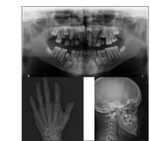
Impacted teeth 11 and 21 were diagnosed based on panoramic radiographs. Impacted supernumerary upper lateral incisors were observed, along with impacted supernumerary upper canines. There was a supernumerary permanent lateral incisor and supernumerary permanent canine per side of the maxillary arch. Malformed second premolars were present in the upper jaw, which prevented the eruption of the supernumerary second premolars. Tooth 15 was impacted.
In the lower jaw, there was an impacted supernumerary canine, first premolar, and second premolar in each of the sides. Only the right lower second premolar was impacted with its supernumerary one.
A skeletal Class III relationship was diagnosed (Sella–Nasion–A point angle: 87°, Sella–Nasion–B point angle: 90°, A point–Nasion–B point angle: −3°) upon lateral cephalometric analysis. A hypodivergent vertical pattern was observed (Sella–Nasion/Gonion–Gnathion: 22°), along with the presence of retrusive upper lip and protrusive lower lip. The maxillary skeletal size (condylion to point A) was diminished. The distance between the mandible and cranial base was increased (Pogonion–Nasion perpendicular).
Figure 1: Intra-and extra-oral photographs of the patient
Torun and Akbulut: Crouzon syndome and supernumerary teeth
263
Nigerian Journal of Clinical Practice • February 2017 • Vol 20 • Issue 2 According to the results of the hand‑wrist radiography
analysis, the skeletal age of the patient was 17 years. The distal phalanxes of the digits were short and had fusiform endings [Figure 2].
Discussion
Our case report describes a patient with CS, who presented with asymmetrical orbits, beaked nose, high forehead, and multiple supernumeraries and impacted teeth. In this case, the orbital characteristics were more dominant than the maxillary hypoplastic issues. The case was diagnosed as type 2, based on the presence of finger malformations. In this case, despite the diminished maxillary skeletal size, the forward position of the maxilla masked the maxillary hypoplasia. The most important issue was the presence of multiple supernumerary teeth. Previous reports indicate that the incidence of missing teeth is 6–8% and that of supernumerary teeth is 1.5–3.5%, in craniofacial syndromes.[10]
The presence of supernumerary teeth in each of the quadrants of the upper and lower jaws has not been reported in literature. The presence of bilateral supernumerary teeth in the upper jaw was reported by Santos et al.[8] However,
other studies have reported only unilateral supernumerary teeth.[11,12] This report describes a case with bilateral
supernumerary teeth in both the upper and lower jaws.
Treatment plan
The dental caries in teeth 16, 26, 35, 36, 46, and 47 will be treated. The rudimentary upper bilateral second premolars as well as teeth 53 and 63 will be extracted.
Following this, the impacted supernumerary upper left and right canines and lateral incisors will be extracted. In the lower jaw, teeth 32, 73, and 83, as well as the supernumerary bilateral impacted canines and left lower first and second premolars will be extracted. On the right side, the supernumerary impacted second premolar will be removed. The skeletal Class III malocclusion will be treated by orthodontic treatment. Surgical‑assisted rapid maxillary expansion and orthodontic treatment will be performed before mandibular setback orthognathic surgery. A multidisciplinary approach involving dental surgeons, oral and maxillofacial surgeons, and orthodontists is required for this treatment.
Declaration of patient consent
The authors certify that they have obtained all appropriate patient consent forms. In the form the patient(s) has/have given his/her/their consent for his/her/their images and other clinical information to be reported in the journal. The patients understand that their names and initials will not be published and due efforts will be made to conceal their identity, but anonymity cannot be guaranteed.
Financial support and sponsorship
Nil.
Conflicts of interest
There are no conflicts of interest.
References
1. Fries PD, Katowitz JA. Congenital craniofacial anomalies of ophthalmic importance. Surv Ophthalmol 1990;35:87‑119.
2. Bowling EL, Burstein FD. Crouzon syndrome. Optometry 2006;77:217‑22. 3. Hlongwa P. Early orthodontic management of Crouzon syndrome: A case
report. J Maxillofac Oral Surg 2009;8:74‑6.
4. Proudman TW, Moore MH, Abbott AH, David DJ. Noncraniofacial manifestations of Crouzon’s disease. J Craniofac Surg 1994;5:218‑22. 5. Horbelt CV. Physical and oral characteristics of Crouzon syndrome, Apert
syndrome, and Pierre Robin sequence. Gen Dent 2008;56:132‑4.
6. Melero S, Leite M, Carvalho I. Dental anomalies in patients carrying the Apert syndrome and the Crouzon syndrome. Salusvita 2005;24:183‑93.
7. Orvidas LJ, Fabry LB, Diacova S, McDonald TJ. Hearing and otopathology in Crouzon syndrome. Laryngoscope 1999;109:1372‑5.
8. Santos AP, Ammari MM, Moliterno LF, Júnior JC. First report of bilateral supernumerary teeth associated with both primary and permanent maxillary canines. J Oral Sci 2009;51:145‑50.
9. Stavropoulos D, Tarnow P, Mohlin B, Kahnberg KE, Hagberg C. Comparing patients with Apert and Crouzon syndromes – Clinical features and cranio‑maxillofacial surgical reconstruction. Swed Dent J 2012;36:25‑34. 10. Nieminen P. Genetic basis of tooth agenesis. J Exp Zool B Mol Dev
Evol 2009;312B: 320‑42.
11. Türkkahraman H, Yilmaz HH, Cetin E. A non‑syndrome case with bilateral supernumerary canines: Report of a rare case. Dentomaxillofac Radiol 2005;34:319‑21.
12. Shetty R, Sandler PJ. Keeping your eye on the ball. Dent Update 2004;31:398‑402.
Figure 2: Multiple supernumerary teeth are observed on orthopantomography; the hand-wrist radiography image shows short distal phalanges in the digits; cephalometric radiography
image of the patient
![Figure 1: Intra-and extra-oral photographs of the patient[Downloaded free from http://www.njcponline.com on Thursday, March 19, 2020, IP: 85.111.55.76]](https://thumb-eu.123doks.com/thumbv2/9libnet/5424198.103280/2.918.187.734.729.1069/figure-intra-photographs-patient-downloaded-njcponline-thursday-march.webp)