DETECTION OF EPILEPTIC INDICATORS ON CLINICAL SUBBANDS OF EEG
Zeynep Y¨ucel, A. B¨ulent ¨
Ozg¨uler
Electrical and Electronics Engineering, Bilkent University Bilkent, 06800, Ankara, Turkey
phone: + (90) 312 290 1219, fax: + (90) 312 266 4192, email: zeynep@ee.bilkent.edu.tr web: www.ee.bilkent.edu.tr/ zeynep
ABSTRACT
Symptoms of epilepsy, which is characterized by abnor-mal brain electrical activity, can be observed on electroen-cephalography (EEG) signal. This paper employs models of chaotic measures on standard clinical subbands of EEG and aims to help detection of epilepsy seizures and diagnosis of epileptic indicators in interictal signals.
1. RELATED WORK
Lehnertz divides EEG analysis techniques into two cate-gories, [19], as linear and nonlinear methods. The algorithms described in [11], [17], [15] are regarded as linear methods. Juling et. al., [17] and Jahankhani et. al., [15], consider wavelet based methods to extract time-frequency character-istics of EEG to discriminate between ictal and interictal phases. Among nonlinear analysis techniques neural net-works is a widely used approach, [20], [9]. In [8], authors employ short term Lyapunov exponent to classify “normal” and “abnormal” EEG signals. The time evolution of the tra-jectory is derived from recurrence plots to anticipate seizures in [21]. Hamadene et al., [13], interpret recurrence plots for prediction of epileptic seizures similar to [21]. Entropy re-lated features are used in predicting seizures, [22], detecting patient-specific pre-cursors, [5], and discriminating between seizure and pre-seizure periods, [6]. Independent component analysis [16], phase locked loops [12] and combination of several of the above approaches [23] are among other EEG signal processing techniques.
In this research, we aim to model chaotic measures of a standard subband of EEG and distinguish between differ-ent characteristics concerning epilepsy. The outline of the paper is as follows. Section 2 describes the dataset discussed in this work. Section 3 explains details of the proposed method. In Sections 4 and 5, details about the classification schemes, performance rates and conclusions are presented.
2. EEG DATASET
The dataset prepared by the Clinique of Epileptology of Bonn University is utilized in this research, [3]. Single chan-nel EEGs are recorded from people with different brain elec-trical potential characteristics at a sampling rate of 173.61 Hz for 23.6 sec. These EEG recordings are grouped into three sets denoted by H, E, and S. Set H contains 200 EEG recordings from healthy people, while sets E and S involve recordings from epileptic patients. The 200 recordings in set
E are taken in the interictal period, i.e. between seizures. Set S is comprised of 100 recordings in ictal period, i.e. during
seizures.
3. METHODOLOGY
The challenge of this research relies on the assumption that the chaotic characteristics of an EEG signal in various fre-quency bands provide more descriptive information about the physiological condition of the person. In this respect, wavelet transform enables us to handle the signal in different resolution levels and within several frequency bands. Sec-tion 3.1 expounds details about standard clinical subbands of EEG and derivation of them by multiresolution analysis. The construction of phase space and derivation of its distinc-tive chaotic features are explained in Sections 3.2 and 3.3, respectively.
3.1 Standard Clinical Subbands of EEG and Multireso-lution Analysis
EEG signals are handled in five standard frequency bands, namely, delta(0 − 4Hz), theta (4 − 8Hz), alpha (8 − 12Hz),
beta(13 − 30Hz), and gamma (30 − 60Hz), [2]. Therefore frequencies between 0− 60Hz provide significant informa-tion about the brain electrical potential. Let x0be any time
series from one of the sets indicated in Section 2. Since
x0 is recorded at a sampling rate of 173.6Hz, frequencies
higher than 60Hz appear in its spectral analysis. Obviously a low pass filtering operation is needed to focus on the significant frequency bands. A 10th order Butterworth low pass filter with a suitable cut-off frequency is employed in the extraction of this band.
Herrmann et. al., [14] state that gamma activity is closely correlated with cognitive functions and propose that epileptic indicators of EEG are a direct consequence of increase in gamma activity. Moreover, Willoughby et. al., [24] show that interictal EEG signals from epileptic patients and healthy people differ enormously in terms of
gamma activity. Hence, we focus on the gamma subband
and employ chaotic measures to represent its discriminating characteristics. The gamma subband is emphasized by applying a single stage wavelet decomposition using a third order Daubechies filter on the low pass filtered signal. The resulting detail coefficients cover the information in the gamma subband and hence we choose to use such a filter in our analysis.
3.2 Construction of Time Delay Vectors
As a nonlinear dynamical system evolves in time, it could get sufficiently close to a set of states and remain within close neighborhood of that set, even if slightly disturbed. Such states are called attractors. Complexity and chaotic characteristics are the two main descriptors of an
attrac-tor. Complexity is related to geometric properties of the attractor, where chaoticity indicates the rate of divergence or convergence of nearby trajectories in phase space.
As the EEG signal is viewed as the output of a nonlin-ear dynamical system, it is observed that chaos related features differ between normal and epileptic brain activity. In this research, we focus on the chaotic behavior so as to discriminate between healthy, interictal and ictal EEG. In the analysis of chaotic behavior, recurrence rate is employed as a measure of chaos. The computation of recurrence rate requires formation of time delay vectors. Let x0be any time
series from one of the sets presented in Section 2 and d1be
the detail coefficients of x0. Time delay vectors for d1are
formed in the following manner.
βi
d1(d0) = {d1(i), d1(i + m0), . . . , d1(i + (d0− 1)m0)},
1≤ i ≤ nd1, nd1= Nd1− (d0− 1)m0,
(1) where d0 denotes minimum embedding dimension, m0
denotes optimum lag and Nd1 is the size of the time series
d1.
In order to form time delay vectors, optimum lag m0
and minimum embedding dimension d0 need to be
deter-mined. Details about calculations of m0and d0are presented
in Sections 3.2.1 and 3.2.2.
3.2.1 Determination of Optimum Lag
Optimum lag is the amount of shift between two portions of the time series, which yields minimum overlapping infor-mation. Mutual information function, I, which indicates the amount of mutual dependence between two variables, is em-ployed in the computation of optimum lag. For two discrete random variables X and Y , mutual information function is calculated in the following manner,
I(X;Y ) =
∑
y∈Yx∈X∑
p(x, y)log( p(x, y) p1(x)p2(y) ). Let di1 contain the data points of d1 between time instants
i and Nd1− m. Similarly, d
i+m
1 contains data points of d1
between time instants i+ m and Nd1:
di1= {d1(i), d1(i + 1), ...d1(Nd1− m)},
di+m1 = {d1(i + m), d1(i + m + 1), ...d1(Nd1)}.
We denote the amount of mutual dependence between d1i and d1i+m by I(m). As I(m) is computed for several values of m, the evolution of mutual dependence between two time series with respect to various values of lag can be observed. Obviously, a larger lag leads to little overlapping information, where a smaller lag provides the number of data points in di
1and d1i+mto be large enough to make plausible
inferences. Therefore the first local minimum of I(m) is proposed to be the optimum lag m0, [1].
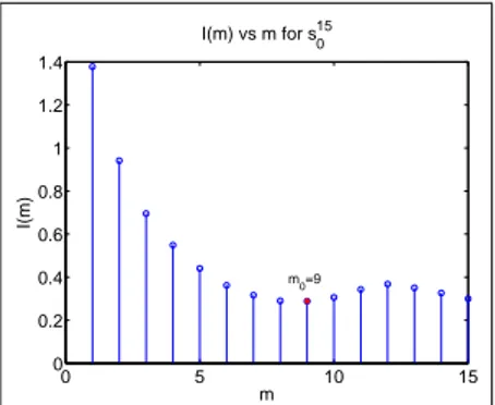
Figure 1 depicts an example of evolution of mutual in-formation for an element of set S with respect to increasing values of m. Optimum lag is found to be m0= 9 for this
particular time series.
0 5 10 15 0 0.2 0.4 0.6 0.8 1 1.2 1.4 I(m) vs m for s015 m I(m) m0=9
Figure 1: Evolution of Mutual Information for a sample time series from set S
3.2.2 Determination of Minimum Embedding Dimension
Cao describes a requirement for an embedding dimension d to be accepted as a true embedding dimension and a method for the calculation of this number, [7]. Assume the time de-lay vectorβdi
1(d) is formed from d1using some arbitrary
em-bedding dimension d, as in Equation 1. According to Cao, a true embedding dimension, d0, satisfies the requirement that
two time delay vectors, βi
d1(d0) andβ
j
d1(d0), that lie close
to each other in d0-dimensional space, will still be close to
each other in(d0+ 1)-dimensional space. In order to
inves-tigate whether a certain embedding dimension d fulfills this requirement or not, we check the proximity of two time delay vectors, which are nearest neighbors in d-dimensional space, in(d + 1)-dimensional space. Letβdn(i,d)
1 (d) denote the
near-est neighbor ofβdi
1(d) in d-dimensional space and ad1(i, d)
denote the ratio of distance between these two time delay vectors in d-dimensional to(d + 1)-dimensional spaces, i.e.,
ad1(i, d) = β i d1(d) −β n(i,d) d1 (d) β i d1(d + 1) −β n(i,d) d1 (d + 1) ,
wherek.k denotes Euclidean distance. Let Ed1(d) denote the
mean value of ad1(i, d)’s:
Ed1(d) = 1 Nd1− dm0 Nd1−dm0
∑
i=1 ad1(i, d), and Ed1 1(d) be equal to Ed1(d + 1)/Ed1(d). E 1 d1 is expectedto settle around a certain value for embedding dimensions larger than some particular d0− 1. As a rule of thumb, d0
is called the minimum embedding dimension. However in practical computations, Ed1
1 could yield misleading results
due to limited number of elements of phase space. To over-come this problem, Cao redefines the quantification of neigh-borhood condition by Ed⋆ 1(d), Ed⋆ 1(d) = 1 N− dτ N−dτ
∑
i=1 |d1(i + dτ) − d1(n(i, d))| .In this case, the variation between successive embedding dimensions is investigated by Ed2 1 = E ⋆ d1(d + 1)/E ⋆ d1(d).
for each time series by applying the same rule of thumb on
Ed⋆
1 and a phase space could be constructed accordingly. In
such a case, the number of time delay vectors contributing to the phase space from a particular time series will be
Nd1− m0(d0− 1).
Here one should note that, although Nd1 is fixed in our
case, m0 and d0 can change the number of constructed
time delay vectors pretty much. In order to have a fair comparison, it is preferred to calculate a separate m0 for
each d1 and to keep d0 fixed. To determine the optimum
constant for minimum embedding dimension, we calculate
d0’s for all possible time series d1 and pick the one which
is most voted. It is observed that d0= 7 is the minimum
embedding dimension for most of the time series. 3.3 Modeling Recurrence Rate Behavior
After determining optimum lag and minimum embedding dimension, one can construct time delay vectors for each
gamma subband as in Equation 1. The collection of time
delay vectors form a lagged phase space with elements
βi
d1(d0). The discriminative features of a particular EEG
recording x0is derived from the recurrence properties of this
lagged phase space.
Section 3.3.2 gives details about modeling recurrence rate and describes the derivation of feature vectors. The distribution of feature vectors is illustrated in Section 3.3.3.
3.3.1 Measures of Chaoticity and Recurrence Plots
Chaos can be measured by correlation dimension, recurrence rate, determinism percentage, Hurst exponent, or largest Lyapunov exponent, [18], among other methods. Here we make use of recurrence rate. Any two states, which lie in some proximity smaller than ε, are called recurrence states. Recurrence plot is a graphical tool to visualize the recurrence states and recurrence rate is a simple recurrence quantifier derived from the recurrence plot.
Let R denote the N × N recurrence plot of a phase space with elementsσi, where 1≤ i ≤ N, as N denotes the
number of elements of the phase space. The value of a point (i, j) on the recurrence plot R is computed by the following equation,
R(i, j) =Θ(ε− σi−σj ),
whereΘis the Heaviside step function andεis the distance threshold. It is obvious from the above equation, that if there are any two states in the phase space,σiandσj, which
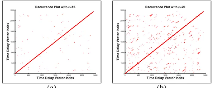
are in some proximity smaller than ε, the value of R(i, j) is 1 and otherwise 0. Figure 2 presents two recurrence plots of a sample time series from set H for ε= 15 and
ε= 20 cases. As expected increasing the threshold makes the neighborhood condition looser and hence number of recurrence points increases.
3.3.2 Recurrence Quantification and Derivation of Feature Vectors
In recurrence quantification, recurrence rateΨ, which basi-cally denotes the density of recurrence points in R, is
em-0 500 1000 1500 2000 2500 3000 0 500 1000 1500 2000 2500 3000
Recurrence Plot with ε=15
Time Delay Vector Index
Time Delay Vector Index
0 500 1000 1500 2000 2500 3000 0 500 1000 1500 2000 2500 3000
Recurrence Plot with ε=20
Time Delay Vector Index
Time Delay Vector Index
(a) (b)
Figure 2: Recurrence plots of a sample time series from set
H for (a)ε= 15 and (b)ε= 20.
ployed. Ψ= 1 N2 N
∑
i, j=1 R(i, j).Feature vectors are derived by examining the evolution of recurrence rate against different values of distance threshold. For a particular distance threshold,εk, the recurrence rate of
lagged phase space of time series d1is given by
Ψk d1= 1 N2 d1 Nd1
∑
i, j=1 i6= j Θ(εk− β i d1(d0) −β j d1(d0) ).To observe the evolution of recurrence rate against dis-tance threshold, we calculate Ψkd
1 for various values of
εk∈ {ε1,ε2, ...,εK} and obtain a series of recurrence rates,
Ψd1 = {Ψ 1 d1,Ψ 2 d1, ...,Ψ K
d1}. As seen in Figure 3-(a), these
series are observed to exhibit a different nature for sets H,
E and S and therefore could be used to represent features.
However, using raw recurrence rate series is not handy, since the size of Ψd1 could be large depending on the number
of distance thresholds, K. In order to provide a dimension reduction, a simple model is developed forΨd1 such that the
feature vector of a particular time series d1is composed of
the parameters of the adopted model forΨd1.
After examining the graph of Ψkd
1 against εk, it is
ob-served that it exhibits almost a linear increase for certain values of εk. Moreover, it is clear from Figure 3 that the
linear regions have different slopes in general and occur mostly at different values ofεkfor sets H, E and S. Thus the
0 2 4 6 8 10 12 14 16 18 20 0 0.1 0.2 0.3 0.4 0.5 0.6 0.7 0.8 0.9 1 Evolution of Ψd1 ε Ψd1 H E S 0 2 4 6 8 10 12 14 16 18 20 0 0.1 0.2 0.3 0.4 0.5 0.6 0.7 0.8 0.9 1
Approximation for Linear Regions
ε Ψd1 H E S (a) (b)
Figure 3: (a) Evolution ofΨkd
1 againstεkfor several sample
time series, (b) Approximations for the linear regions.
the linear approximation a0ε+ b0and the value of distance
thresholdε0at the center of the linear region. In this manner,
parameters a0, b0 and ε0 are assumed to summarize the
evolution ofΨd1 with respect to distance threshold and are
considered to provide a distinction.
3.3.3 Distribution of Feature Vectors
After determining a model for the evolution of recurrence rate and solving for the model parameters, we should check whether these parameters provide a distinction between un-derlying brain electrical potentials. Figure 4 depicts the dis-tribution of parameters a0, b0 andε0for sets H, E, S and
verifies that the parameters occupy mostly different regions.
0 0.2 0.4 0.6 0.8 1 0 0.2 0.4 0.6 0.8 1 0 0.2 0.4 0.6 0.8 1 a0 Distribution of Features b0 ε0 H E S
Figure 4: Distribution of features
4. CLASSIFICATION
K-nearest neighbor classification is employed in
discrimi-nating the physiological differences between the scalp EEG recordings described in Section 2. This section is dedicated to the details of the classification scheme. Section 4.1 ex-plains the cross-correlation schemes used in the testing and training of linear discriminant classifier. Performance rates are presented in Section 4.2. Comparison with the existing techniques is handled in Section 4.3.
4.1 Cross-Correlation Scheme
The test performance is investigated via a series of classifi-cation experiments. Test performance shows how well the classifier performs when new patterns are investigated for class membership. While measuring test performance, the classifier is trained with a number of training patterns and then tested by new patterns. The number of training patterns is increased step by step and the classifier is tested by the rest of the dataset at each step. As we increase the number of training examples, we expect to see the classification performance to increase and settle down around a steady state value. In this manner, we can see how large a data set suffices to describe the classes thoroughly.
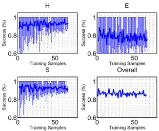
The graphs below show the success rates while num-ber of training samples is increased from 5 to 70. For each case ten experiments are made. The solid line shows the mean of those, while the vertical ones indicate their maximum and minimum.
4.2 Test Performance
From Figure 5, it is observed that the classifier performs bet-ter in distinction of sets H and S. As number of training sam-ples gets over 20, success rates reach 88% and 94% respec-tively. For the set E, performance is around 72% regardless of the number of training samples.
0 50 0.6 0.8 1 Training Samples Success (%) H 0 50 0.6 0.8 1 Training Samples Success (%) E 0 50 0.6 0.8 1 Training Samples Success (%) S 0 50 0.6 0.8 1 Training Samples Success (%) Overall
Figure 5: Evolution of success rate in testing
4.3 Comparison with Existing Techniques
Andrzejak provides a list of papers which use the same dataset in analysis and classification of EEG signals, [4]. Most of these papers consider different groups of EEG signals by either eliminating some classes, dividing some classes into further sub-classes or formulate a different prob-lem by comparing the symptoms of epilepsy to symptoms of other physiological disorders. Among these papers, [10] and [1] consider a similar problem formulation to ours. Our scheme employs a phase space, which is built similar to the one in [1], however, we introduce the modeling of recurrence rate of the phase space and classification using the model pa-rameters unlike [1]. Gautama et. al. [10] treat the same prob-lem considered in this work by employing delay vector vari-ance (DVV) method. They achieve 74.4% overall success rate. With our method, overall success rate is always around 85% as seen in Figure 5. Thus our method outperforms DVV in overall performance.
5. CONCLUSION
In this paper, we propose a method for discrimination of EEG recordings from people with different epileptic characteris-tics. A model is developed for the recurrence rate derived from gamma band of the EEG signals. It is shown to exhibit different natures for healthy, ictal and interictal cases. The proposed classification scheme performs well for healthy and ictal EEGs but only fairly good for interictal EEGs and needs to be improved. As a future work, we will consider apply-ing hierarchical classification with a more effective classifier. Moreover, the multiresolution analysis could be improved by employing a filterbank which results in less aliasing than the Daubechies wavelets.
REFERENCES
[1] Adeli, H., Ghosh-Dastidar, S., Dadmehr, N., “A Wavelet-Chaos Methodology for Analysis of EEGs and EEG Sub-bands to Detect Seizure and Epilepsy”, IEEE
Transac-tions on Biomedical Engineering, Volume 54, Issue 2,
Feb. 2007, Page(s):205 - 211.
[2] Assaleh, K., Al-Nashash, H., Thakor, N., “Spectral Subtraction and Cepstral Distance for Enhancing EEG Entropy”, Engineering in Medicine and Biology
Soci-ety, 2005. IEEE-EMBS 2005. 27th Annual International Conference of the, 2005, 2751-2754.
[3] Andrzejak, R. G., Lehnertz, K., Mormann, F., Rieke, C., David, P., Elger, C.E., “Indications of Nonlinear Deter-ministic and Finite-Dimensional Structures in Time Se-ries of Brain Electrical Activity: Dependence on Record-ing Region and Brain State”, Phys. Rev. E, Volume 64, No 6, Nov. 2001, Pages 061907-8.
[4] Klinik f¨ur Epileptologie, http://www.meb.uni-bonn.de/epileptologie/cms/frontcontent.php
[5] D’Alessandro, M., Esteller, R., Vachtsevanos, G., Hin-son, A., Echauz, J., Litt, B., “Epileptic seizure prediction using hybrid feature selection over multiple intracranial EEG electrode contacts: a report of four patients”, IEEE
Transactions on Biomedical Engineering, Volume 50,
Is-sue 8, Aug. 2003 Page(s):1041 - 1041.
[6] Bianchi, A.M., Panzica, F., Tinello, F., Franceschetti, S., Cerutti, S., Baselli, G., “Analysis of multichannel EEG synchronization before and during generalized epilep-tic seizures”, Proceedings of First International IEEE
EMBS Conference on Neural Engineering, 20-22 March
2003, Page(s):39 - 42
[7] Cao, L., “Practical Methods for Determining The Min-imum Embedding Dimension of a Scalar Time Series”.
Physica D, 1997, Page(s) 43-50.
[8] Chaovalitwongse W. A. , Fan Y.-J. , Sachdeo R. C. , “On the Time Series K-Nearest Neighbor Classification of Abnormal Brain Activity”, IEEE Transactions on
Sys-tems and Man and Cybernetics-Part A: SysSys-tems and Hu-mans:Accepted for future publication, Issue 99, 2007.
[9] Fischer, P., Tetzlaff, R., “Pattern detection by cellu-lar neuronal networks (CNN) in long-term recordings of a brain electrical activity in epilepsy”, Proceedings
of IEEE International Joint Conference on Neural Net-works, 2004, Volume 1, 25-29 July 2004.
[10] Gautama, T., Mandic, D.P., Van Hulle, M.M., “Indica-tions of nonlinear structures in brain electrical activity”,
Physical Review E, Volume 67, Issue 4, April 2003.
[11] G¨uler, I., Kiymik, M.K., Akin, M., Alkan A., “AR Spectral Analysis of EEG Signals by Using Maxi-mum Likelihood Estimation”, Computers in Biology and
Medicine, Volume 31, 2001, pp. 441-450.
[12] Gysels, E., Le Van Quyen, M., Martinerie, J., Boon, P., Vonck, K., Lemahieu, I., Van De Walle, R., “Long-term evaluation of synchronization between scalp EEG signals in partial epilepsy”, Proceedings of the 9th
In-ternational Conference on Neural Information Process-ing, 2002. ICONIP ’02. , Volume 3, 18-22 Nov. 2002
Page(s):1495 - 1498.
[13] Hamadene, W., Peyrodie, L., Seidiri, H.,
“Interpreta-tion of RQA variables: Applica“Interpreta-tion to the predic“Interpreta-tion of epileptic seizures”, The 8thInternational Conference on Signal Processing, Volume 4, 16-20 2006.
[14] Herrmann, C.S., Demiralp, T., “Human EEG gamma oscillations in neuropsychiatric disorders”, Clinical
Neu-rophysiology, Volume 116, 2005, Page(s): 2719-2733.
[15] Jahankhani, P., Revett, K., Kodogiannis, V., “Data Min-ing an EEG Dataset with an Emphasis on Dimensionality Reduction”, IEEE Symposium on Computational
Intel-ligence and Data Mining, 2007, CIDM 2007, March 1
2007-April 5 2007 Page(s):405 - 412.
[16] James, C.J., Lowe, D.,“Using independent component analysis & dynamical embedding to isolate seizure activ-ity in the EEG”, Proceedings of the 22ndAnnual Interna-tional Conference of the IEEE Engineering in Medicine and Biology Society, 2000, Volume 2, 23-28 July 2000
Page(s):1329 - 1332.
[17] Junling, Z., Dazong, J., “A linear epileptic seizure pre-dictor based on slow waves of scalp EEGs”, 27thAnnual International Conference of the Engineering in Medicine and Biology Society, 2005, IEEE-EMBS 2005, 01-04
Sept. 2005, Page(s):7277 - 7280.
[18] Kannathal, N., Acharya, U. R., Lim, C.M., Sadasivan, P. K., “Characterization of EEG-A comperative study”,
Computer Methods and Programs in Biomedicine, 2005,
Page(s) 17-23.
[19] Lehnertz, K., “Seizure prediction techniques: robust-ness and performance issues”, Proceedings of the Second
Joint EMBS/BMES Conference, 2002, Volume 3, 23-26
Oct. 2002, Page(s):2037 - 2038.
[20] Niederhoefer, C., Tetzlaff, R.,, “Prediction Error Pro-files allowing a Seizure Forecasting in Epilepsy? ”, 10th
International Workshop on Cellular Neural Networks and Their Applications, 2006. CNNA ’06, 28-30 Aug.
2006 Page(s):l - 6.
[21] Ouyang, G., Xie, L., Chen, H., Li, X., Guan, X., Wu, H., “Automated Prediction of Epileptic Seizures in Rats with Recurrence Quantification Analysis”, 27th
Annual International Conference of the Engineering in Medicine and Biology Society, IEEE-EMBS 2005, 2005,
Page(s):153 - 156.
[22] Srinivasan, V., Eswaran, C., Sriraam, N., “Approximate Entropy-Based Epileptic EEG Detection Using Artificial Neural Networks”, IEEE Transactions on Information
Technology in Biomedicine, Volume 11, Issue 3, May
2007, Page(s):288 - 295.
[23] Qu, H., Gotman, J.,“A patient-specific algorithm for the detection of seizure onset in long-term EEG monitoring: possible use as a warning device”, IEEE Transactions on
Biomedical Engineering, Volume 44, Issue 2, Feb. 1997
Page(s):ll5 - 122.
[24] Willoughby, J.O., Fitzgibbon, S.P., Pope, K.J., Macken-zie, L., Medvedev, A.V., Clark, C.R., Davey, M.P., Wilcox, R.A., “Persistent abnormality detected in the non-ictal electroencephalogram in primary generalized epilepsy”, Journal of Neurology Neurosurgery and


