Can flecainide totally eliminate bidirectional ventricular tachycardia
in pediatric patients with Andersen-Tawil syndrome?
Flekainid Andersen-Tawil sendromlu pediyatrik hastalarda bidireksiyonel
ventriküler taşikardiyi tamamen ortadan kaldırabilir mi?
1Department of Pediatric Cardiology and Arrhythmia, Mehmet Akif Ersoy Thoracic and Cardiovascular Surgery
Training and Research Hospital, İstanbul, Turkey
2Department of Pediatric Cardiology, Bağcılar Training and Research Hospital, İstanbul, Turkey 3Department of Pediatric Cardiology and Arrhythmia, Medipol University Hospital, İstanbul, Turkey
Yakup Ergül, M.D.,1 Senem Özgür, M.D.,1 Sertaç Hanedan Onan, M.D.,2 Volkan Tuzcu, M.D.3
Özet– Andersen-Tawil sendromu (ATS), kas güçsüzlüğü (periyodik paralizi), ritm bozuklukları ve gelişim bozuk-luklarına neden olan bir hastalıktır. QT uzaması ve bidi-reksiyonel ventriküler taşikardi (VT) ve polimorfik VT’yi de içeren ventriküler aritmiler ortaya çıkabilir. Tüm olgu-ların yaklaşık %60’ı, KCNJ2 genindeki mutasyonlardan kaynaklanmaktadır. On üç yaşında kız hasta, sık vent-riküler erken atımlar nedeniyle hastanemize sevk edildi. Hasta, morfolojik bozuklukları, EKG değişiklikleri ve pe-riyodik kas güçsüzlüğü nedeniyle ATS olarak düşünüldü ve tanı genetik olarak doğrulandı. Bidireksiyonel VT ve sık polimorfik erken ventriküler atımları için ilk basa-mak tedavi olarak beta bloker başlandı. Ancak tedaviye rağmen, hastanın VT atakları kontrol altına alınamadı. Bunun üzerine Flekainid tedaviye eklendi. Flekainid ile prematüre ventriküler atımların sayısı çarpıcı bir şekilde azaldı. Ayrıca VT atakları tamamen kayboldu. Bu has-ta, ülkemizde nadir görülen ATS’li hastalardan biridir. Bu makalede ATS’li bir hastada ritm bozukluğunun başarılı yönetimi anlatılmıştır.
Summary– Andersen-Tawil syndrome (ATS) is a disorder that causes episodes of muscle weakness (periodic paraly-sis), changes in heart rhythm, and developmental abnormal-ities. QT prolongation and ventricular arrhythmias, including bidirectional ventricular tachycardia (VT) and polymorphic VT, may occur. About 60% of all cases of the disorder are caused by mutations in the KCNJ2 gene. A 13-year-old fe-male patient was referred for frequent premature ventricular contractions. Suspicion of ATS due to dysmorphic findings, electrocardiogram changes, and periodic muscle weakness was genetically confirmed. Beta-blocker therapy was initi-ated as a first-line treatment for bidirectional VT and frequent polymorphic premature ventricular contractions. Despite proper treatment, the VT attacks were not brought under control. Flecainide was added to the treatment regime. The number of premature ventricular contractions was dramati-cally reduced with flecainide and the VT attacks completely disappeared. This patient is a rare example of ATS in our country. This article provides a description of successful management of rhythm disturbance in a patient with ATS.
A
ndersen-Tawil syndrome (ATS) is a very raredisorder that is characterized by ventricular ar-rhythmias and prolonged QT intervals, periodic flac-cid muscle weakness, and anomalies consisting of a small mandible, low-set ears, hypertelorism, syn-dactyly, clinosyn-dactyly, and scoliosis.[1] Mutations in the KCNJ2 gene, which encodes the alpha subunit of the potassium channel Kir2.1, have been identified in
50% of patients with ATS.[1] De novo mutations were observed in the remaining cases.
Cardiac manifestations of ATS include frequent premature ventricular contractions (PVCs), QU in-terval prolongation, prominence of U waves, and polymorphic ventricular tachycardia (VT) known as bidirectional VT.[2] Despite the high burden of PVCs, Received: September 21, 2017 Accepted: December 12, 2017
Correspondence: Dr. Yakup Ergül. Mehmet Akif Ersoy Göğüs Kalp ve Damar Cerrahisi Eğitim ve Araştırma Hastanesi, Pediatrik Kardiyoloji Kliniği, İstanbul, Turkey.
Tel: +90 212 - 692 20 00 e-mail: [email protected]
© 2018 Turkish Society of Cardiology
patients may be asymptomatic. As a result of the low speed, even in cases of bidirectional VT, the occurrence of clinically impor-tant events has been
reported to be approximately 3%.[2,3] The possibil-ity of life-threatening arrhythmias is relatively low compared with other types of genetic arrhythmia.[3,4] However, a large ventricular arrhythmia burden over a long period can be detrimental to left ventricular function and lead to cardiomyopathy.[4]
In this article, the case of a 13-year-old girl with frequent polymorphic ventricular premature beats and bidirectional VT is presented. Clinically, the patient was thought to have ATS due to findings of the typical dysmorphic features and episodes of periodic paral-ysis and ventricular arrhythmias. The clinical diag-nosis was confirmed by molecular genetic analysis. Ventricular arrhythmias, which are very resistant to beta-blockers, completely subsided with flecainide treatment. To the best of our knowledge, this is the first presentation of successful treatment of ventric-ular arrhythmias with flecainide in a pediatric patient with genetically confirmed ATS in Turkey.
CASE REPORT
A 13-year-old female patient visited our clinic for fre-quent PVCs. Polymorphic PVCs were detected at an-other center where the patient had initially presented with chest pain. It was learned from her documenta-tion that she had complaints of intermittent palpita-tions but that she had never fainted. There were no significant features in her medical history other than
prematurity and periodic episodes of muscular weak-ness that repeated every 2 to 3 months and lasted for a week. These episodes were so severe that the patient could not go to school. There was no familial history of sudden death, syncope, or seizures.
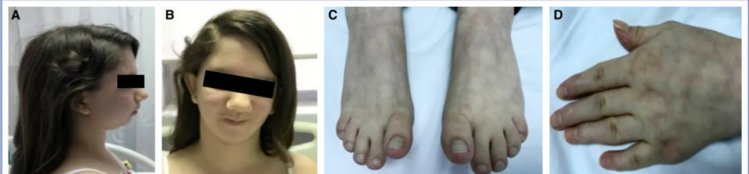
A physical examination revealed a syndromic ap-pearance, growth failure with short stature (height: 138 cm [<3rd percentile] and weight 35 kg [<3rd per-centile]), microretrognathia, syndactyly between the second and third toes and fingers, a broad forehead, and clinodactyly of the fifth finger of both hands (Fig. 1a-d). Her heart sounds were very arrhythmic, and there were very frequent premature contractions. Her neurological status was consistent with mild cognitive developmental retardation.
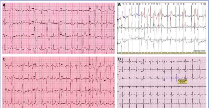
Upon admission, a 12-lead electrocardiogram (ECG) showed frequent and bidirectional PVCs (Fig. 2a). At the initial assessment, the bidirectional PVCs were so severe that a corrected QT interval (QTc) could hardly be measured. Although a QTc was mea-sured as 445 milliseconds, prominent U waves and a long TU distance were significant (QTc measure-ments including U wave were >600 milliseconds). A 24-hour period of Holter monitoring demonstrated frequent polymorphic PVCs, bidirectional VT, and non-sustained polymorphic VT (Fig. 2b). The base-line 24-hour monitoring results demonstrated a very high burden of PVCs — about 28,500 (28%) isolated polymorphic PVCs — with nonsustained VT occur-ring 4 times duoccur-ring the recording period. Echocardio-graphy revealed mitral valve prolapse with normal left ventricular function.
A treadmill exercise test could not be conducted due to the lower extremity muscle weakness of the patient. However, bidirectional VT was detected at doses of 0.2 microgram/kg/minute infusion during an Abbreviations:
ATS Andersen-Tawil syndrome CPVT Catecholaminergic polymorphic VT ECG Electrocardiogram
ICD Implantable cardioverter-defibrillator PVC Premature ventricular contraction QTc Corrected QT interval
RFCA Radiofrequency catheter ablation VT Ventricular tachycardia
Figure 1. (A) Typical dysmorphic facial features of Andersen-Tawil syndrome: microretrognathia, and (B) broad forehead; (C) Syndactyly between the second and third toes; (D) Clinodactyly of the fifth finger.
adrenaline stimulation test (Fig. 2c). The biochemi-cal values, including potassium, were normal. Based on the findings, Anderson-Tawil syndrome was sus-pected. The diagnosis was confirmed by molecular genetic analysis, which demonstrated a p.V200M (c.598G >A) mutation in the KCNJ2 gene. The pa-tient had no other siblings and her mother’s and ther’s QTc values were normal. Her mother’s and fa-ther’s genetic testing results were still expected at the time of writing.
Beta-blocker therapy (propranolol 4 mg/kg/day) was started after the initial evaluation. Despite appro-priate therapy, the PVC burden was not controlled. Thereafter, flecainide treatment (100 mg/m2/day) was initiated, and the PVC burden immediately decreased dramatically. Within 48 hours after administration of the drug, almost all of the PVCs had disappeared. Fle-cainide was also very effective in the suppression of bidirectional VT attacks. An implantable cardiovert-er-defibrillator (ICD) device was not considered be-cause the ventricular arrhythmias were controlled with medical treatment. During a 6-month follow-up period, ECG and 24-hour ambulatory ECG tests were repeated, and a treadmill exercise test was con-ducted. Two weeks after beginning combination ther-apy, there was no PVC recorded on a surface ECG.
The QT interval shortened to 440 milliseconds, but U waves persisted prominently some precordial leads, especially V2-V3 (Fig. 2d). Diazoxide was added to the antiarrhythmic treatment for the periodic muscu-lar paralysis, and the frequency of episodes of paral-ysis decreased.
DISCUSSION
ATS is characterized by a triad of episodic flaccid muscle weakness, ventricular arrhythmias, and a pro-longed QT interval, as well as anomalies including low-set ears, widely spaced eyes, a small mandible, fifth-digit clinodactyly, syndactyly, short stature, and scoliosis.[1,5] Most patients have a mutation in the KCNJ2 gene, which encodes the inward rectifier K+(IK1) channel.[3–5] This causes prolongation in the terminal phase of the cardiac action potential, produc-ing early afterdepolarization.[6,7] The depressed IK1 function also leads to cellular calcium overload, caus-ing late afterdepolarization and leads to triggered ac-tivity.[6,7] These cardiac action potential changes result in a prolonged QT interval, which is originally related to a prolonged QU interval, bidirectional VT, and Tor-sades de pointes.[3,6] Although the arrhythmia burden is usually high, patients are rarely symptomatic.[4,7] That does not mean that symptoms are never seen,
Figure 2. (A) Surface electrocardiogram recorded at admission showed bigeminy and bidirectional premature ventricular con-tractions (PVCs); (B) Bidirectional ventricular tachycardia was seen on Holter monitoring; (C) Polymorphic PVCs became visible during an adrenaline infusion test; (D) After flecainide treatment, there were no PVCs, but prominent U waves persisted.
A
C
B
however. Cardiac arrest occurs in about 3% of pa-tients.[7,8] Although a risk factor for cardiac arrest was not previously identified, one recent study claimed that micrognathia, periodic paralysis, and prolonged Tpeak-Tend time may be associated with a higher risk of arrhythmia, syncope, and cardiac arrest.[9]
The primary cardiac manifestations of ATS include frequent PVCs, QU interval prolongation, prominent U-waves, and a special type of polymorphic VT called bidirectional VT. Bidirectional VT is rarely observed in children, but it is the hallmark of a handful of dis-ease states, including ATS, catecholaminergic poly-morphic VT (CPVT), and digitalis toxicity.[1–3] Each of these clinical entities shares a common underlying abnormality in calcium homeostasis that can trigger delayed afterdepolarization-dependent arrhythmias. In ATS, polymorphic VT and/or bidirectional VT is relatively slow, well tolerated, and usually asymp-tomatic, with a heart rate of about 130 to 150 bpm. Frequent ventricular ectopy at rest is helpful in distin-guishing ATS from typical CPVT. In typical CPVT, rapid bidirectional VT is usually associated with exer-cise. In addition, asymptomatic PVCs are frequently observed in ATS patients, whereas in CPVT patients, PVCs and asymptomatic non-sustained VT are un-common.[2,8] Finally, dysmorphic facial features and periodic paralysis in ATS patients are very helpful in distinguishing these cases from CPVT.[1–3,5]
The classification of Andersen-Tawil syndrome as a type of long QT interval is still controversial.[7,10] Some authors have emphasized that it should be seen as TU or QU prolongation rather than QT interval.[3,6,7] There is also U wave prominence. But, as opposed to the unaffected population, U waves become more prominent during sympathetic status in patients with ATS.[11]
Beta-blockers are used as a first-line treatment. However, there are many reports about partial effi-cacy of beta-blockers.[12,13] Therefore, calcium chan-nel blockers, especially verapamil, were of interest.[12] Direct inhibition of cellular calcium overload, which is potentially responsible for arrhythmias, was con-sidered reasonable.[7,12] However, they are only frac-tionally as successful in preventing life-threatening arrhythmias as beta-blockers.[13] At this point, the su-periority of flecainide in ATS should be emphasized. The mechanism by which flecainide suppresses ven-tricular ectopy and arrhythmias is not fully understood.
[14] However, a decrease in sarcoplasmic reticulum cal-cium overload and sodium currents enhanced by the sodium/calcium exchanger, may explain the antiar-rhythmic action of flecainide.[4,7,13] In patients with An-dersen-Tawil syndrome, the markedly prolonged QU interval leads to calcium overload. Therefore, intracel-lular calcium overload and triggered activity may be possible mechanisms for arrhythmias in patients with ATS. Flecainide can change the sodium-calcium ex-change, resulting in a decrease in calcium through the exchanger and the calcium content in the sarcoplasmic reticulum.[4,7,13,14] Flecainide has a good safety record and a low incidence of adverse effects, rare reports of end-organ toxicity, and a low risk of ventricular proar-rhythmia in children. Potential cardiac adverse effects of flecainide include proarrhythmia (especially atrial flutter with rapid ventricular response, with aberrant conduction and broad QRS complexes), conduction abnormalities, and negative inotropic effects.[15,16] Nonetheless, in children using flecainide, the QRS width should be monitored and patients should be ob-served for potential proarrhythmic effects.
As in other hereditary primary genetic arrhythmia syndromes, it may seem appropriate to eliminate these arrhythmias by means of radiofrequency catheter ab-lation (RFCA). To our knowledge, there is no publica-tion of successful RFCA in a case of ATS. Delannoy et al.[3] reported that RFCA was unsuccessful in the 5 cases in which it was attempted.
In ATS patients for whom drug treatment is not ef-fective and who often show bidirectional VT despite treatment or who have had previous cardiac arrest, the use of an ICD may be considered.[17] However, the ef-fectiveness of an ICD in ATS patients without severe cardiac symptoms is controversial. Airey et al.[18] have performed a follow-up study of 16 ATS patients with various cardiac symptoms who underwent ICD im-plantation. Of these 16 patients, 3 received an ICD for documented VF arrest. Eight patients (50%) received inappropriate shocks, while 4 (25%) had appropriate device intervention. The latter group consisted of 3 patients with an ICD implanted for documented VF arrest and 1 with bidirectional VT and syncope. Given these results, it may be advisable that an ICD is only used when patients are at high risk for cardiac arrest and death.[17,18]
The present case is an example of one of the rarest genetically diagnosed conditions seen in our country.
[19] This is also the first presentation in Turkey of ef-fective suppression of ventricular ectopy and VT via flecainide in a patient with ATS.
In conclusion, ATS should be kept in mind if there is an association between polymorphic PVC, bidirec-tional VT, and dysmorphic properties. A long QT in-terval associated with significant U-wave (especially in the V2-V3 leads) on a 12-lead ECG is diagnostic. The use of flecainide in combination with beta-block-ers appears to be very effective in the treatment of ventricular arrhythmias in ATS.
Peer-review: Externally peer-reviewed. Conflict-of-interest: None.
Informed Consent: Written informed consent was
ob-tained from the parents of the patient for the publication of the case report and the accompanying images.
Authorship contributions: Concept: Y.E., V.T.;
De-sign: Y.E., S.Ö.; Supervision: Y.E., V.T.; Materials: Y.E., S.H.O.; Data collection: Y.E., S.Ö.; Literature search: Y.E., S.Ö.; Writing: Y.E., S.Ö.
REFERENCES
1. Veerapandiyan A, Statland J, Tawil R. Andersen-Tawil Syn-drome. In: Adam MP, Ardinger HH, Pagon RA, Wallace SE, Bean LJH, Stephens K, et al, editors. GeneReviews®. Seat-tle: University of Washington; 2004 [updated 2018 Jun 7]. p. 1993–2018.
2. Kukla P, Biernacka EK, Baranchuk A, Jastrzębski M, Jagodz-ińska M. Electrocardiogram in Andersen-Tawil Syndrome. New Electrocardiographic Criteria for Diagnosis of Type-1 Andersen-Tawil Syndrome. Curr Cardiol Rev 2014;10:222–8. 3. Delannoy E, Sacher F, Maury P, Mabo P, Mansourati J, Mag-nin I, et al. Cardiac characteristics and long-term outcome in Andersen-Tawil syndrome patients related to KCNJ2 muta-tion. Europace 2013;15:1805–11. [CrossRef]
4. Fox DJ, Klein GJ, Hahn A, Skanes AC, Gula LJ, Yee RK, et al. Reduction of complex ventricular ectopy and improvement in exercise capacity with flecainide therapy in Andersen-Taw-il syndrome. Europace 2008;10:1006–8. [CrossRef]
5. Babu SS, Nigam GB, Peter CS, Peter CS. Andersen-Tawil syn-drome: A review of literature. Neurol India 2015;63:772–4. 6. Miyamoto K, Aiba T, Kimura H, Hayashi H, Ohno S,
Yasuo-ka C, et al. Efficacy and safety of flecainide for ventricular arrhythmias in patients with Andersen-Tawil syndrome with KCNJ2 mutations. Heart Rhythm 2015;12:596–603. [CrossRef]
7. Bökenkamp R, Wilde AA, Schalij MJ, Blom NA. Flecainide for recurrent malignant ventricular arrhythmias in two siblings with Andersen-Tawil syndrome. Heart Rhythm 2007;4:508–11.
8. Zhang L, Benson DW, Tristani-Firouzi M, Ptacek LJ, Tawil R, Schwartz PJ, et al. Electrocardiographic features in Ander-sen-Tawil syndrome patients with KCNJ2 mutations: charac-teristic T-U-wave patterns predict the KCNJ2 genotype. Cir-culation 2005;111:2720–6. [CrossRef]
9. Krych M, Biernacka EK, Ponińska J, Kukla P, Filipecki A, Gajda R, et al. Andersen-Tawil syndrome: Clinical presen-tation and predictors of symptomatic arrhythmias - Possible role of polymorphisms K897T in KCNH2 and H558R in SC-N5A gene. J Cardiol 2017;70:504–10. [CrossRef]
10. Barajas-Martinez H, Hu D, Ontiveros G, Caceres G, Desai M, Burashnikov E, et al. Biophysical and molecular characteri-zation of a novel de novo KCNJ2 mutation associated with Andersen-Tawil syndrome and catecholaminergic polymor-phic ventricular tachycardia mimicry. Circ Cardiovasc Genet 2011;4:51–7. [CrossRef]
11. Sumitomo N, Shimizu W, Taniguchi K, Hiraoka M. Calcium channel blocker and adenosine triphosphate terminate bidi-rectional ventricular tachycardia in a patient with Anders-en-Tawil syndrome. Heart Rhythm 2008;5:498–9. [CrossRef]
12. Erdogan O, Aksoy A, Turgut N, Durusoy E, Samsa M, Altun A. Oral verapamil effectively suppressed complex ventricular arrhythmias and unmasked U waves in a patient with Anders-en-Tawil syndrome. J Electrocardiol 2008;41:325–8. [CrossRef]
13. Van Ert HA, McCune EC, Orland KM, Maginot KR, Von Ber-gen NH, January CT, et al. Flecainide treats a novel KCNJ2 mutation associated with Andersen-Tawil syndrome. Heart-Rhythm Case Rep 2016;3:151–4. [CrossRef]
14. Pellizzón OA, Kalaizich L, Ptácek LJ, Tristani-Firouzi M, Gonzalez MD. Flecainide suppresses bidirectional ventricular tachycardia and reverses tachycardia-induced cardiomyopa-thy in Andersen-Tawil syndrome. J Cardiovasc Electrophysiol 2008;19:95–7.
15. Perry JC, Garson A Jr. Flecainide acetate for treatment of tach-yarrhythmias in children: review of world literature on effica-cy, safety, and dosing. Am Heart J 1992;124:1614–21. [CrossRef]
16. Tamargo J, Capucci A, Mabo P. Safety of flecainide. Drug Saf 2012;35:273–89. [CrossRef]
17. Nguyen HL, Pieper GH, Wilders R. Andersen-Taw-il syndrome: clinical and molecular aspects. Int J Cardiol 2013;170:1–16. [CrossRef]
18. Airey K, Wilde A, Hofman N, Etheridge S, Ptacek L, Abuis-sa H, et al. Incidence of device therapy and complications in patients with andersen-tawil syndrome with ICDS. JAC-C;57:E1233. [CrossRef]
19. Oz Tuncer G, Teber S, Kutluk MG, Albayrak P, Deda G. An-dersen-Tawil Syndrome with Early Onset Myopathy: 2 Cases. J Neuromuscul Dis 2017;4:93–5. [CrossRef]
Keywords: Bidirectional ventricular tachycardia; flecainide; long QT.
Anahtar sözcükler: Bidireksiyonel ventriküler taşikardi; uzun QT;