Case Reports Anatol J Cardiol 2020; 23: 299-306
302
Informed consent: Informed consent was obtained from the patient prior to the creation of the manuscript.
References
1. Baranchuk A, Torner P, de Luna AB. Bayés Syndrome: What Is It? Circulation 2018; 137: 200-2.
2. Bayés de Luna A, Martínez-Sellés M, Bayés-Genís A, Elosua R, Baranchuk A. Surface ECG interatrial block-guided treatment for stroke prevention: rationale for an attractive hypothesis. BMC Car-diovasc Disord 2017; 17: 211.
3. Bayés de Luna A, Escobar-Robledo LA, Aristizabal D, Weir Restrepo D, Mendieta G, Massó van Roessel A, et al. Atypical advanced in-teratrial blocks: Definition and electrocardiographic recognition. J Electrocardiol 2018; 51: 1091-3.
4. Çinier G, Tekkeşin Aİ, Çelik TY, Mercan Ö, Tanboğa Hİ, Günay MB, et al. Value of Interatrial Block for the Prediction of Silent Ischemic Brain Lesions. J Atr Fibrillation 2018; 11: 2037.
5. Wu MH, Lu CW, Chen HC, Chiu SN, Kao FY, Huang SK. Arrhythmic burdens in patients with tetralogy of Fallot: a national database study. Heart Rhythm 2015; 12: 604-9.
6. Orczykowski M, Borowiec K, Biernacka E, Bodalski R, Urbanek P, Derejko P, et al. Ablation of atrial tachyarrhythmias late after surgi-cal correction of tetralogy of Fallot: long-term follow-up. Kardiol Pol 2018; 76: 1097-105.
Address for Correspondence: Lucian Muresan, MD, Department of Cardiology,
“Emile Muller” Hospital;
20 Avenue du Docteur René Laennec, 68100, Mulhouse-France
Phone: 00 33 389 646464 Fax: 00 33 389 647635 E-mail: [email protected]
©Copyright 2020 by Turkish Society of Cardiology - Available online at www.anatoljcardiol.com
DOI:10.14744/AnatolJCardiol.2020.16064
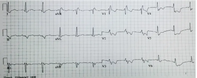
Figure 4. Twelve-lead ECG recorded with correctly-placed ECG electrodes, revealing sinus rhythm with a different morphology of the P wave and QRS complexes compared with Figure 3, with positive P waves in leads II, III, and aVF; positive in leads I and aVL; left axis deviation; right bundle branch block
Acute left main coronary artery
occlusion following transcatheter aortic
valve replacement without obvious risk
factors of coronary obstruction
Beytullah Çakal, Sinem Çakal, Oğuz Karaca, Bilal Boztosun
Department of Cardiology, İstanbul Bağcılar Medipol Mega University Hospital; İstanbul-Turkey
Introduction
Transcatheter aortic valve implantation (TAVI) has emerged as a prominent alternative for patients with severe aortic ste-nosis, who are deemed to be at a greater risk for surgical inter-ventions (1). However, extreme caution is recommended against the lethal complications, such as coronary obstruction (CO) or annulus rupture, associated with this procedure that need to be overcome urgently (1). Herein, we report an occlusion of acute left main coronary artery (LMCA) after TAVI, which was treated with triple stenting, due to immediate stent recoils.
Case Report
A 79-year-old woman, who was diagnosed with severe aortic stenosis, was referred to our clinic for the evaluation of TAVI. Because she had accompanying diseases including anemia and chronic obstructive pulmonary disease, the heart team con-cluded that the patient was at a high risk for surgery (a logistic
Case Reports
Anatol J Cardiol 2020; 23: 299-306
303
EuroSCORE of 25.7%, an STS score of 14.6%, and a EuroSCORE II of 8.1%); therefore, TAVI was scheduled.
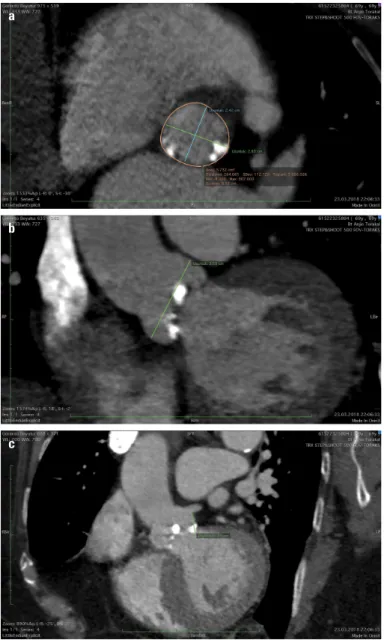
Contrast-enhanced computed tomography (CT) showed an aortic annulus size of 24.2/28 mm, an annular area of 573 mm2,
the diameter of sinus valsalva to be 36 mm, and the height of LMCA to be 13.5 mm (Fig. 1a-1c). The heart team planned com-bined percutaneous coronary intervention (PCI) of critical (90%) proximal left anterior descending (LAD) lesion and TAVI as a single session for reducing the patient’s suffering and the length of hospital stay. First, the team performed stenting of proximal LAD with 3
×
23 mm Xience Pro (Abbot Vascular), followed by 25-mm balloon predilatation and a successful deployment of 29-mm Edwards Sapien XT valve (Edwards Lifesciences, CA, USA). Although the displacement of bulky calcification toward LMCA was observed during the balloon predilatation (Video 1), initially at least, the LMCA flow appeared to be normal during aortogra-phy despite the fact that the mass was completely in front of the takeoff of the LMCA (Video 2). During the process of suturing with Proglide, the patient was extremely hypotensive (50/30 mm Hg) and further showed ventricular fibrillation. Following that, successful cardiopulmonary resuscitation was achieved in less than 20 minutes. Angiography revealed the case of LMCA ostial calcific obstruction (Video 3). Given the fact that the ostium of LMCA was above the Sapien XT frame struts, 6 F EBU 3.5 guide catheter was engaged easily and Pt 2 wire was passed through the lesion. A 4.0*18 mm everolimus-eluting stent was implanted in LMCA and postdilated at a high pressure with 4.0- and 4.5-mm non-compliant (NC) balloons (Fig. 2a, Video 4). Because acute stent recoil occurred due to calcified bulky leaflet, 4.5*15-mm bare metal stent was deployed in LMCA. Repeated high pres-sure up to 26 Atm in 4.5-mm NC balloon dilatations resulted into good stent apposition followed by persistent stent compression (Fig. 2b). In order to achieve a good radial expansion force, 4.0*20 mm graft stent (PK Papyrus; Biotronik, Bülach, Switzerland) was implanted within the previous two stents and expanded at a high pressure with a NC balloon. A final angiography still revealed ful-ly unexpanded stents in the osteal LMCA caused by heavy cal-cification (Fig. 2c, Video 5) with better coronary blood flow. The patient was transferred to the intensive care unit. She showed a promising recovery and was discharged under the treatment of ticagrelor and aspirin on the fifth day after the TAVI procedure.a
b
c
Figure 1. Computed tomography images before transcatheter aortic valve implantation; (a) An annulus size of 24.2/28 mm and an annulus area of 573 mm2; (b) Height of left main coronary artery: 13.5 mm; c)
Diameter of sinus valsalva: 36 mm
a b c
Figure 2. (a) Heavy calcification compressing stent; (b) Stent recoil was observed after the second stent, (c) Final image after the graft stent within two stents
Case Reports Anatol J Cardiol 2020; 23: 299-306
304
Discussion
The common complications that can occur during TAVI are vascular access complications, aortic root rupture, paravalvular regurgitation, the need for a permanent pacemaker, and coro-nary obstruction (2). Corocoro-nary occlusion has a low incidence rate of about 1%; however, a 30-day mortality rate of 40.6% was reported at its occurrence (3).
Two possible explanations for CO were provided: the dis-placement of the calcification from the native valve, which is more common, and the obstruction of the coronary ostium by a portion of the transcatheter valve frame (4). Ribeiro et al. (5) identified low coronary ostial height (<12 mm), sinus of valsalva diameter of below 30 mm, female gender, older age, balloon ex-pandable valve, and valve-in-valve procedures as the most im-portant predictors of risk of CO. Although the abovementioned factors are logical for the prediction of CO, none of them were present in our case, thereby suggesting that there is a lot to ex-plore about CO. In addition, the relationship between the LMCA and the "new place" of the bulky calcification is very essential. On the event of any disturbing relationship, repeat aortography can be performed after waiting for a while before finishing the procedure.
There was a higher mortality rate, even after successful stenting (22%) or CABG (50%), which increased to as much as 100% in case of unsuccessful PCI (4). Some researchers suggest the implantation of a second stent when the first stent is not suf-ficient due to extrinsic compression (6).
Conclusion
As shown in our case, coronary occlusion during TAVI is not always predictable. Once CO takes place, the deployment of one stent along with multiple balloon dilatations could not prevent the stent compression. Therefore, we suggest the use of a sec-ond or even a third stent with a greater radial force to improve the stent expansion, if needed.
Informed consent: Written informed consent was obtained from this patient.
Video 1. Balloon predilatation. Notice displacement of the calcification towards LMCA take-off
Video 2. Aortography after TAVI. LMCA had normal blood flow despite the exisitence of calcification adjacent to the LMCA.
Video 3. Angiography following successful resuscitation. Heavy calcification caused occlusion of the LMCA.
Video 4. LMCA was stented with 4.0*18 mm everolimus-elut-ing stent. Stent recoil in the setteverolimus-elut-ing of bulky calcification was still persisted after multiple post-dilatations.
Video 5. Better stent expansion with good coronary blood flow was maintained after triple stenting.
References
1. Leon MB, Smith CR, Mack M, Miller DC, Moses JW, Svensson LG, et al.; PARTNER Trial Investigators. Transcatheter aortic-valve im-plantation for aortic stenosis in patients who cannot undergo sur-gery. N Engl J Med 2010; 363: 1597-607. [CrossRef]
2. Arai T, Lefèvre T, Hovasse T, Garot P, Benamer H, Unterseeh T, et al. Incidence and predictors of coronary obstruction following transcatheter aortic valve implantation in the real world. Catheter Cardiovasc Interv 2017; 90: 1192-7. [CrossRef]
3. Hayashida K, Lefèvre T, Chevalier B, Hovasse T, Romano M, Garot P, et al. Transfemoral aortic valve implantation new criteria to predict vascular complications. JACC Cardiovasc Interv 2011; 4: 851-8. 4. Ribeiro HB, Nombela-Franco L, Urena M, Mok M, Pasian S, Doyle
D, et al. Coronary obstruction following transcatheter aortic valve implantation: a systematic review. JACC Cardiovasc Interv 2013; 6: 452-61. [CrossRef]
5. Ribeiro HB, Webb JG, Makkar RR, Cohen MG, Kapadia SR, Kodali S, et al. Predictive factors, management, and clinical outcomes of coronary obstruction following transcatheter aortic valve implan-tation: insights from a large multicenter registry. J Am Coll Cardiol 2013; 62: 1552-62. [CrossRef]
6. Saia F, Marrozzini C, Marzocchi A. Displacement of calcium nod-ules of the native valve as a possible cause of left main occlusion following transcatheter aortic valve implantation. J Invasive Car-diol 2011; 23: E106-9.
Address for Correspondence: Dr. Beytullah Çakal, İstanbul Bağcılar Medipol Mega Üniversite Hastanesi, Kardiyoloji Kliniği,
Tem Otoyolu Göztepe Çıkışı No: 1, Bağcılar, İstanbul-Türkiye
Phone: +90 506 284 55 88 E-mail: [email protected]
©Copyright 2020 by Turkish Society of Cardiology - Available online at www.anatoljcardiol.com
DOI:10.14744/AnatolJCardiol.2020.91535
Successful percutaneous treatment of
pulsatile tinnitus, a rare symptom of
carotid artery stenosis
Fatih Yılmaz*, Ahmet Karaduman*, İsmail Balaban*, Murat Velioğlu**, Nuri Havan**
Departments of *Cardiology, and **Radiology, Kartal Koşuyolu Training and Research Hospital; İstanbul-Turkey
Introduction
Carotid artery stenosis is one of the primary reasons of cere-brovascular events. Additionally, carotid artery stenosis can lead to dizziness, imbalance, and sudden severe headaches. Further-more, a rare and noteworthy symptom of carotid artery stenosis is pulsatile tinnitus (1, 2). The purpose of this case report is to