Address for correspondence: Hazım Alper Gursu, MD, Paediatric Cardiologist, Baskent University Medical Faculty, Department of Pediatric Cardiology, Ankara, Turkey, tel: +90 505 5618799, fax: +90 312 2237333, e-mail: [email protected] Received: 03.11.2015 Accepted: 26.12.2015
Analysis of right ventricle function with
strain imaging before and after
pulmonary valve replacement
Hazım Alper Gursu
1, Birgul Varan
1, Elif Sade
2, Ilkay Erdogan
1, Murat Ozkan
3 1Department of Pediatric Cardiology, Baskent University Medical Faculty, Ankara, Turkey2Department of Cardiology, Baskent University Medical Faculty, Ankara, Turkey 3Department of Cardiovascular Surgery, Baskent University Medical Faculty, Ankara, Turkey
Abstract
Background: Pulmonary valve insufficiency may develop after surgical treatment of tetralogy
of Fallot (ToF). Severe pulmonary valve insufficiency may result in right ventricular
dysfunc-tion. We aimed to compare cardiac magnetic resonance (CMR), with echocardiography.
Methods: Patients who developed severe pulmonary valve insufficiency after total correction for
ToF, were included in the study. CMR was used to measure end-diastolic, end-systolic volumes
and ejection fraction of the right ventricle before and 6 months after replacement, and
echocar-diographic strain imaging was obtained before, and 1, 3, and 6 months after replacement.
Results: There were significant differences between pre- and post-replacement QRS durations,
and right ventricle end-diastolic and end-systolic volumes measured with CMR (p < 0.05).
However, right ventricular ejection fraction (RVEF) did not change. Therefore, CMR
deter-mined that right ventricle size and volume increased, and right ventricular function
deterio-rated before replacement. After replacement, no significant improvement was seen in RVEF.
Lower-than-normal right ventricle strain and strain rate before replacement indicated that
healthy and dysfunctional myocardium could be differentiated by this method. Pre-replacement
strain and strain rate of asymptomatic and symptomatic patients were similar. Strain and
strain rate values increased 6 months after replacement (p < 0.05).
Conclusions: We suppose that increased experience with strain imaging, and further studies
on a larger patient group with a longer follow-up period would show that this method is quite
advantageous, and it will take its place in the literature as a non-invasive technique that may
be used instead of magnetic resonance. (Cardiol J 2016; 23, 2: 195–201)
Key words: cardiac surgery, echocardiography, magnetic resonance,
right ventricular dysfunction, strain
Introduction
In this study, we aimed at determining right ventricular (RV) function and volumes with strain, strain rate, and cardiac magnetic resonance imaging
(MRI) before and after pulmonary valve replace-ment in patients who had surgery due to tetralogy of Fallot (ToF), and were diagnosed with severe pulmonary valve insufficiency, RV dilatation and dysfunction on follow-up. We planned to compare
ORIGINAL ARTICLE
Cardiology Journal 2016, Vol. 23, No. 2, 195–201
DOI: 10.5603/CJ.a2016.0007 Copyright © 2016 Via Medica ISSN 1897–5593
gold standard diagnostic method, MRI, to deter-mine the amount of pulmonary valve insufficiency, with echocardiography particularly in children and young patients. In addition, we aimed at investi-gating whether valve replacement resulted in any improvement in RV size and performance, and evaluate RV functions with strain imaging after replacement.
Pulmonary valve dysfunction after total cor-rection for ToF is one of the most important prob-lems in the long-term follow-up of those patients. Pulmonary valve insufficiency develops due to loss of pulmonary valve integrity when a transannular patch is used during surgery. Pulmonary valve insufficiency causes RV volume load, dysfunction, arrhythmia, and sudden death. Early recognition of RV functional deterioration is quite important in routine follow-up of ToF patients that had to-tal correction. Pulmonary valve replacement is needed to protect RV function, and remodeling [1]. Replacement has positive effects including an increase in functional class and exercise capacity, and a decrease in QRS duration. However, there is no consensus on indications and timing of re-placement.
Methods
Study population
A total of 15 patients that developed severe pulmonary valve insufficiency after total correc-tion for ToF and had replacement, were included in the study. Clinical Research Ethical Committee approved the study (decree no.: KA11/172). The families of the patients younger than 18 years of age, and the patients ≥ 18 years of age provided their informed consents. The patients had physical examinations, and their symptoms such as palpita-tion, fatigue, dyspnea, and syncope were noted.
Inclusion criteria
The patients diagnosed with pulmonary atre-sia, pulmonary stenosis or absent pulmonary valve had been excluded from the study. The patients with moderate or severe pulmonary valve regurgi-tation, accompanied by one or more of the following criteria were included in the study:
— RV end-diastolic volume ≥ 160 mL/m2;
— RV end-systolic volume ≥ 70 mL/m2;
— RV ejection fraction (RVEF) ≤ 45%; — aneurysm of RV outflow tract;
— moderate or severe tricuspid regurgitation; — exercise intolerance, heart failure, syncope; — arrhythmias: sustain ventricular tachycardia.
Electrocardiographic examination
“Hewlett-Packard PageWriter 200 CE, Germa-ny” device was used to obtain a 12-channel electro-cardiography at a rate of 25 mm/s and an amplitude of 10 mm/mV. Cardiac rhythm was analyzed, QRS duration was measured, and QTc interval that was standardized in relation with the heart rate using Bazett’s formula (QTc = QT/√R-R) was estimated. Twenty-four hour Holter recording was obtained with a 3-channel Holter device (Delmar Reynolds, Irvine, California, USA). Records were analyzed for heart rate, rhythm, and conduction disturbances.
Cardiac magnetic resonance
A 1.5 Tesla MRI device was used in the study (Avanto, Siemens Medical Systems, Erlangen, Ger-many). T2-weighted cine, and T1-weighted images were obtained parallel to the short axis of heart, at 2- and 4-chamber positions. Measurements were performed for RV functions on the images paral-lel to the short axis of the heart. Right ventricle end-diastolic and end-systolic volumes, and RVEF were measured with MRI, before and 6 months after replacement.
Echocardiographic examination
Echocardiographic examination was per-formed while the patient was lying on left decubitus position (2-dimensional, M-mode, color Doppler echocardiography) using Vivid i device (GEMS Tirat Carmel, Israel) and a 3-MHz transducer. The mechanical myocardial functions of all patients were examined by echocardiographic imaging per-formed before and 1, 3, 6 months after replacement. Routine 2-dimensional, M-mode, color and tissue Doppler echocardiography, 2-dimensional strain, and strain rate imaging were performed. The degree of pulmonary insufficiency was regarded as “mild” in absence of significant regurgitating jet in the RV outlet or retrograde diastolic flow in pulmonary artery; as “moderate” in presence of retrograde diastolic flow in main pulmonary artery; and “severe” in presence of retrograde diastolic flow in branches of pulmonary artery. Tricuspid valve insufficiency was graded in relation with the proportion of tricuspid insufficiency jet area to the area of right atrium.
Strain imaging
Longitudinal regional myocardial functions were determined in the basal, middle, and apical segments of free wall of RV, and in the inter-ventricular septum for middle region, on apical 4-chamber view before, and 1, 3, and 6 months
after replacement. Velocity, displacement, strain, and strain rate measurements were performed for every region. EchoPac PC (GE Healthcare, Wauke-sha, Wisconsin) software was used for analysis. Us-ing this software, strain and strain rates of septum, and basal, middle and apical segments of RV free wall, and their average values were calculated. The measurements were performed on apical 4-cham-ber view. The display sector was adjusted to “nar-row” to see the structures better. The smallest gain available was adjusted to discriminate the border between blood and myocardium, and to obtain clear signals. Doppler signals were recorded at a rate of at least 100 mm/s. Four cycles were recorded, and their average values were calculated. Myocardial velocity and strain measurements were based on Doppler technique. The maximum angle between myocardium and ultrasound beam was 15 degrees.
Ethical standards
Our study complies with appropriate institu-tional and nainstitu-tional guidelines for ethical matters.
Statistical analysis
The statistical analysis of data was performed with SPSS for Windows 15.0 (SPSS Inc., Chicago, IL) package program. Wilcoxon test was used to analyze time-dependent changes in MRI and strain echocardiography. Two-way variance analysis was performed to analyze the effects of age at total correction, and the duration between total cor-rection and valve replacement on the parameters measured. The correlations among variables were determined with Spearman correlation analysis. P < 0.05 was regarded as statistically significant. For intra-observer variability (94%) all echo-cardiographic parameters were analyzed by one researcher at two different times.
Results
A total of 15 patients that developed severe pulmonary valve insufficiency after total correction and had valve replacement were included in the
study. There were 8 (53.3%) males and 7 (46.7%) females. The mean age of the patients was 14.5 ± ± 4.5 (7–22) years. The mean age at total cor-rection was 2.3 ± 1.3 (1–5) years. The mean age of pulmonary valve replacement was 14.3 ± 4.5 (6.5–21.5) years.
All patients had significant pulmonary valve insufficiency with different severities. One patient had moderate, and 14 (93.3%) patients had severe valve insufficiency. All of the 15 patients included had tricuspid valve insufficiency; it was mild in 8 (53.3%), moderate in 6, and severe in 1 patient.
Twelve (80%) patients had arrhythmia on 24-h Holter monitoring. Three patients had normal Holter findings. The most frequent arrhythmia type was ventricular premature beats (40%). Couplet, bigeminy beats, ventricular tachycardia, and atrial premature beats were less frequent.
Six (40%) patients were asymptomatic. Other pa-tients had fatigue (20%), palpitations, and chest pain. Before replacement, the mean RV end-dias-tolic volume was 170.2 ± 32.2 mL/m2, mean RV
end-systolic volume was 98.8 ± 21.5 mL/m2, and
RVEF was 40.5 ± 12.8% on MRI. After replace-ment, mean RV end-diastolic volume was 98.5 ± ± 21.3 mL/m2, mean RV end-systolic volume was
62.9 ± 19.2 mL/m2, and mean RVEF was 36.2 ±
± 10.9% (Table 1).
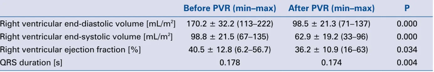
Pre- and 6-month post-replacement RV end-diastolic volume, RV end-systolic volume, and QRS durations showed significant differences (p < 0.05). RV volume and QRS duration decreased after replacement, however ejection fraction did not increase.
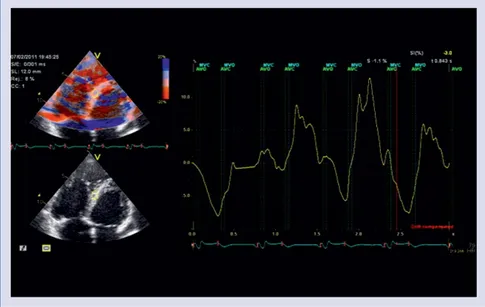
There were significant decreases in middle segment’s strain (p < 0.05) 1 month after replace-ment. Examinations performed 3 months after replacement showed increase in strain and strain rate of septum. However, strain and strain rate of septum increased significantly 6 months after replacement (p < 0.05) (Figs. 1, 2).
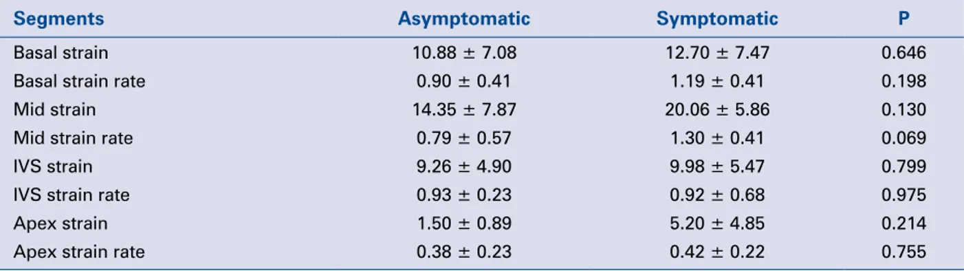
There were no differences for pre-replacement strain or strain rate values of any segments between symptomatic and asymptomatic patients (Table 2).
Table 1. Comparison of parameters before and after pulmonary valve replacement (PVR).
Before PVR (min–max) After PVR (min–max) P Right ventricular end-diastolic volume [mL/m2] 170.2 ± 32.2 (113–222) 98.5 ± 21.3 (71–137) 0.000
Right ventricular end-systolic volume [mL/m2] 98.8 ± 21.5 (67–135) 62.9 ± 19.2 (33–96) 0.000
Right ventricular ejection fraction [%] 40.5 ± 12.8 (6.2–56.7) 36.2 ± 10.9 (16–63) 0.034
This indicated that myocardial deformation was present in asymptomatic patients as well as in the symptomatic ones, and myocardial deformation might present with low myocardial strain and strain rate values in the asymptomatic period.
Discussion
Complex geometric structure of RV limits evaluation of its systolic and diastolic functions
with standard techniques including 2-dimensional and M-mode echocardiography. MRI has been indicated as the gold standard method to measure ejection fraction, and volumes and global systolic functions of both ventricles [2]. Reduced exercise capacity, which is a strong indicator of deteriora-tion of the disease, is weakly correlated with RV performance capacity determined by MRI. There-fore, a more sensitive method is needed for early determination of deteriorated RV performance.
Figure 2. Strain imaging of middle septum 6 months after replacement. Figure 1. Strain imaging of middle septum before replacement.
Table 2. Differences between asymptomatic (n = 6) and symptomatic (n = 9) patients before
pulmo-nary valve replacement.
Segments Asymptomatic Symptomatic P
Basal strain 10.88 ± 7.08 12.70 ± 7.47 0.646 Basal strain rate 0.90 ± 0.41 1.19 ± 0.41 0.198 Mid strain 14.35 ± 7.87 20.06 ± 5.86 0.130 Mid strain rate 0.79 ± 0.57 1.30 ± 0.41 0.069
IVS strain 9.26 ± 4.90 9.98 ± 5.47 0.799
IVS strain rate 0.93 ± 0.23 0.92 ± 0.68 0.975 Apex strain 1.50 ± 0.89 5.20 ± 4.85 0.214 Apex strain rate 0.38 ± 0.23 0.42 ± 0.22 0.755 IVS — interventricular septum
Recently, strain and strain rate values obtained on the basis of tissue Doppler imaging has emerged as an advanced diagnostic procedure to determine regional myocardial function [3, 4].
Tsang et al. [5] found mean pre-replacement RV end-diastolic volume as 173 ± 44 mL/m2, RV
end-systolic volume as 102 ± 26 mL/m2, and RVEF
as 42 ± 9%, however mean RV end-diastolic volume was found as 103 ± 19 mL/m2, RV
end-systolic volume was found as 54 ± 11 mL/m2,
and RVEF was found as 47 ± 6% 1–2 years after replacement. Although there were significant dif-ferences for pre- and post-replacement RV end-diastolic and RV end-systolic volumes, there was no significant difference for RVEF. Knirsch et al. [6] determined pre-replacement RV end-diastolic volume as 201 ± 47 mL/m2, and RVEF as 45 ± 6%,
while those values were found as 109 ± 15% and 42 ± 8%, 6 months after replacement. Although the studies in the literature found a statistically significant reduction in end-diastolic volume after replacement, they did not find any change in ejec-tion fracejec-tion which reflected global systolic func-tion [2, 5, 7, 8]. In our study, although we found highly significant differences in end-diastolic and end-systolic volumes after replacement, we did not find any significant difference for ejection fraction (Table 1). Therefore, we thought that the RV func-tion should be evaluated with another diagnostic method, and we compared the pre-op and post-op strain values.
Recently, strain and strain rate modalities have got ahead of segmental wall motion analy-sis on tissue Doppler imaging since the latter technique is operator dependent, subjective, and semi-quantitative. Color Doppler myocardial im-aging enables calculation of regional longitudinal
myocardial strain and strain rates of RV. Weidemann et al. [9] performed a study on 23 healthy children, and reported that RV strain and strain rate values were quite heterogeneous, and the maximum value was determined in the middle zone of RV free wall. The same study reported mean strain and strain rate as –36 ± 11 and –2.4 ± 0.6 in the basal zone, as –43 ± 13 and –2.8 ± 0.7 in the middle zone, and as –34 ± 11 and –2.5 ± 0.6 in the apical zone of RV free wall. Weidemann et al. [9] performed another study on asymptomatic 30 patients that had total correction, and showed that strain and strain rates of basal, middle, and apical segments of RV free wall were lower than the normal values. The high-est values were obtained in the middle segment. Similarly, Eyskens et al. [10] found regional strain and strain rates lower than normal in basal, middle, and apical segments of RV free wall in asympto-matic patients that had total correction. In this way, they calculated strain and strain rates, and showed presence of regional systolic myocardial dysfunc-tion in absence of any symptoms. Knirsch et al. [6] investigated myocardial functions of 16 pediatric patients that had severe pulmonary valve insuffi-ciency with 2-dimensional strain echocardiography. They used apical 4-chamber position to measure longitudinal functions of apical, middle, and ba-sal segments of RV free wall before, and 1 and 6 months after replacement. They determined that pre-replacement RV regional longitudinal systolic strain values decreased particularly in RV free wall. Knirsch et al. [6] found pre-replacement myocardial function of septum similar with the healthy control group, and it did not change after replacement. Solarz et al. [11] calculated strain and strain rates of RV free wall and septum on apical 4-chamber position, and found that myocardial function of
septum was preserved before replacement despite global RV myocardial dysfunction. Solarz et al. [11] reported that strain and strain rate decreased in all segments of RV free wall, however they did not change in septum. Scherptong et al. [12] supposed that myocardial function of septum was preserved as a compensatory mechanism against decreased RV free wall function. In our study, in accordance with the literature, we observed that strain and strain rates of all RV free wall segments were lower than normal values before replace-ment. Contrary to other studies in the literature, we found that myocardial function of septum was deteriorated, and strain and strain rate of septum were low before replacement. Strain and strain rate of septum continued to to be low 1 month after replacement, however we found significant increase in both strain and strain rate of septum 3 months and 6 months after replacement (p < 0.05). However, there was no significant increase in other segments. Therefore, we supposed that septum was the segment that improved by replacement the best. The highest pre-replacement strain and strain rate were found in the middle segment of RV free wall, and the lowest ones were found in the apical segment where there was least myocardial deformation, and those results were in accordance with the results of Weidemann et al. [9]. However, our results were contrary to the findings of Knirsch et al. [6] and Solarz et al. [11] who reported that functions of septum were preserved before replace-ment. Scherptong et al. [12] calculated strain and strain rates of septum, apex, and RV free wall on 4-chamber window in 18 adult patients. They found strain and strain rate lower in the apical segment of RV free wall compared to other segments; and in contrary to our study, those values were higher in the basal segment compared to other segments, but they lower than the normal values. The au-thors stated that, although longitudinal systolic strain values were lower, and end-diastolic and end-systolic volumes on MRI were higher before replacement, ejection fraction did not change. They found that strain parameters were more sensitive than MRI parameters when RV performance was taken into consideration. Similar to our findings, Scherptong et al. [12] found reduction in longitudi-nal systolic strain and strain rates in all segments before replacement. Different strain and strain rates were calculated in different segments of RV. In accordance with the literature, those findings indicated that strain and strain rates of RV were quite heterogeneous in both healthy children and the patients that had total correction, and there
were functional differences among the segments. Therefore, strain and strain rate have emerged as a modality that could demonstrate regional myo-cardial functions of RV.
The values found in our study were quite lower than the ones obtained in healthy children by Weidemann et al. [9]. Right ventricle strain and strain rates helped to differentiate healthy myo-cardium from near-deteriorated and deteriorated myocardium. In our study, we also determined that strain and strain rate decreased in asymptomatic patients as well as in the symptomatic ones before replacement. We found significantly lower strain and strain rates in all segments except septum in asymptomatic patients when compared to the symptomatic ones, however the differences were not significant (Table 2). Therefore, we supposed that RV dysfunction could be diagnosed earlier by examining the functions of myocardial segments in asymptomatic patients that had total correction, before global myocardial function is deteriorated and the patients have symptoms. This may only be possible by serial examinations of strain and strain rates.
Limitations of the study
Despite its contribution to the literature, our study has some limitations. Firstly, the number of patients is rather low to really determine mean-ingful outcome. Secondly, detecting a radial and circumferential deformation, in addition to longi-tudinal deformation would be better.
Conclusions
In conclusion, the patients that had total correction and developed pulmonary valve insuf-ficiency must be followed up closely. The efinsuf-ficiency of replacement may be low when it is performed late, after development of irreversible RV dysfunc-tion. Researchers tried to detect this critical period by using different methods. MRI is particularly used to determine RV functions. In our study, we used new echocardiography techniques for early diagnosis of RV dilatation and dysfunction, and tried to determine efficiency of treatment. Although we admit that MRI is still the gold standard technique, we suppose that increased experience on strain and strain rate imaging, and further studies on larger patient cohorts with a longer follow-up will show that those modalities are quite beneficial in the diagnosis, and they will take their place in the literature.
References
1. Warner KG, O’Brien PK, Rhodes J, Kaur A, Robinson DA, Payne DD. Expanding the indications for pulmonary valve replacement after repair of tetralogy of Fallot. Ann Thorac Surg, 2003; 76: 1066–1072.
2. Buechel ER, Dave HH, Kellenberger CJ et al. Remodelling of the right ventricle after early pulmonary valve replacement in children with repaired tetralogy of Fallot: assessment by cardio-vascular magnetic resonance. Eur Heart J, 2005; 26: 2721–2727. 3. Weidemann F, Eyskens B, Mertens L et al. Quantification of
re-gional right and left ventricular function by ultrasonic strain rate and strain indexes after surgical repair of tetralogy of Fallot. Am J Cardiol, 2002; 90: 133–138.
4. Leitman M, Lysyansky P, Sidenko S et al. Two-dimensional strain: A novel software for real-time quantitative echocardio-graphic assessment of myocardial function. J Am Soc Echocardi-ogr, 2004; 17: 1021–1029.
5. Tsang FH, Li X, Cheung YF, Chau KT, Cheng LC. Pulmonary valve replacement after surgical repair of tetralogy of Fallot. Hong Kong Med J, 2010; 16: 26–30.
6. Knirsch W, Dodge-Khatami A, Kadner A et al. Assessment of myocardial function in pediatric patients with operated tetralogy of Fallot: Preliminary results with 2D strain echocardiography. Pediatr Cardiol, 2008; 29: 718–725.
7. Oosterhof T, van Straten A, Vliegen HW et al. Preoperative thresholds for pulmonary valve replacement in patients with corrected tetralogy of Fallot using cardiovascular magnetic reso-nance. Circulation, 2007; 116: 545–551.
8. Oosterhof T, Vliegen HW, Meijboom FJ, Zwinderman AH, Bouma B, Mulder BJ. Long-term effect of pulmonary valve replacement on QRS duration in patients with corrected tetralogy of Fallot. Heart, 2007; 93: 506–509.
9. Weidemann F, Eyskens B, Jamal F et al. Quantification of re-gional left and right ventricular radial and longitudinal function in healthy children using ultrasound-based strain rate and strain imaging. J Am Soc Echocardiogr, 2002; 15: 20–28.
10. Eyskens B, Brown SC, Claus P et al. The influence of pulmonary regurgitation on regional right ventricular function in children after surgical repair of tetralogy of Fallot. Eur J Echocardiogr, 2010; 11: 341–345.
11. Solarz DE, Witt SA, Glascock BJ, Jones FD, Khoury PR, Kim-ball TR. Right ventricular strain rate and strain analysis in patients with repaired tetralogy of Fallot: Possible interven-tricular septal compensation. J Am Soc Echocardiogr, 2004; 17: 338–344.
12. Scherptong RW, Mollema SA, Blom NA et al. Right ventricular peak systolic longitudinal strain is a sensitive marker for right ventricular deterioration in adult patients with tetralogy of Fallot. Int J Cardiovasc Imaging, 2009; 25: 669–676.