Neurology India | Volume 66 | Issue 3 | May‑June 2018 865 Letters to Editor
Intracranial chondroma without meningeal
attachment
Sir,
Chondromas are benign tumors composed of mature hyaline cartilage. They generally have limited growth potential and are not locally aggressive. They usually occur in the small bones of the hands and feet, and intracranial locations of these tumors are rare. Although intracranial chondromas are usually located at the skull‑base, there are cases reported in the literature where the tumor originated from the dura mater of the convexity, falx cerebri, tentorium, or choroid plexus.[1,2] An intracranial
chondroma without any meningeal attachment is an extremely rare condition.
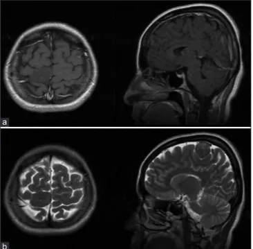
A 34‑year old female patient reported to our clinic with complaints of progressive and severe headache for the past 3 months. Her physical examination was normal. There was no significant family history, but she had a history of severe head trauma due to a car accident 10 years ago. Due to the progressive nature of her severe headache, neuroimaging was performed. Cranial computed tomography (CT) showed a hypodense, solitary, right‑sided parasagittal mass. Magnetic resonance imaging (MRI) of the brain revealed a right‑sided parasagittal, extra‑axial lesion, which was hypointense on T1‑weighted and heterogeneously hypo‑ to hyperintense on T2‑weighted images. A slight enhancement was observed after gadolinium administration [Figure 1]. She was prepared for surgery with the provisional diagnosis of a meningioma, based upon her preoperative imaging. She underwent a
right‑sided parietal craniotomy under general anesthesia. The dura mater was opened and an ivory colored, solid, extra‑axial tumor was exposed [Figure 2]. It was dissected easily from the adjacent parenchyma and had no meningeal attachment. The intraoperative preliminary diagnosis was a meningioma or a chondroma. Total tumor resection was performed with the microsurgical technique. The postoperative course was uneventful, and her headache resolved. She was discharged on the fourth postoperative day. Gross examination of the resected tumor revealed a solid, 3 cm × 3 cm × 2.5 cm, ivory‑white colored, cartilaginous mass [Figure 2]. After serial sectioning, hematoxylin and eosin staining confirmed the lobular architecture of the well‑differentiated cartilaginous tissue. The cells showed no nuclear atypia or mitotic activity, and the diagnosis of a benign chondroma was made [Figure 2]. Intracranial chondromas are extremely rare tumors. They represent 0.2%–0.5% of all intracranial tumors.[3] There are
multiple hypotheses about the pathogenetic mechanisms of these tumors in the literature, but the most commonly prevailing theory is that these tumors originate from embryonic residues, possibly in connection with inflammation or trauma.[3]
In addition, there is often a history of severe head trauma, as seen in our case. Surgical resection is the treatment modality for these tumors, and total resection should be the aim because of their benign characteristics. If there is a meningeal attachment, the involved dura or falx should be resected, too. Total resection of tumors in cases such as ours is much easier because of the avascularity of the tumor and the absence of adhesions to the adjacent tissues.[4] There is no role for radiotherapy in these
cases. Some authors suggest that radiotherapy may inititiate or increase the process of malignant degeneration within these tumors. Therefore, radiotherapy is not recommended for residual or non‑operable tumors.[3]
Declaration of patient consent
The authors certify that they have obtained all appropriate patient consent forms. In the form the patient(s) has/have given his/her/their consent for his/her/their images and other clinical information to be reported in the journal. The patients understand that their names and initials will not be
Figure 2: (a) Image of the resected tumor. (b) Light microscopy of the tumor
revealing the lobular architecture of the well‑differentiated cartilaginous tissue (hematoxylin and eosin staining, ×200) b a Figure 1: Preoperative MRI scan revealing a right parasagittal, extra‑axial mass, which is (a) hypointense on T1‑weighted images and (b) heterogeneously hypo‑ to hyperintense on T2‑weighted images. A slight enhancement was seen after gadolinium administration b a
866 Neurology India | Volume 66 | Issue 3 | May‑June 2018
Letters to Editor
published and due efforts will be made to conceal their identity, but anonymity cannot be guaranteed.
Financial support and sponsorship Nil.
Conflicts of interest
There are no conflicts of interest.
References
1. Abeloos L, Maris C, Salmon I, Baleriaux D, Sadeghi N, Lefranc F. Chondroma of the dural convexity: A case report and literature review. Neuropathology 2012;32:306‑10. 2. Fountas KN, Stamatiou S, Barbanis S, Kourtopoulos H. Intracranial falx chondroma: Literature review and a case report. Clin Neurol Neurosurg 2008;110:8‑13. 3. Colpan E, Attar A, Erekul S, Arasil E. Convexity dural chondroma: A case report and review of the literature. J Clin Neurosci 2003;10:106‑8.4. Erdogan S, Zorludemir S, Erman T, Akgul E, Ergin M, Ildan F, et al. Chondromas of the falx cerebri and dural convexity: Report of two cases and review of the literature.J Neurooncol 2006;80:21‑5.
Kadir Oktay, Umit Akin Dere
1, Murat Arslan
2,
Sevcihan Kesen
3, Tufan Ciftci
4Department of Neurosurgery, Gaziantep Medical Park
Hospital, Gaziantep, 1Department of Neurosurgery, Baskent
University School of Medicine, Ankara, 2Department of
Neurosurgery, Kepez State Hospital, Antalya, Departments
of 3Radiology and 4Pathology, Mehmet Akif Inan Training and
Research Hospital, Sanliurfa, Turkey
Address for correspondence: Dr. Kadir Oktay, Department of Neurosurgery, Gaziantep Medical Park Hospital, Gaziantep ‑ 27000, Turkey. E‑mail: [email protected]
How to cite this article: Oktay K, Dere UA, Arslan M, Kesen S, Ciftci T.
Intracranial chondroma without meningeal attachment. Neurol India 2018;66:865‑6.
This is an open access journal, and articles are distributed under the terms of the Creative Commons Attribution-NonCommercial-ShareAlike 4.0 License, which allows others to remix, tweak, and build upon the work non-commercially, as long as appropriate credit is given and the new creations are licensed under the identical terms.
Access this article online Website:
www.neurologyindia.com
Quick Response Code
DOI:
10.4103/0028‑3886.232290
PMID:
xxxx
Calvarial intraosseous leiomyoma: Another
consideration in children with suspected
esosinophilic granuloma
Figure 1: Magnetic resonance imaging (MRI) revealed a 20 mm×15 mm mass
arising from the frontal bone extending outwards. (Transverse plane)
Sir,
An 11‑year old boy complained of a painless, growing mass on the forehead for the past 3 months. He had no headache or visual symptoms. On examination, the mass was firm, non‑tender, and non‑pulsatile. His medical history was unremarkable. The magnetic resonance imaging (MRI) of the brain showed a 20‑mm × 15 mm lesion, on the right frontal bone [Figures 1 and 2].
Due to its rapid course and destruction of the bone, a diagnosis of eosinophilic granuloma (EG) was the first consideration. Total resection of the mass was performed. The mass was firm and hypo‑vascular. It eroded the whole layer of the frontal bone but spared the dura. We drilled the adjacent bone 5 mm beyond the tumor edge. The bony defect was repaired with a small titanium plate.
Histological analysis showed that the specimen mainly contained spindle cells with positive immunohistochemistry