Gastric cancer is the eighth most common malig-nancy in the United States, and has an annual inci-dence of 1 per 10 000 people. The inciinci-dence in Japan is approximately 78 per 10 000 [1], and it is one of the commonest types of gastrointestinal tumor. Survival of patients with gastric cancer is poor, with an overall 5-year survival rate of 15%. The survival rate declines linearly with advanced stage. While patients with early gastric carcinoma have a 90% 5-year survival rate, only 10% of patients with advanced gastric cancer survive for 5 years [2].
From 60–65% of gastric cancer patients staged according to conventional methods have local disease.
However, 50–60% of these patients experience recur-rence and die from disseminated disease. Despite advances in surgical techniques, these ratios have remained stable over the last 50 years. In general, it is accepted that a number of gastric carcinoma cases have disseminated disease that cannot be diagnosed by conventional screening methods. The diagnosis of the patients with advanced disease is usually estab-lished during surgical exploration. It emphasizes the importance of the identification of useful diagnostic and prognostic markers for gastric cancers.
Disseminated cells from primary solid tumors are considered the cause of metastases formation and relapse of the disease. Consequently, its detection is of great importance for staging, prognosis and decisions about therapy. Transcripts of the tumor-associated marker carcinoembryonic antigen (CEA) and the epi-thelial tumor marker, cytokeratin (CK)-19 have been used successfully to detect disseminated cancer cells. We have therefore aimed to investigate the combined use of CEA and CK19 to detect micrometastases in advanced gastric carcinoma patients before operation. We have detected circulatingcancer cells in patients with gastric cancer using reverse transcriptase-poly-merase chain reaction (RT-PCR) for CK-19 and CEA
EXPRESSION OF CK-19 AND CEA MRNA IN PERIPHERAL BLOOD
OF GASTRIC CANCER PATIENTS
S. Kutun
1,*, A. Celik
2, M. Cem Kockar
3, U. Erkorkmaz
4, A. Eroğlu
5, A. Cetin
1, B. Erkosar
6, C. Yakicier
61
Department of General Surgery, Ankara Oncology Hospital, Demetevler, Ankara 06530, Turkey
2Department of General Surgery, Faculty of Medicine, Gaziosmanpasa University,Tasliciftlik Kampus,
Tokat, 60250, Turkey
3
Department of Gastroenterology, Ankara Oncology Hospital, Demetevler, Ankara 06530, Turkey
4Department of Biostatistics, Faculty of Medicine, Gaziosmanpasa University, Tasliciftlik Kampus, Tokat,
60250, Turkey
5
Department of General Surgery and Surgical Oncology Ankara University Medical School, Cebeci
Kampus, Ankara,06590, Turkey
6
Department of Molecular Biology and Genetics, Bilkent University, Bilkent, 06800, Turkey
Aim: To investigate the clinical and pathological relevance of detection of circulating tumor cells (CTC) in the peripheral blood of gastric carcinoma patients before operation. Patients and Methods: Fifty patients with gastric adenocarcinoma were analysed prospectively. Patients were divided into two groups according to the extent of the tumor. Group I (unresectable) consisted of 22, and group II (resectable) consisted of 28 patients. Peripheral blood samples were collected pre-operatively from all 50 patients as well as from ten healthy controls and analyzed for carcinoembryonic antigen (CEA) and cytokeratin-19 (CK-19) messenger ribonucleic acids (mRNAs). Tumor localisation, stage, presence of signet cell formation, nodal metastases, serousal and lympho-vascular invasion were recorded for all patients. Results: Expression of CK-19 was detected in 24 (48%), and CEA in 10 (20%) cases. Nine patients (40%) in group I and 15 (53.6%) in group II were positive for CK-19 expression. CEA expression was more frequent among group I patients (6 vs. 4 cases). There was no significant difference between the groups in the expression of CK-19 and CEA mRNA, tumor localisation, presence of signet formation, and presence and extent of nodal metastases. Patients with major vascular invasion (MVI) expressed significantly higher levels of CTC mRNA compared to those without MVI (p = 0.023 for CEA, and p = 0.009 for CK-19). The median 1 and 2-year survival was 9.5 and 10.5 months for group I, and 20 and 28.5 months for group II, respectively (p = 0.001). The mean survival was 6.7 months for patients with MVI, and 30.2 months for those without MVI (p = 0.0001). Conclusions: High levels of CTCs were observed in patients with MVI invasion, rather than other causes of unresectability. It can be suggested that expression of both CEA and CK-19 in the peripheral blood of gastric cancer patients are strong predictors of MVI and significantly worse survival rates.
Key Words: gastric carcinoma, micrometastases, mRNA, cytokeratin-19, carcinoembryonic antigen, vascular invasion.
Received: October 25, 2010.
*Correspondence: Fax: 00–90–312–3454979 E-mail: [email protected] Abbrevations used: CEA — carcinoembryonic antigen; CTC — cir-culating tumor cell; CK-19 — cytokeratin-19; cDNA-complementery deoxyribonucleic acid; CT — computed tomography; DNA — deoxy-ribonucleic acid; EUS — endoscopic ultrasound; GAPDH — glyceral-dehyde 3-phosphate dehydrogenase; H&E — hematoxylin and eosin; IHC — immunohistochemistry; LN — lymph node; LNM — lymph node metastases; LVI — lymphovascular invasion; mRNA — messenger ribonucleic acid; MVI — major vascular invasion; N — lymph node; PET — positron emission tomography; RT-PCR — reverse transcrip-tase polymerase chain reaction; T — tumor size; US — ultrasound. Exp Oncol 2010
transcripts and have explored their possible correla-tions with prognostic parameters.
PATIENTS AND METHODS
Patients. We analysed prospectively 50 patients
with gastric adenocarcinoma treated at Ankara On-cology Hospital between 2002 and 2005. Abdominal ultrasound (US) and chest-X-ray were done for each patient. The Ethical Committee of Ankara Oncology Hospital gave approval for this study, and written informed consent was obtained from all patients. Survival periods were obtained from hospital records or by phone contact. In patients unavailable for phone contact, the last contact was recorded as the survival period. Peripheral blood was obtained pre-operatively from 50 patients, as well as from ten healthy controls.
The patients were divided into two groups: those with unresectable tumors (group I) and those with resectable tumous (group II). There were 22 (44%) patients in group I and 28 (56%) patients in group II. Age, gender, tumor localisation, stage and presence of signet cell formation were recorded for all patients. The patients who underwent surgical resection (group II), lymph node (LN) status, serousal and lymphovas-cular invasion were recorded if present.
Semi-quantitative expression of micrometastases by polymerase chain reaction (PCR) was as follows: 1) no expression; 2) baseline (minimal) expression; 3) 1+ expression; 4) 2+ expression; 5) 3+ expression; 6) 4+ expression.
RNA preparation and RT-PCR. Total RNA was
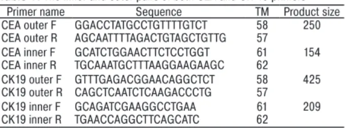
isolated from 300 μl of whole blood using the QIAamp RNA Blood Mini Kit (QIAGEN) according to the manu-facturer’s instructions. Reverse transcription was performed using 100 ng to 1 μg of RNA, 50 pmol of oligo(dT)18 primer, 200 U ofOMNISCRIPT reverse transcriptase (QIAGEN, USA), 40 units of rRNasin RNase inhibitor (Promega), and the deoxynucleotide triphosphates (final concentration 500 μmol/L) in a total volume of20 μL. Samples were incubated at 37 °C for 1 h andthen heated at 95 °C for 3 min. For PCR, 1 μL ofthe reverse transcription sample was used. The in-tegrity of RNA specimens was verified by performing RT-PCR with the housekeeping gene glyceraldehyde 3-phosphate dehydrogenase (GAPDH). Nested PCR was conducted by addition of 1 μL of complemen-tary deoxyribonucleic acid (cDNA) to 20 μl of reaction mixture containing 10 mM Tris–hydrochloric acid (pH 9.0), 50 mM potassium chloride, 2.5 mM magnesium chloride, 250 nM deoxynucleotid triphosphate 10 pmol of each outer primer, and 2.5 units of Taq DNA poly-merase (QIAGEN, USA). The reaction mixtures were subjected to 35 cycles of amplification in a program-mable thermal cycler (Perking-Elmer Cetus, USA) us-ing the followus-ing sequence after a denaturation step at 94 °C for 5 min: 94 °C for 30 sec, 56 °C for 30 sec and 72 °C for 1 min, and a final extension step at 72 °C for 10 min. A sample of 2 μl of 20-fold diluted first ampli-fication product was further amplified using an inner
pair of primers. The inner and outer pairs of primers are given in Table 1.
Table 1. The inner and outer pairs of both CEA and CK-19 primers
Primer name Sequence TM Product size
CEA outer F GGACCTATGCCTGTTTTGTCT 58 250
CEA outer R AGCAATTTTAGACTGTAGCTGTTG 57
CEA inner F GCATCTGGAACTTCTCCTGGT 61 154
CEA inner R TGCAAATGCTTTAAGGAAGAAGC 62
CK19 outer F GTTTGAGACGGAACAGGCTCT 58 425
CK19 outer R CAGCTCAATCTCAAGACCCTG 57
CK19 inner F GCAGATCGAAGGCCTGAA 61 209
CK19 inner R TGAACCAGGCTTCAGCATC 62
PCR primer sequences for each gene were designed to span atleast one intron region to avoid amplification of genomic DNA. According to this design, the PCR products of 154 and 209 base-pairs were amplified from CEA and CK19 cDNAs with nested PCR, respectively. The total RNA extracted from col15 and SW620 cells was used for optimization of the PCR and as a positive control. Blood cells from healthy volunteers were used as negative controls for every PCR reaction. Each sam-ple was subjected to electrophoresis in 1.5% agarose gels and stained with ethidium bromide. Samples from each patient were considered to have a positive score if any fragment showed a band of the expected size as positive control both for CEA and CK-19. Some PCR amplification products were sequenced to confirm the specificity of CK19 and CEA cDNA amplification products. The samples were judged to be positive (from + to ++++) or negative on the basis of the intensity value for the PCR product on the gel. Quantification of the expressions was based on the hypothesis that advanced stage tumors might present with greater amounts of micrometastatic tumor cells. Comparison between the groups was performed using this scale.
Statistical analysis. The χ2 test or Fisher’s exact test was used for categorical variables. Comparison according to age between groups was done using Student’s t-test. The 1 year and 2 year survival rates were evaluated by the Kaplan-Meier method. The significance of differences between the groups was calculated by the log-rank test. All statistical analy-ses were performed using the programme package number Crucher statistical system 2004 (Kaysville, UT, USA), and a p value < 0.05 was considered as significant.
RESULTS
Characteristics of patients and surgical proce-dures. A total of 50 patients with gastric cancer were
included in our study; 22 (44%) females and 28 (56%) males, with a mean age of 60.9 years (range 42–84). The tumors were localized in the cardia in 16 (32%), in the corpus in 11 (22%), and in the antrum in 22 (44%) patients. One patient (2%) presented with diffuse gastric cancer. Signet cell formation was detected in 18 (36%) patients.
Eleven patients in group II underwent subtotal gastrectomy, and total gastrectomy was applied to the remaining 17 patients. Total gastrectomy combined with partial colon resection was applied to one patient, and three patients underwent total gastrectomy
com-bined with splenectomy. Fourteen patients underwent D2, and 5 patients underwent D3 lymph node dissec-tion. All palpable lymph nodes were removed in the remaining 9 cases.
In group I, we detected peritonitis carcinomatosis in 12 patients, pancreatic invasion in 14, invasion of celiac truncus in 7, hepatic pedincle invasion in 6, Cru-ckenberg tumor in 2 and liver metastases in 2 patients. The reasons for unresectability consisted of pancre-atic invasion, celiac truncus and or heppancre-atic pedincle invasion. Treatment options for unresectable tumors were gastroenterostomy in 10, jejunostomy in 4, and gastrostomy in 3 patients. Explorative laparotomy and biopsy were applied to 5 patients. All patients re-ceived chemotherapy consisted of 5-fluorouracil and leucovorin at four weeks for 6 cycles.
Detection of CK-19 and CEA expression in pe-ripheral blood. No amplified product was obtained
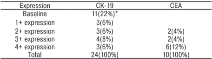
after RT-PCR when evaluating CK19 and CEA expres-sion in the 10 healthy controls. CK-19 expresexpres-sion was detected in 24 gastric cancer patients (48%). Expres-sion was not observed in 26 cases (52%). In patients with positive expression, 11 (22%) had baseline activity, 3 (6%) had 1+ expression, 3 (6%) had 2+ expression, 4 (8%) had 3+ expression, and the remaining 3 (6%) had 4+ expression. CEA expression was detected in 10 patients (20%), and was not observed in 40 cases (80%). In patients with positive expression, 2 (4%) had 2+ expression, 2 (4%) had 3+ expression, and the remaining 6 (12%) had 4+ expression (Table 2). Semi-quantitative expression of micrometastases by PCR are given in Table 3.
Table 2. CK-19 and CEA expressions among groups
Groups Cytokeratin-19 Total
Carcinoembryonic
antigen Total
Negative Positive Negative Positive
Group I 13(59%) 9(41%) 22(44%) 16(79%) 6(21%) 22(44%)
GroupII 13(40%) 15(60%) 28(56%) 24(90%) 4(10%) 28(56%)
Total 26(52%) 24(48%) 50(100%) 40(80%) 10(20%) 50(100%)
Table 3. Semi-quantitative expression of CK-19 mRNA (in 24 patients)
and CEA mRNA (in 10 patients) micrometastases by RT- PCR in patients with gastric cancer
Expression CK-19 CEA Baseline 11(22%)* 1+ expression 3(6%) 2+ expression 3(6%) 2(4%) 3+ expression 4(8%) 2(4%) 4+ expression 3(6%) 6(12%) Total 24(100%) 10(100%)
Note: * number of the patients.
There was no significant difference for either of the markers studied (χ2 = 4.097, p = 0.536 for CK-19 and χ2 = 0.791, p = 0.374 for CEA). Expressions of CEA and CK-19 without quantification did not also reveal a significant difference (χ2 = 2.052, p = 0.562). The difference in expressions of CK-19 and CEA mRNA ac-cording to age, gender, tumor location, and presence of signet cell formation were not significant (p = 0.631; χ2 = 0.034, p = 0.854; χ2 =2.037, p = 0.565; χ2 = 2.063, p = 0.151, respectively). The degree of expression did
not differ significantly between the groups (χ2 = 5.396, p = 0.145).
Serousal invasion was observed in 25 of 28 patients who underwent surgical resection. Lymphovascular invasion was detected in 18 (36%) individuals. Lym-phatic metastases were detected in 21 (42%) of the 28 (56%) patients in group II. In the remaining 7 patients, dissected lymph nodes (LNs) were free of metastases.
CK-19 was positive in 4 of 7 patients without lym-phatic involvement. However, this difference was not significant (p = 0.28). CEA was negative in all patients without lymphatic involvement. In cases with lymphatic invasion CEA expression was not different from those without LN metastasis (χ2 = 5.483, p = 0.140). We compared the difference between groups without quantifying the number of metastatic lymph nodes, and the difference was also not significant (χ2 = 2.775,
p = 0.521 for CK-19 and χ2 = 7.885, p = 0.098 for CEA).
Consideration of parameters in group II revealed no significant difference according to serousal inva-sion (p = 0.556 for CK-19 and p = 0.618 for CEA) and lymphovascular invasion (p = 0.247 for CK-19 and
p = 0.335 for CEA).
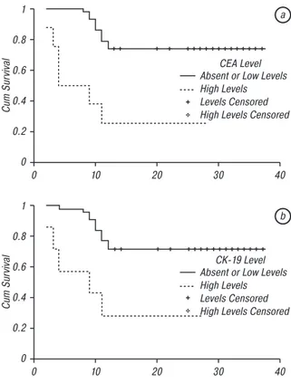
Median 1 year survival in group I and group II was 9.5 months (range 2–24) 20 months (range 11–29 months) respectively (p = 0.001). Median 2 years survival in group I was 10.5 months (range 2–24 months), and was 28.5 months (range 11–29) in group 2 (p = 0.001). MVI was detected in 7 cases. CK-19 was positive in 6 of these seven patients (85.7%) and CEA was positive in 5 patients (71.4%) with ce-liac truncus involvement, and both were positive in 4 (57.1%) of them. We detected both CK-19 and CEA positivity in 5 patients, and in 4 of them (80%) celiac truncus was invaded by the tumor. Furthermore, pa-tients with celiac truncus invasion expressed higher levels of micrometastases: for CEA; two patients had +++ expression, and three cases had ++++ expression. For CK-19; two patients had ++, two had +++, and the remaining two had ++++ micrometastatic expression. Patients with MVI expressed significantly higher levels of micrometastases compared to those without MVI (χ2 = 12.311, p = 0.023 for CEA, χ2 = 9.844, p = 0.009 for CK-19). All the patients with MVI have died within one year. We have evaluated the survival of patients according to expression patterns of CEA and CK-19. Mean survival of patients with high levels of CEA and CK-19 were 11.1 months and 11.9 months, respec-tively. Those of the remaining cases with minimal or no expression for CEA and CK-19 were 30.1 months and 29.4 months, respectively. The difference for both was significant (p < 0.001 for CEA and p = 0.002 for CK-19). Mean survival periods of patients due to the expression patterns were given in Table 4, survival curves are depicted in Fig. 1, a, b.
Table 4. Mean survival of patients according to expression patterns of
CEA and CK-19
Time (months) (mean ± SD) Log rank (χ2) p value
Death Alive
CEA Absent or low level 9.75 ± 2.18 27.52 ± 5.70 14.851 <0.001
High level 5.80 ± 3.96 20.50 ± 9.19
CK-19 Absent or low level 10.27 ± 1.27 27.48 ± 5.69 9.807 0.002
1 0.8 0.6 0.4 0.2 0 0 10 20 30 40 Cum Sur
vival Absent or Low LevelsCEA Level
High Levels Levels Censored High Levels Censored
1 0.8 0.6 0.4 0.2 0 0 10 20 30 40 Cum Sur
vival Absent or Low LevelsCK-19 Level
High Levels Levels Censored High Levels Censored
a
b
Fig. 1. One year survival curves of patients according to the
expression levels of CEA (a) and CK-19 (b); χ2 (log-rank) = 14.851,
p < 0.001 for CEA and χ2 (log-rank) = 9.807, p = 0.002 for CK-19 The sensitivity and specificity of CK-19 in detecting MVI was 26.08 and 96.15%, respectively. For CEA the sensitivity and specificity was 50 and 95%, respec-tively. When we consider only high titres of markers, sensitivity increased to 85.7% and 62.5% for CK-19 and CEA, respectively. In combined usage of CEA and CK-19 for detecting MVI, sensitivity was 80%. Considering combined usage of high titres in detecting MVI, sensitivity was 100%.
Median sur vival of patients with MVI was 6.7 months (95% CI: 3.554–9.875 months), and those of the remaining was 30.2 months (95% CI: 26.672–33.653 months). Patients with MVI had a sig-nificantly lower survival rates (χ2 (log-rank) = 23.324, p = 0.0001), (Fig. 2). 1 0.8 0.6 0.4 0.2 0 0 10 20 30 40 Cum Sur
vival PositiveMVI
Negative Positive Censored Negative Censored
Fig. 2. One year survival curves of patients according to the
presence of major vascular invasion. (χ2 (log-rank) = 23.324,
p = 0.0001)
DISCUSSION
Malignant process initiates from neoplastic prolif-eration of a cell leading to local enlargement and as the
progression advances, lymphatic and hematogenous metastases occur [3]. There is still a dilemma concern-ing the significance of detectconcern-ing circulatconcern-ing tumor cells (CTC), since only a portion of CTC have the ability to perform adhesions and invasion. This raises the ques-tion of whether there is a correlaques-tion between CTC and tumor stage, and what portion of patients with CTC will develop distant metastases. Detection of distant metastases plays a vital role in accurate staging and treatment of malignant tumor. For this reason, the goal of a screening method should be to identify individuals who are at high risk of developing distant metastases. It is common to observe cases with micrometastases in which conventional screening methods failed to demonstrate the course [4]. From this point of view, we investigated the role of PCR in detecting patients with locally advanced disease, and the correlation of prognostic parameters with PCR expression.
Detection of CTC by conventional haematological methods is both difficult and inaccurate [5, 6]. Using improved methods like immunohistochemistry (IHC) and PCR, not only the metastatic focus but only one metastatic cell can be identified [7–9]. Due to its great sensitivity, PCR can detect trace amounts (1 per 106) of cells in peripheral blood, lymph nodes, cerebrospinal fluid, and bone marrow [10]. Furthermore, microme-tastases undetected by IHC can be detected by PCR [11–13]. We applied nested RT-PCR to determine the CEA and CK19 mRNA molecules in combination, in the peripheral blood of gastric cancer patients. CEA is commonly used as a tumor marker and CK-19 is expected to display some specificity for epithelial cells.
Some markers like CEA, CK-19, CK-20 and α-fetoprotein have been used to detect CTC and mi-crometastases. In different series micrometastasis and CTC were detected in 20 to 70 percent of patients with gastric cancer, and stated as favourable markers of distant spread [10, 14, 15]. Conflicting results have been reported concerning the use of CK19 mRNA in the peripheral blood of patients with solid tumor due to the presence of pseudo genes and DNA con-tamination. To avoid these problems, RNA samples were incubated with DNase before cDNA synthesis, and primers were designed on the exon boundaries for both genes. Our results suggest that this method seems to be highly specific for micrometastatic cell de-tection because we have not detected any amplifica-tion product for both genes in healthy donor samples. But expression of both genes was observed in gastric cancer patients (CEA in 20% and CK-19 in 48% of the cases). Our results emphasize the heterogeneity of gene expression in tumors and justify the use of more than one mRNA marker for tumor cell detection.
Unlike some previous studies with different tumor types, we were not able to find any correlation between CK-19 and CEA mRNA expression with prognostic parameters. The positivity for PCR s in our samples from peripheral blood was lower than those of bone marrow samples and lymph nodes, when compared with published data. A possible explanation of this
controversy is the phenomenon described, particularly in patients with breast [16] and gastric [17] cancers, caused by the peculiar adhesive interaction between metastatic epithelial cells and stromal cells. In the report by Bonavina and co-workers [17], the role of micrometastatic expression by PCR in patients with non-metastatic tumors of the esophagus or gastric cardia was evaluated. The authors found PCR positive expression in peripheral blood in 3 of 18 (16.6%) pa-tients, but they could not demonstrate any correlation with tumor type or lymph node status. Micrometastatic expressions in bone marrow were detected in 14 of 18 patients (77.7%), and it was concluded that this ratio agrees with data in the literature [17]. Decreased ratios of peripheral micrometastatic expression might result from the degradation caused by circulating lymphocytes, and disseminated tumor cells in the cir-culation detected by RT-PCR might represent the sub-group that are able to preserve cell-surface antigens from lymphocytes. Comparison of survival curves of patients with different micrometastatic focuses might lead to a better understanding of the micrometastatic process.
Kodera et al. [18] have investigated the role of PCR in detecting peritoneal disseminated tumor cells, and found that positive PCR results were predictive of sur-vival even more strongly than LN metastasis (LNM) in patients who underwent curative surgical procedure for gastric carcinoma. Their results suggested that LN involvement correlated with peritoneal micrometas-tases. LN micrometastases in gastric cancer patients have been investigated. Even if it does not parallel with survival, CK-20 immunostaining has been detected in 20.7% of node negative gastric cancer patients [19], although the micrometastatic expression pattern af-fected the prognosis. Moreover, detection of multiple individual isolated tumor cells had the worst prognosis [19]. Single cells and small clusters of LNM are not uncommon in gastric cancer. Especially in diffuse type gastric cancer, the neoplastic cells lose their intercellular adhesion molecules, and unpredictable lymphatic spread can occur. Therefore, micrometa-static involvement of LN in gastric cancer is not well defined. Wu et al. [20] detected LNM in 10.8% of node negative patients assessed by routine hematoxylin and eosin (H&E) staining. They concluded that detection of cytokeratin by RT-PCR was useful for the detection of LNM, and that it correlated with diffuse histologi-cal subtype and depth of tumor invasion. If we could detect a correlation between conventional prognostic parameters and micrometastases, we might be able to detect patients with locally advanced tumors. In other words, if there had been a correlation, patients with early gastric carcinoma who do not have micrometa-static expression might undergo non-invasive surgi-cal resections. Otherwise, adjuvant chemotherapy and antiangiogenic treatment may be necessary for patients with micrometastases.
The major important point of our study is the high percentage of micrometastases in patients with celiac
invasion. In these patients, the levels of micrometa-static expression were much higher than others. To the best of our knowledge, this is the first report showing that high levels of micrometastatic expression sug-gested by the expression of both CK-19 and CEA might be a clue to predicting MVI in gastric cancer patients.
The imaging modalities used commonly in clinical staging of gastric cancer are gastroscopy, abdominal US, endoscopic US (EUS), computed tomography (CT) and occasionally positron emission tomography-CT (tomography-CT-PET). The combination of these methods is crucial in making the right diagnosis as well as the stage and follow-up after multimodal treatment. EUS and CT are especially beneficial for identifying invasion of gastric tumor within and beyond the gastric wall to adjacent structures. There are a number of reports indicating that EUS and CT have the ability to detect the tumor size and involvement of lymph nodes up to 80 and 60%, respectively [21–23]. However, disappoint-ing reports of CT on pre-operative stagdisappoint-ing for gastric cancer with low accuracy in tumor size (T) and node (N) staging exist in the literature [24, 25]. Magnetic resonance imaging (MRI) yielded discouraging results in gastric tumor staging as compared to CT or EUS [26]. Even with modern multislice CT, it may not be possible to evaluate infiltration of the vascular struc-tures with any certainty. There are some nonspecific signs, like the degree of deformity in the circumference of the vascular structure or more than 90° of contact between the tumor and the vessel wall [27]. From this point of view our study permitted us to conclude that appropriate vascular invasion might be predicted by the combined detection of CEA and CK-19 mRNA in peripheral blood of gastric carcinoma patients.
We did not observe a significant correlation be-tween the presence of circulating tumor cells and stage, LNM and serousal invasion. We had only one case of early gastric carcinoma, all the remaining patients had at least subserousal invasion. In the pres-ent series early gastric cancer is infrequpres-ent. If we had more patients with early gastric carcinoma, we might be able to exclude the significance of the expression of micrometastases within this subgroup, and we believe that our results might then be different.
In the future, staging systems will have to provide specific information on the biological properties of residual cancer cells in order to provide more ex-act prognostic estimates and guide patients to an individually tailored multimodal treatment. However, it should not be forgotten that any cancer cell detected in peripheral blood may not be able to metastasize, and the significance of micrometastatic cell detec-tion remains to be proven by multicentre prospective randomized trials with long-term follow-up.
In conclusion, pre-operative combined nested RT-PCR for CK-19 and CEA (if both are positive) might predict MVI and thereby avoid the morbidity of invasive interventions like angiography. We believe that this procedure is minimally invasive and cost-effective,
and could be the method of choice for pre-operative evaluation of local invasion in gastric cancer.
ACKNOWLEDGEMENTS:
The authors would like to thank International Science Editing for their assistance with revision of this manuscript.
REFERENCES
1. DeVita Jr VT. Cancer. In: DeVita Jr VT, S Hellman, ST Rosenberg, eds. Cancer of the stomach. Philadelphia: JB Lippincott Comp, 1993; 765–96 pp.
2. Ming SC. Pathology of the gastrointestinal tract. In: Ming SC, Goldman H, eds. Adenocarcinoma and other ma-lignant epithelial tumors of the stomach. Philadelphia: WB Saunders, 1992; 584–617 pp.
3. Diel IJ, Kaufman M, Goerner R, et al. Detection of tumor cells in bone marrow of patients with primary breast cancer: a prognostic factor for distant metastasis. J Clin Oncol 1992; 10: 1534–9.
4. Kell MR, Winter DC, O’Sullivan GC. Biological behaviour and clinical implications of micrometastases. Br J Surg 2000; 87: 1629–39.
5. Ridell B, Landsy K. Incidence and histopathology of metastases of mammary carcinoma in biopsies from the pos-terior iliac crest. Cancer 1979; 44: 1782–8.
6. Ingle JN, Tormey DC, Tan HK. The bone marrow examination in breast cancer. Cancer 1978; 41: 670–74.
7. Coombes RC, Dearnaley DP, Redding WH. Protides of the biological fluids. In: Peeters H, ed. Micrometastases in breast cancer. Oxford: Pergamon, 1983; 31: 317–23.
8. Dearnaley DP, Sloane JP, Ormerod MG, et al. Increased detection of mammary carcinoma cells in marrow smears using antisera to epithelia membrane antigen. Br J Cancer 1981; 44: 85–90.
9. Dearnaley DP, Ormerod MG, Sloane JP. Detection of isolated mammary carcinoma cells in marrow of patients with primary breast cancer. JR Soc Med 1983; 76: 359–64.
10. Sakakura C, Takemura M, Hagiwara A. Overexpression of dopa decarboxylase in peritoneal dissemination of gastric cancer and its potential as a novel marker for the detection of peritoneal micrometastases with real-time RT-PCR. British J Cancer 2004; 90: 665–71.
11. Noguchi S, Aihara T, Nakamori S, et al. The detection of breast carcinoma micrometastases in axillary lymph nodes by means of reverse transcriptase-polymerase chain reaction. Cancer 1994; 74: 1595–600.
12. Mattano LA, Moss TJ, Emerson SG. Sensitive detec-tion of rare circulating neuroblastoma cells by the RT-PCR. Cancer Res 1992; 52: 4701–5.
13. Zigeuner RE, Riesenberg R, Pohla H. Immunomag-netic cell enrichment detects more disseminated cancer cells than immunohistochemistry in vitro. J Urol 2000; 164: 1834–7.
14. Schmidt P, Thiele M, Rudroff C, et al. Detection of tumor cells in peritoneal lavages from patients with gastro-intestinal cancer by multiplex reverse transcriptase PCR. Hepatogastroenterology 2001; 48: 1675–9.
15. Kakeji Y, Maehara Y, Shibahara K. Clinical significance of micrometastases in bone marrow of patients with gastric cancer and its relation to angiogenesis. J Gast Can 1999; 2: 46–51.
16. Datta JH, Adams PT, Drobyski WR. Sensitive detection of occult breast cancer by the reverse transcriptase polymerase chain reaction. J Clin Oncol 1994; 12: 475–82.
17. Bonavina L, Soligo D, Quirici N. Bone marrow-disse-minated tumor cells in patients with carcinoma of the esopha-gus or cardia. Surgery 2001; 129: 15–22.
18. Kodera Y, Nakanishi H, Ito S. Quantitative detection of disseminated free cancer cells in peritoneal washes with real-time reverse transcriptase polymerase chain reaction. Ann Surg 2002; 235: 499–506.
19. Lee HS, Kim MA, Yang HK. Prognostic implica-tion of isolated tumor cells and micrometastases in regional lymph nodes of gastric cancer. World J Gastroenterol 2005;
11: 5920–25.
20. Wu ZY, Zhan WY, Li JH. Expression of E-cadherin in gastric carcinoma and its correlation with lymph node micrometastasis. World J Gastroenterol 2005; 11: 3139–43.
21. Ang TL, Ng TM, Fock KM, et al. Accuracy of endo-scopic ultrasound staging of gastric cancer in routine clinical practice in Singapore. Chin J Dig Dis 2006; 7: 191–6.
22. Kim JH, Eun HW, Goo DE, et al. Imaging of various gastric lesions with 2D MPR and CT gastrography performed with multidetector CT. Radiographics 2006; 26: 1101–18.
23. Hur J, Park MS, Lee JH, et al. Diagnostic accuracy of multidetector row computed tomography in T- and N staging of gastric cancer with histopathologic correlation. J Comput Assist Tomogr 2006; 30: 372–7.
24. Kirk SJ, Moorehead RJ, McIlrath E, et al. Does preoperative computed tomography scanning aid assessment of oesophageal carcinoma? Postgrad Med J 1990; 66: 191–4.
25. Andaker L, Morales O, Hojer H, et al. Evaluation of pre-operative computed tomography in gastric malignancy. Surgery 1991; 109: 132–5.
26. Palmowski M, Grenacher L, Kuntz C, et al. Magnetic resonance imaging for local staging of gastric carcinoma: re-sults of an in vitro study. J Comput Assist Tomogr 2006; 30: 896–90.
27. Fiore D, Baggio V, Ruol A, et al. Multimodal imaging of esophagus and cardia cancer before and after treatment. Radiol Med 2006; 111: 804–17.