DOI: 10.5455/annalsmedres.2019.03.143 2019;26(7):1222-6
Multidetector computed tomography and magnetic
resonance imaging findings in pulmonary hydatid cysts
Hakan Cebeci1, Mustafa Yasir Ozlu1, Abidin Kilincer1, Emine Uysal1, Mehmet Sedat Durmaz1, Ramazan Ucar2, Guven Sadi Sunam3
1Selcuk University Medical Faculty, Department of Radiology, Konya, Turkey
2Necmettin Erbakan University, Meram Medical Faculty, Department of Internal Medicine, Division of Allergy and Clinical Immunology, Konya, Turkey 3Selcuk University Medical Faculty, Department of Thoracic Surgery, Konya, Turkey
Copyright © 2019 by authors and Annals of Medical Research Publishing Inc.
Abstract
Aim: Computed tomography (CT) and magnetic resonance imaging (MRI) are diagnostic modalities in pulmonary hydatid disease.
We aimed to present CT and MRI findings in pulmonary hydatid disease.
Material and Methods: CT and MRI findings of pulmonary hydatid cysts from January 2011 through June 2018 were evaluated
retrospectively. CT images of 112 patients and MRI images of 27 patients were evaluated. Patients’ gender and ages, lesion number and distribution, concomitant organ involvement, complications, HU density of lesions on CT and signal characteristics of lesions on MRI were analyzed. T2 hypointense rim, T1 and T2 signal intensity, daughter vesicle and detached membranes were the evaluated parameters on MRI.
Results: 147 hydatid cyst lesions of 119 (66 female, 53 male) patients with a mean age of 39.4±19.6 years were included in the study
62 (52.1%) of patients had isolated lung involvement. 79 of 119 patients (66.4%) had solitary lung lesion. Mean CT density of cysts were 16 HU. 25 of 32 cysts showed T2 hypointense rim on MRI.
Conclusion: Imaging plays important role in diagnosis and presurgical evaluation of pulmonary hydatid disease. CT is mostly used
in diagnosis; MRI should be a choice particularly in pediatric population.
Keywords: Computed tomography; pulmonary; hydatid cyst; magnetic resonance.
Received: 18.03.2019 Accepted: 15.05.2019 Available online: 25.06.2019
Corresponding Author: Hakan Cebeci, Selcuk University Medical Faculty, Department of Radiology, Konya, Turkey E-mail: [email protected]
INTRODUCTION
Hydatid cyst disease is a parasitic disease and an important health problem all around the world. Echinococcusgranulosus, is responsible for hydatid disease in human population (1). Chest roentgenography, multidetector computed tomography (MDCT) and magnetic resonance imaging (MRI) are the modalities used for the radiological diagnosis of pulmonary hydatid disease. CT provides images with high geometric resolution in fast image acquisition times for the evaluation of hydatid cyst. However, MRI has the advantage of the multiplanar and multiparameter imaging capabilities avoiding ionizing radiation exposure. Longer image acquisition time is the disadvantage of MRI. Indirect hemagglutination test (IHA) is commonly used laboratory test in patients with suspected hydatid disease, but sensitivity of this test is limited, particularly in pulmonary hydatid disease (2).
The purpose of this study was to evaluate the CT findings and MRI signal characteristics of pulmonary hydatid cysts.
MATERIAL and METHODS
The study involved 147 lesions in 119 patients with pulmonary hydatid cysts. This retrospective study was approved by institutional review board. Informed consent was waived. CT and MRI findings of pulmonary hydatid cyst patients, who were operated in our hospital between January 2011 and June 2018 were evaluated retrospectively. Two radiologist evaluated images together with consensus agreement (1 radiologist with 6 year experience and 1 radiology resident at the third year of education). Patients’ gender and ages, lesion number and distribution, concomitant organ involvement, complications, Hounsfield unit (HU) density of lesions
on CT and signal characteristics of lesions on MRI were analyzed. T2 hypointense rim, T1 and T2 signal intensity, daughter vesicle and detached membranes were the evaluated parameters on MRI.
CT imaging were obtained with a 256 (Siemens, Somatom Definition Flash) and 16 (Siemens, Somatom Scope) multidetector-CT. MRI examinations were obtained with a 1.5 Tesla scanner (Siemens, MagnetomAera). MRI protocol included T2-weighted fat suppressed axial (TR: 1400 ms, TE: 91 ms, slice thickness: 6 mm), T2-weighted haste coronal (TR: 461 ms, TE: 28 ms, slice thickness: 6 mm), T1-vibe fat suppressed axial (TR: 4.36 ms, TE: 1.98 ms, slice thickness: 3 mm) and coronal (TR: 2.96 ms, TE: 1.1 ms, slice thickness: 1.8 mm), T2-weighted haste fat suppressed sagittal (TR: 700 ms, TE: 91 ms, slice thickness: 6 mm), T2-blade fat suppressed axial (TR: 3060 ms, TE: 118 ms, slice thickness: 6 mm), and contrast enhanced T1-vibe fat suppressed axial (TR: 4.36 ms, TE: 1.98 ms, slice thickness: 3 mm) and coronal (TR: 2.96 ms, TE: 1.1 ms, slice thickness: 1.8 mm).
SPSS, version 16.0, was used for statistical analysis. Descriptive statistics were used for calculating means and standard deviations.
RESULTS
One hundred seven hydatid cyst lesions in 119 (66 females, 53 males) patients with a mean age of 39.4±19.6 years were included in the study. All patients had undergone surgery and all lesions had a pathological diagnose consistent with hydatid disease. CT images of 112 patients and MRI images of 27 patients were evaluated. The hydatid cysts were evaluated for size, number, localization, HU density, complication on CT images. On MRI, signal characteristics of cyst fluid, T2-hypointense capsule, involving daughter cysts, germinal membrane detachment were evaluated. 86 (58.5%) of lesions were in right lung and 61(41.5%) of lesions were left sided. Distribution of pulmonary hydatid cysts were demonstrated in table 1.
Sixty-two (52.1%) of patients had isolated lung involvement, while 57 patients had accompanied liver lesions, 3 had spleen and 1 patient had brain lesions. 79 of 119 patients (66.4%) had solitary lung lesion while others had multiple hydatid cyst. Sizes of lesions were between 8mm and 153 mm with the mean of 49.5 mm. In 2 lesions located and ruptured into the pleural cavity, we could not measure lesion diameter. Mean HU density of cysts were 16 HU (range: -43 and 89 HU). 25 of 32 cysts showed T2 hypointense rim on MRI. Figure 1 reveals hypointense capsule on T2 weighted axial image of a hydatid cyst in a 41 year-old patient. MRI signal characteristics of lesions were listed in Table 2.
Table 1. Distributions of pulmonary hydatid cyst lesions
Right lower lobe 47 (32%) Right middle lobe 15 (10%) Right upper lobe 22 (15%) Left lower lobe 38 (26%) Left upper lobe 23 (15%)
Pleural 2 (1.3%)
Figure 1. A 41 year-old male patient. T2 weighted axial image
shows hyperintense cystic lesion in right lung lower lobe with hypointense capsule (arrow)
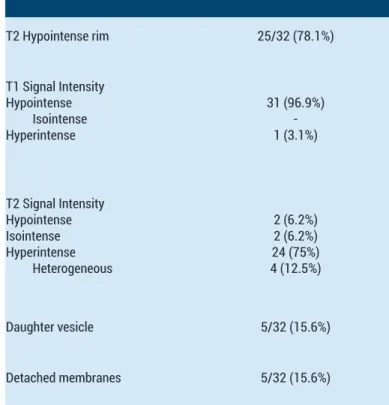
Table 2. MRI signal characteristics of hydatic cyst lesions
T2 Hypointense rim 25/32 (78.1%) T1 Signal Intensity Hypointense Isointense Hyperintense 31 (96.9%) -1 (3.-1%) T2 Signal Intensity Hypointense Isointense Hyperintense Heterogeneous 2 (6.2%) 2 (6.2%) 24 (75%) 4 (12.5%) Daughter vesicle 5/32 (15.6%) Detached membranes 5/32 (15.6%)
Ninety of 147 pulmonary hydatid cysts were uncomplicated. Figure 2 reveals a CT image of an uncomplicated hydatid cyst. While 55 of 147 pulmonary hydatid cysts were ruptured and complicated cysts. Figure 3 reveals a CT image of a cyst hydatid lesion ruptured to pleural cavity. Infectious complications were revealed in only 4 pulmonary hydatid cysts.
Figure 2. A 43 year-old female patient. Uncomplicated hydatid
cyst in right lung lower lob is shown on CT image. The region of interest (ROI) was placed inside the lesion and measurement of density revealed 1 HU
Figure 3. A 29 year-old female patient. Axial non-contrast CT
image shows a ruptured cyst hydatid and germinatid membranes in right pleural cavity
DISCUSSION
Hydatid cyst disease is a parasitic disease all over the world, particularly in the Mediterranean area. Disease is still endemic in Turkey (3,4). Pulmonary hydatid disease usually presents with solitary cystic lesion. Ozmen et al.
reported 70% solitary lung hydatid cysts in their series (5). 66.4% of our patients revealed solitary cysts. Pulmonary hydatid cyst is more common in children and often located in the right lobe (6-8). Mostly, pulmonary hydatid cysts are unilateral, bilateral involvement reported in 2% to 30% (7,9-11). Right lung lower lobe was the mostly involved location in our patients with 47 cystic lesions (31.9%). The results of this study regarding lesion location and multiplicity of pulmonary hydatid disease were consistent with the previous reports in the literature (1,5,12,13). Previous reports revealed higher incidence of pulmonary hydatid disease in males (13-15). Ozmen et al. found 62% male predominance in their series involving pulmonary hydatid cysts. They indicated that there is no study in the literature reporting female predominance (5). However, in our patients, there was a slight female predominance with a ratio of 55%.
Concomitant organ involvement is frequently seen in hydatid disease. However, any organ can be infected by Echinococcusgranulosus, mostly liver hydatid cysts associate with the hydatid disease of lungs. Concomitant involvement of pulmonary and hepatic hydatid disease varies from 16–23.1%. In our study, 57 of 119 patients (47%) had accompanied liver lesions. Spleen and brain are other organs associated with pulmonary hydatid disease in this study. In 3 patients, hydatid cysts in the spleen were noted and 1 patient had associated hydatid cyst in the brain. No other organ involvement was noted.
Pulmonary hydatid cysts may appear in various sizes from 1 to 20 cm in diameter. Cysts of the lungs that are 10 cm in diameter or larger than 10 cm are defined as giant hydatid cysts.The lungs are the only organ in which hydatid cysts can grow so large and this is attributed to compressibility of lung tissue. The high prevalence of pulmonary hydatid cyst in childhood can also be attributed to this feature (16,17). In a study conducted by Kuzucu et al., 41 of 169 patients had giant cysts and 63% of these giant cysts were aged 30 years or younger. In our series, 11 lesions were larger than 10 cm and 3 of these cysts have the radiologic signs of rupture. There was no statistically significant difference among complications between giant cysts and non-giant cysts. Ages of patients with giant pulmonary hydatid cyst were between 8 and 52 years, and 8 of 11 patients were younger than 30 years.
Most frequent complication detected in pulmonary hydatid cysts is the rupture of the cyst to endobronchial system or into the pleural cavity. Cyst rupture may lead to significant clinical and radiological consequences (5). Spontaneous rupture may be seen in cysts, however, trauma or secondary infections may also cause cyst rupture (9). There have been various studies reporting different incidence of ruptured pulmonary hydatid cysts. Tor et al. reported a ratio of 27% complicated hydatid cysts for their series (18). However, Aribas et al. detected a ratio of 88.4% ruptured cystic hydatidosis(19). We found a ratio of 37% (55 of 147 lesions) for complicated cysts. 98 of 147 hydatid cyst were uncomplicated. Rupture into the pleural
cavity also reported in previous studies with a range of 3% to 9%. Only 2 lesions in this study were ruptured to pleural cavity.
CT is the mostly used radiological modality in diagnosis and presurgical evaluation of hydatid cysts. CT was used in 112 of 119 patients in this study. CT imaging of hydatid cysts may differ from each other depending on size, degeneration and complication status of cyst. CT characteristic of uncomplicated hydatid cysts: well demarcated, oval shaped, fluid containing lesions. Unlike hydatid cysts of the liver, calcification and daughter cyst formation are rare in lung hydatids. Differentiation of uncomplicated pulmonary hydatid cysts from other cysts of lungs can be a challenging problem in many times. If the patient had also a cyst involving peripheral calcification or daughter cysts, it can help to diagnose a pulmonary hydatid cyst (1) HU density of pulmonary hydatid cysts on CT commonly is not a reliable parameter for diagnosis. HU density of hydatid cyst lesions in this study varies between -43 and 89 HU. On CT, there are common imaging findings in pulmonary hydatid cysts. If there is air density between pericyst and laminated membrane of the cyst, then it is called as ‘Meniscus sign. This sign occurs when the growing hydatid cyst produce erosions in the bronchioles. Another sign which is less frequently observed is the ‘onion peel sign’ that seen when the air is between the endocyst and pericyst. Most common sign in complicated pulmonary hydatid cysts is the ‘water lily sign’, it is observed when the air enters the cyst cavity, the endocyst and pericyst completely separate from each other and the collapsed degenerated membranes floats freely in the dependent part the cyst cavity. The other unusual finding in complicated cysts is a thin-walled cavitary lesion, which is more common in pulmonary hydatid disease under medical treatment. But it can be also observed during the natural course of hydatid cysts without medical treatment (16).
MRI is less frequently used in diagnosis and presurgical evaluation of hydatid cysts but it provides useful information and particularly in pediatric population it should be a choice. Absence of ionizing radiation exposure is the major advantage of MRI. Twenty-seven patients in this study were evaluated with MRI. The MRI signal characteristics of hydatid cyst may differ depending on the developmental phase. Uncomplicated pulmonary hydatid cyts appear hypointense on T1-weighted images and hyperintense on T2-weighted images, in most cases. T1 and T2-weighted images can be heterogeneous or homogeneous. In parent cysts with daughter cysts, the parent cyst fluid has been found to be usually of higher signal intensity than daughter cyst fluid on T1- and T2-weighted images. Daughter cysts are the most characteristic features of hydatid cysts. It is easier to diagnose a hydatid cyst including daughter cysts. Daughter cysts mean a live hydatid cyst;, however, their absence does not mean the sterility (2, 20). In the evaluation of MRI examinations of our patients, we detected daughter
cysts on MRI of 5 patients. Depending upon the content of daughter cysts, the signal intensity on MRI may vary (21). In the natural course of a pulmonary hydatid disease, the host produces a fibrous capsule around the parasitic membranes to isolate the cyst (22). This fibrous capsule is usually seen hypointense on T2-weighted images (2, 22). On T1-weighted images, the capsule has isointense signal to the fluid and shows mild enhancement with intravenous injection of gadolinium based contrast agent, which reflects the vascularity of the pericyst. The presence of a hypointense rim on T2 -weighted images in a lung cyst is helpful in differentiating unilocular uncomplicated hydatid cysts from simple cysts (23). In our series, the hypointense capsule was seen on T2-weighted images in 25 patients. If the cyst is degenerated or complicated, air and detached parasitic membranes may be seen (24). Detached membranes also help differential diagnose. In MRI examinations included in this study, we observed detached parasitic membranes in MRI of 5 patients. This study has some limitations. Retrospective study design is the first limitations. Second, the observers evaluated images with consensus agreement and inter-observer variability did not researched.
CONCLUSION
In conclusion, imaging plays important role in diagnosis and presurgical evaluation of pulmonary hydatid disease. Solitary cystic lesions are more common in pulmonary hydatid disease and lower lobes are more involved. Liver is the most accompanying organ to pulmonary hydatid cysts. However, CT is mostly used in diagnosis; MRI should be a choice particularly in pediatric populati
on.
Competing interests: The authors declare that they have no competing interest.Financial Disclosure: There are no financial supports
Ethical approval: This work has been approved by the Institutional Review Board.
Hakan Cebeci ORCID: 0000-0002-2017-3166 Mustafa Yasir Ozlu ORCID: 0000-0003-4940-7592 Abidin Kilincer ORCID: 0000-0001-6027-874X Emine Uysal ORCID: 0000-0001-8533-4939
Mehmet Sedat Durmaz ORCID: 0000-0002-1340-2477 Ramazan Ucar ORCID: 0000-0003-1314-4444 Guven Sadi Sunam ORCID: 0000-0001-9655-0012
REFERENCES
1. Garg MK, Sharma M, Gulati A, et al. Imaging in pulmonary hydatid cysts. World J Radiol 2016;8:581.
2. Singh S, Gibikote SV. Magnetic resonance imaging signal characteristics in hydatid cysts. Australas Radiol 2001;45:128-33.
3. Işıtmangil T. Evaluation of 308 patients surgically treated for thoracic hydatidosis. Turkish J Thorac Cardiovasc Surg 2010;18:27-33.
4. Arroud M, Afifi MA, El Ghazi K, et al. Lung hydatic cysts in children: comparison study between giant and non-giant cysts. Pediatr Surg Int 2009;25:37-40.
5. Ozmen CA, Onat S. Computed tomography (CT) findings of pulmonary hydatid cysts in children and the factors related to cyst rupture. Med Sci Monit 2017;23:3679-86.
6. Moro PL, Budke CM, Schantz PM, et al. Economic impact of cystic echinococcosis in Peru. PLoS neglected tropical diseases 2011;5:e1179.
7. Doğan R, Yüksel M, Cetin G, et al. Surgical treatment of hydatid cysts of the lung: report on 1055 patients. Thorax 1989;44:192-9.
8. Kurul IsC, Topcu S, Altinok T, et al. One-stage operation for hydatid disease of lung and liver: principles of treatment. J Thorac Cardiovasc Surg 2002;124:1212-5.
9. Aribas OK, Kanat F, Turk E, et al. Comparison between pulmonary and hepatopulmonary hydatidosis. Eur J Cardiothorac Surg 2002;21:489-96.
10. Karaoglanoglu N, Kurkcuoglu IC, Gorguner M, et al. Giant hydatid lung cysts. Eur J cardiothorac surg 2001;19:914-7. 11. Solak H, Özgen G, Yüksek T, et al. Surgery in hydatid cyst of
the lung: A report of 460 cases. Scand J Thorac Cardiovascr Surg 1988;22:101-4.
12. Pedrosa I, Saiz A, Arrazola J, et al. Hydatid disease: radiologic and pathologic features and complications 1: (cme available in print version and on rsna link). Radiographics 2000;20:795-817.
13. Onal O, Demir OF. The relation between the location and the perforation rate of lung hydatid cysts in children. Asian J Surg 2018;41:422-6.
14. Katrancıoğlu Ö, Şahin E, Karadayı Ş, et al. Diagnosis and treatment approaches of the lung hydatid cysts in childhood. Current Thoracic Surg 2017;2:11-4.
15. Montazeri V, Sokouti M, Rashidi MR. Comparison of pulmonary hydatid disease between children and adults 2007.
16. Polat P, Kantarci M, Alper F, et al. Hydatid disease from head to toe. Radiographics 2003;23:475-94.
17. Kuzucu A, Ulutas H, Celik MR, Yekeler E. Hydatid cysts of the lung: lesion size in relation to clinical presentation and therapeutic approach. Surg Today 2014;44:131-6.
18. Tor M, Atasalihi A, Altuntas N, et al. Review of cases with cystic hydatid lung disease in a tertiary referral hospital located in an endemic region: a 10 years’ experience. Respirat 2000;67:539-42.
19. Aribas OK, Kanat F, Gormus N, et al. Pleural complications of hydatid disease. J Thorac Cardiovasc Surg 2002;123:492-7. 20. Marani S, Canossi G, Nicoli F, et al. Hydatid disease: MR
imaging study. Radiol 1990;175:701-6.
21. Von Sinner W, Rifai A, Te Strake L, et al. Magnetic resonance imaging of thoracic hydatid disease: correlation with clinical findings, radiography, ultrasonography, CT and pathology. Acta Radiol 1990;31:59-62.
22. von Sinner WN. New diagnostic signs in hydatid disease; radiography, ultrasound, CT and MRI correlated to pathology. Eur J Radiol 1991;12:150-9.
23. Guthrie J, Lawton J, Chalmers A. Case report: the MR appearances of primary intramuscular hydatid disease. Clin Radiol 1996;51:377-9.
24. Apt WL, Fierro JL, Calderón C, et al. Vertebral hydatid disease: clinical experience with 27 cases. J Neurosurgery 1976;44:72-6.