28
Geliş(Recevied) :07/02/2018Kabul(Accepted) :13/03/2018
Research Article DOI:10.30708/mantar.391682
High-Throughput Genomic Simple Sequence Repeat (SSR) Marker
Development and Construction of a High Resolution Physical Map in
Button Mushroom (Agaricus bisporus) Genome
Ali Tevfik UNCU*
1,
Ayşe Özgür UNCU
2*Corresponding author: [email protected]
1Necmettin Erbakan University, Department of Molecular Biology & Genetics, Meram, Konya, 42090, TURKEY
2Necmettin Erbakan University, Department of Biotechnology, Meram, Konya, 42090, TURKEY
Abstract: Button mushroom [Agaricus bisporus (J.E. Lange) Imbach], is both a model organism and a worldwide cultivated edible mushroom species. Despite its scientific and economic relevance, efforts to develop molecular markers in A. bisporus genome are scarce and relatively recent. The present research reports the development of 3943 novel simple sequence repeat (SSR) markers specific to the A. bisporus genome. The marker set is well-distributed among the 13 A. bisporus chromosomes, saturating the entire genome with sequence-specific markers. The large number of novel, A. bisporus specific DNA markers introduced in the present work constitute a valuable resource for molecular genetic research in button mushroom, including strain identification and protection, and gene/QTL (Quantitative Trait Locus) mapping.
Key words: Molecular genetics, Bioinformatics, Sequence-specific markers, DNA markers
Kültür
Mantarı (Agaricus bisporus) Genomuna Özel Yüksek Miktarda Basit Dizi
Tekrarı (SSR) Markörünün Geliştirilmesi ve Yüksek Çözünürlükte Fiziksel Genom
Haritasının Oluşturulması
Öz: Kültür mantarı [Agaricus bisporus (J.E. Lange) Imbach], bir model organizma olmasının yanı sıra dünya genelinde yetiştirilen bir yenilebilir mantar türüdür. Bilimsel ve ekonomik önemine rağmen A. bisporus genomuna özel moleküler markör geliştirme çalışmaları ancak yakın zamanda gerçekleştirilmeye başlanmış olup, oldukça kısıtlı sayıdadır. Bu çalışma kapsamında A. bisporus genomuna özel 3943 adet yeni basit dizi tekrarı (SSR) markörü geliştirilmiştir. Geliştirilen markörler A. bisporus kromozomlarına dengeli şekilde dağılmakta olup, genomun sekans-spesifik markörler ile doyurulmasını sağlamaktadır. Çalışma kapsamında tanıtılmakta olan yüksek sayıdaki A. bisporus’a özel yeni DNA markörü, bu türde gerçekleştirilecek moleküler genetik çalışmalar için kıymetli bir kaynak teşkil edecektir.
Anahtar kelimeler: Moleküler genetik, Biyoinformatik, Sekans-spesifik markörler, DNA markörleri Introduction
Button mushroom [Agaricus bisporus (J.E. Lange) Imbach], also referred to as common mushroom, is the most commonly cultivated edible mushroom species (Barroso et al., 2000; Foulongne-Oriol et al., 2012). In addition, it is the model fungus species for fungal adaptation, persistence and growth in humic-rich
environments (Morin et al., 2012; Heneghan et al., 2016). Despite its role as a model organism and economic value due to its widespread cultivation as the predominant edible mushroom in European countries and USA, molecular genetic research in A. bisporus has been quite limited. While gene and QTL (Quantitative Trait Loci) mapping became the standard approach in modern plant and
29
animal breeding, mapping traits in fungal genomes is a relatively recent research objective (Foulongne-Oriol et al., 2012). Molecular markers are unquestionably the indispensable tools of modern breeding (Jones et al., 2009). Thus, an abundance of molecular markers specific for plant and animal genomes were developed and have been utilized for the improvement of cultivated crops and livestock. On the other hand, DNA markers specific to the A. bisporus genome are limited. Because linkage drag is a prominent problem that impairs breeding of improved button mushroom cultivars (Gao et al., 2016), saturating A. bisporus chromosomes with molecular markers would be highly useful to monitor the segregation of linked loci in high resolution and determine the desired recombinants. Yet, molecular breeding of white button mushroom toward improved yield, resistance to bacterial and fungal diseases, and technological properties (e.g. amenability to mechanical harvest) has not been properly established. For example, bacterial and fungal diseases are among major causes of yield loss, shortened shelf life and decreased quality in button mushroom cultivation (Samuels et al., 2002; Sokovic and Van Griensven, 2006; Berendsen et al., 2010). Major pathogens of button mushroom are listed as the bacterium Pseudomonas tolaasii Paine; the fungal species Lecanicillium fungicola (Preuss) Zare and W. Gams, and Trichoderma harzianum Rifai. On the other hand, there is lack of molecular genetic research to identify genes/QTLs that confer resistance to the above listed pathogens. Similarly, work that identified yield related molecular markers is extremely scarce. In addition to traits related to disease resistance and yield, it has to be noted that button mushroom is reported as a source of health beneficial dietary fibers and antioxidants (Jeong et al., 2010; Liu et al., 2013), therefore, traits regarding nutritional quality are also potential targets of breeding. Thus, molecular genetic research to dissect the genetic basis of nutritionally important traits in button mushroom is also required.With the recent advances in high throughput DNA sequencing technologies, vast amount of genomic data is continuously being generated and deposited in publicly available databases (Uncu et al., 2015a), enabling in silico design of genome-specific molecular markers using bioinformatic approaches. Entire genomes can be surveyed for the high-throughput identification of molecular markers that represent the genomes of the relevant species in high-resolution. A draft genome
assembly of A. bisporus is available since 2012 (Morin et al., 2012), and the latest assembly that correctly represents the 13 chromosomes was recently published (Sonnenberg et al., 2016). Thus, it is now feasible to develop sequence-specific molecular markers and saturate the A. bisporus genome, which will greatly contribute to studies on chromosomal recombination and segregation patterns, as well as strain selection, conservation and molecular breeding efforts.
According to the common definition, SSRs (Simple Sequence Repeats) are iterations of short DNA motifs (Jones et al., 2009), where motif size ranges from one to eight bases. SSRs found wide applications in molecular genetics since they are highly polymorphic, reproducible and provide extensive genome coverage (Uncu et al., 2015b). In the present work, SSR (Simple Sequence Repeat) markers specific to the A. bisporus genome were developed via bioinformatics, by mining the recently released genome assembly for tandem repeat sequences. As a result, 4138 SSR loci were identified within the A. bisporus genome. In order to convert the SSR loci to markers, primers were designed that flank the repeat sequences. e-PCR was performed to confirm the amplificability of the marker loci in silico, and it was feasible to further verify the e-PCR results with wet-lab experiments.
Material and Method
Simple sequence repeat identification
Chromosome assemblies of A. bisporus var. bisporus strain H39 (GenBank: CP015470.1 - CP015482.1) were retrieved from NCBI Genome Database (https://www.ncbi.nlm.nih.gov/genome/) as individual files in FASTA format. GMATA (Genome-wide Microsatellite Analyzing Tool Package) (Wang and Wang, 2016) software was utilized for mining the chromosome sequences for simple sequence repeats and, designing and mapping flanking amplification primers. Because GMATA requires Perl, R and Java running environments, the three programs were installed prior to running the software. For simple sequence repeat identification, FASTA files for individual chromosomes were imported to the ‘SSR identification’ module of the GMATA software. Parameters used for mining mononucleotide repeats were; Min-length (nt): 1, Max-length (nt): 1, Min. repeat-times: 8. Parameters for oligonucleotide repeat
30
identification were; Min-length (nt): 2, Max-length (nt): 8, Min. repeat-times: 5. SSR loci information files (.ssr) were generated individually for each chromosome that contained the SSR start & end positions, motifs and number of repeats.Marker design and mapping
‘Marker designing’ module of GMATA was utilized in order to design PCR markers for the identified SSR loci. For each chromosome, assembly files in FASTA format and output files generated by the ‘SSR identification’ module (.ssr) were imported to the module as the ‘Sequence file’ and the ‘SSR loci file’, respectively. Marker design parameters were adjusted as: Min. amplicon size: 100 bp, Max amplicon size: 300 bp, Optimal annealing Tm: 60 ⁰C, Flanking sequence length: 400 bp, Max template length: 2000 bp. Two output files with .mk and .sts extensions were generated for each chromosome, which contained left and right primer sequences, calculated melting temperatures, primer locations and anticipated product sizes. ‘e-mapping’ module of the GMATA software was utilized to run the e-PCR algorithm (Schuler, 1997). The algorithm conducted in silico PCR, generating the amplicons and mapping the markers. For each chromosome, the FASTA file that contains the entire chromosome sequence and the .sts file that contains primer sequences were imported as the ‘Sequence file’ and the ‘Marker file’, respectively. Default parameters of GMATA were used for e-mapping. Marker position information for each chromosome was obtained in .emap format. MapChart 2.3 computer program (Voorrips 2002) was used to visualize the physical map of the A. bisporus genome constructed based on the absolute positions of mapped simple sequence repeat loci.
DNA extraction, simple sequence repeat amplification and genotyping
Fresh fruiting bodies of A. bisporus were prepared for DNA extraction as described by Loftus et al. (1988), with minor modifications. DNA was extracted using a CTAB protocol. Briefly, fruiting bodies were cut into coarse pieces and ground in liquid nitrogen using a mortar and pestle. Ground tissue (200 mg) was taken to a 2 ml centrifuge tube, mixed with 800 μL of CTAB buffer [100 mM Tris-HCl (pH 8.0), 20 mM EDTA (pH 8.0), 1.4 M NaCl, 2% (w/v) CTAB, 1% PVP] and incubated at 65 °C for 1 h. Following incubation, 600 μL of chloroform:isoamyl alcohol
(24:1) was added and the samples were centrifuged at 14,000 rpm for 10 min at room temperature. DNA in the supernatant phase was precipitated by incubating at 4 °C for 60 min with 200 μL of isopropanol. DNA pellets were obtained by centrifugation at 4 °C for 10 min at 14,000 rpm. Pellets were washed with 100 μL of 70% ethanol, then dried, and suspended in 100 μL of Tris-EDTA buffer (pH 8.0).
SSR marker fragments were amplified in 20 μL reaction mixtures that contained 1× Colorless GoTaq Flexi PCR buffer (Promega Corp., Madison, WI, USA), 1.5 mM MgCl2, 0.25 mM of each deoxyribonucleotide triphosphate (dNTP) (Promega Corp.), 1 U GoTaq G2 Flexi DNA Polymerase (Promega Corp.), 0.25 μM of each primer, and 10 ng of DNA as template. Thermal cycling conditions consisted of one cycle of initial denaturation for 30 s at 98 °C, followed by 40 cycles of 98 °C for 10 s, 58 °C for 20 s, 72 °C for 30 s, with a final extension step of 2 min at 72 °C.
Amplified marker fragments were subjected to capillary electrophoresis analysis. PCR amplicons were directly loaded to the Qiaxcel Advanced capillary electrophoresis system (Qiagen, Valencia, CA) and run using a Qiaxcel DNA High Resolution Kit (Qiagen). QX DNA Size Marker 25–500 bp, v2.0 (Qiagen), was used as the size standard, and QX Alignment Marker 15 bp/600 bp (Qiagen) was used for size standard alignment. The high-resolution run method OM800 was applied with a sample injection time of 10 s. Marker amplicons were visualized using the QIAxcel ScreenGel Software (Qiagen).
Results and Discussion
Simple sequence repeat identification
A. bisporus genome assembly is comprised of 30.8 megabases (Mb) of sequence distributed over the 13 chromosomes (Sonnenberg et al., 2016). In the present work, simple sequence repeat mining in the A. bisporus genome resulted in the identification of 4138 SSR loci, corresponding to an average marker density of 7.31 kilobases (kb) per marker interval (Table 1). The highest number of SSRs (439 SSRs) was identified in chromosome 1 whereas the lowest number of repeat loci (233 SSRs) was identified in chromosome 10. When individual chromosomes were evaluated for simple sequence repeat frequency, chromosome 13 displayed the highest SSR frequency of 5.16 kb/marker interval and chromosome 2 displayed the lowest SSR density of 9.13
31
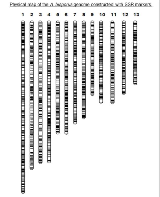
kb between adjacent markers (Table 1). Physical map of the A. bisporus genome, constructed based on the absolute positions of the SSR loci along the 13 chromosomes, is displayed in Figure 1. Data regarding the physical map (simple sequence repeat motifs, amplification primers for each motif and absolute marker positions on each chromosome) are provided in Excel format as ‘Electronic Supplementary Material’. Figure 1 displays the homogeneity of marker distribution along physical chromosomes.There is a single report on genome wide simple sequence repeat identification in button mushroom, where the previous, unrepaired version of the A. bisporus genome assembly was surveyed for simple sequence repeat loci (Foulongne-Oriol et al., 2013). While some chromosomal segments were mislocated in the assembly and later corrected (Sonnenberg et al., 2016), the study provided valuable information regarding repeat abundance in the A. bisporus genome. On the other hand, SSR loci were not converted to PCR markers in the study. Foulongne-Oriol et al. (2013) reported an SSR density of 7.4 kb/marker interval with 4062 microsatellite loci, which indicate similar overall SSR abundance values with the present study. In addition, in concordance with the findings of the present research, Foulongne-Oriol et al. (2013) reported the highest and lowest SSR densities for chromosomes 13 and 2, respectively.
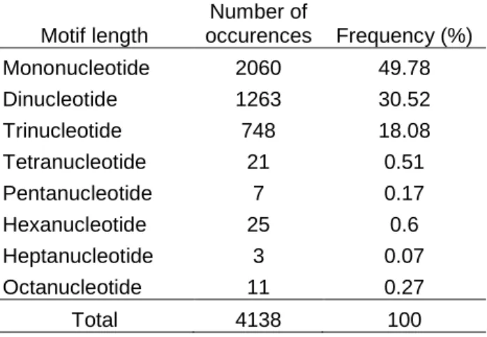
In the present research, among the 4138 SSR loci identified in the A. bisporus genome, mononucleotide repeats were predominant and represented 49.78% of the total number of SSRs (Table 2). Because mononucleotide repeats do not exert a functional constraint on most genomic sequences (introns and intergenic sequences), it is likely that SSR searches in genomes will yield mononucleotide SSRs as the most abundant motif type (Gu et al., 2010). In contrast, when SSR searches are restricted to coding sequences, it is reasonable to anticipate trinucleotide repeats as the predominant SSR type, since repeats that cause frameshift mutations are likely to get eliminated (Celik et al., 2014). According to the results of our SSR survey, dinucleotide motifs constituted the second most abundant simple sequence repeat type in the A. bisporus genome, corresponding to 30.52% of the identified SSRs (Table 2). The sum of mono-, di- and
trinucleotide SSRs was 4071, representing 98.38% of the total number of SSRs. Thus, simple sequence repeats in the A. bisporus genome were mainly iterations of one to three nucleotides and repeats of motifs longer than trinucleotide represented only 1.62% of the identified SSRs (Table 2). Our results were in agreement with Foulongne-Oriol et al. (2013), who reported a similar pattern of SSR motif abundance in the A. bisporus genome. In agreement with the relevant literature regarding genome wide SSR surveys (Uncu et al. 2015b), majority of the mononucleotide repeats were stretches of A/T nucleotides and constituted 46.45% of the identified SSRs. The second most abundant repeat motif was TC/GA with a relative abundance of 8.72% among A. bisporus genomic SSRs (Table 3).
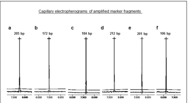
Marker design and experimental validation Marker design through GMATA software was performed on the 4138 recently identified SSR loci. Simple sequence repeats that were located on chromosomal ends were not suitable for primer design, therefore eliminated by the marker design algorithm. As a result, a total of 3943 primer pairs that amplify SSR fragments from the A. bisporus genome were generated. Marker distribution over the 13 A. bisporus chromosomes is displayed in Table 1. Amplificability of the newly developed markers was verified in silico by e-mapping. Primer sequences, repeat motifs and genomic locations of the entire set of markers (3943 markers) are provided in Excel format as ‘Electronic Supplementary Material’. Amplificability of a total of 39 randomly selected marker loci (three SSR markers per chromosome) was experimentally evaluated in order to confirm the results obtained by e-mapping. SSR fragments were visualized in high resolution with a capillary electrophoresis system. Capillary electropherograms displaying a representative set of six SSR markers is provided in Figure 2. As a result, the entire set of tested SSR markers yielded amplification products, as expected. In addition, the experimentally obtained product sizes matched those calculated by the e-mapping algorithm. Thus, it was valuable to experimentally evaluate the newly designed markers and confirm the results obtained by primer design and e-mapping.
32
Figure 1. Physical map of the A. bisporus genome constructed with SSR markers. Numbers refer to A.
bisporus chromosomes. Black bars represent simple sequence repeat loci. Primer and genomic location
information for each SSR locus is provided in ‘Electronic Supplementary Material’.
33
Chromosome Located SSRs SSR density (kb) SSR markera
1 439 8.61047836 422 2 355 9.126760563 346 3 387 8.087855297 370 4 393 7.938931298 377 5 310 8 299 6 332 7.469879518 317 7 285 7.824561404 270 8 328 6.585365854 311 9 268 6.044776119 249 10 233 7.682403433 222 11 274 6.496350365 260 12 262 6.030534351 253 13 273 5.164835165 247
aNumber of SSR markers designed per chromosome.
Table 2. Simple sequence repeat (SSR) types in A. bisporus genome.
Motif length Number of occurences Frequency (%) Mononucleotide 2060 49.78 Dinucleotide 1263 30.52 Trinucleotide 748 18.08 Tetranucleotide 21 0.51 Pentanucleotide 7 0.17 Hexanucleotide 25 0.6 Heptanucleotide 3 0.07 Octanucleotide 11 0.27 Total 4138 100
Table 3. Most abundant SSR motifs.
SSR motif Number of SSRs Motif frequencya (%)
A/T 1922 46.45
TC/GA 361 8.72
CT/AG 233 5.63
AT 209 5.05
34
The first tandem repeat sequence was cloned from the A. bisporus genome in 2000 (Barroso et al., 2000), followed by the development of a set of 33 SSR markers (Foulongne-Oriol et al 2009). The high abundance of simple sequence repeats in the A. bisporus genome was displayed later by Foulongne-Oriol et al. (2013), yet, the work did not involve marker design but introduced repeat bearing loci as candidates for future marker development. Thus, a database of A. bisporus specific markers with highgenome coverage has not been reported before the present study. As a result of the present work, it was feasible to develop a total of 3943 novel, sequence-specific SSR markers that provide a high-resolution coverage of the A. bisporus genome. Absolute genomic positions and primer sequences of the 3943 novel SSR markers are available for researchers who conduct molecular genetic research in the important mushroom species.
Figure 2. Capillary electropherograms displaying the amplification profiles of six A. bisporus SSR markers. Marker information is as follows: a, Chromosome 1, MK111, mononucleotide SSR (A/T); b, Chromosome 1, MK193, dinucleotide SSR (AT); c, Chromosome 5, MK139, mononucleotide SSR (A/T); d, Chromosome 5, MK72, mononucleotide SSR (A/T); e, Chromosome 5, MK50, trinucleotide SSR (ATG/CAT); f, Chromosome 12, MK14, dinucleotide SSR (TG/CA).
References
Celik I, Gultekin V., Allmer J., Doganlar S., Frary A., Development of genomic simple sequence repeat markers in opium poppy by next-generation sequencing, Molecular Breeding, 34(2), 323-334 (2014).
Barroso G., Sonnenberg A. S. M., Van Griensven L. J. L. D., Labarѐre J., Molecular cloning of a widely distributed microsatellite core
sequence from the cultivated mushroom Agaricus bisporus, Fungal Genetics and Biology, 31, 115-123 (2000).
Berendsen R. L., Baars J. J. P., Kalkhove S. I. C., Lugones L. G., Wösten H. A. B., Bakker P. A. H. M., Pathogen profile Lecanicillium
fungicola: causal agent of dry bubble disease in white-button mushroom, Molecular Plant Pathology, 11(5), 585-595 (2010).
Foulongne-Oriol M., Spataro C., Savo,e J. M., Novel microsatellite markers suitable for genetic studies in the white button mushroom
Agaricus bisporus, Applied Microbiology and Biotechnology, 84, 1125-1135 (2009).
Foulongne-Oriol M., Rodier A., Rousseau T., Savoie J. M., QTL mapping of yield-related components and oligogenic control of the cap
colour in the button mushroom Agaricus bisporus, Applied and Environmental Microbiology, 78(7), 2422-2434 (2012).
Foulongne-Oriol M., Murat C., Castanera R., Ramírez L., Sonnenberg A. S. M., Genome-wide survey of repetitive DNA elements in the
button mushroom Agaricus bisporus, Fungal Genetics and Biology, 55, 6-21 (2013).
Gao W., Baars J. J. P., Maliepaard C., Visser R. G. F., Zhang J., Sonnenberg A. S. M., Multi-trait QTL analysis for agronomic and quality
35
Gu T., Tan S., Gou X., Araki H., Tian D., Avoidance of long mononucleotide repeats in codon pair usage, Genetics, 186, 1077-1084 (2010).
Heneghan M. N., Burns C., Costa A. M. S. B., Burton K. S., Challen M. P., Bailey M., Foster G. D., Functional analysis of Agaricus
bisporus serine proteinase 1 reveals roles in utilization of humic rich substrates and adaptation to the leaf-litter ecological niche,
Environmental Microbiology, 18(12), 4687-4696 (2016).
Jeong S. C., Jeong Y. T., Yang B. K., Islam R., Koyyalamudi S. R., Pang G., Cho K. Y., Song C. H., White button mushroom (Agricus
bisporus) lowers blood glucose and cholesterol levels in diabetic and hypercholesterolemic rats, Nutrition Research, 30, 49-56
(2010).
Jones, N., Ougham H., Thomas H., Pasakinskiene I., Markers and mapping revisited: Finding your gene, New Phytologist, 183, 935-966 (2009).
Liu J., Jia L., Kan J., Jin C. H., In vitro and in vivo antioxidant activity of ethanolic extract of white button mushroom (Agaricus bisporus), Food and Chemical Toxicology, 51, 310-316 (2013).
Loftus M.G., Moore D., Elliott T. J., DNA polymorphisms in commercial and wild strains of the cultivated mushroom, Agaricus bisporus, Theoretical and Applied Genetics, 76, 712-718 (1988).
Morin E., Kohler A., Baker A. R., Foulongne-Oriol M., Lombard V., Nagy L. G., et al. Genome sequence of the button mushroom Agaricus
bisporus reveals mechanisms governing adaptation to a humic-rich ecological niche, Proceedings of the National Academy of
Sciences, 109(43), 17501-17506 (2012).
Samuels G. J., Dodd S. L., Gams W., Castlebury L. A., Petrini O., Trichoderma species associated with the green mold epidemic of
commercially grown Agaricus bisporus, Mycologia, 94(1), 146-170 (2002).
Schuler G. D., Sequence mapping by electronic PCR, Genome Research, 7, 541-550 (1997).
Sokovic M., Van Griensven L. J. L. D., Antimicrobial activity of essential oils and their components against the three major pathogens of
the cultivated button mushroom, Agaricus bisporus, European Journal of Plant Pathology, 116, 211-224 (2006).
Sonnenberg A. S. M., Gao W., Lavrijssen B., Hendrickx P., Sedaghat-Tellgerd N., Foulongne-Oriol M., et al. A detailed analysis of the
recombination landscape of the button mushroom Agaricus bisporus var. bisporus, Fungal Genetics and Biology, 93, 35-45 (2016).
Uncu A. O, Uncu A. T., Celık, İ, Doganlar S., Frary A., A primer to molecular phylogenetic analysis in plants, Critical Reviews in Plant Sciences, 34, 454-468 (2015a).
Uncu A. O., Gultekin V., Allmer J., Frary A., Doganlar S., Genomic simple sequence repeat markers reveal patterns of genetic relatedness
and diversity in sesame, The Plant Genome, 8, doi: 10.3835/plantgenome2014.11.0087 (2015b).
Voorrips R. E., MapChart: software for the graphical presentation of linkage maps and QTLs, Journal of Heredity, 93, 77-78 (2002). Wang X., Wang L., GMATA: An integrated software package for genome-scale SSR mining, marker development and viewing, Frontiers