Accepted: 2016.01.08 Published: 2016.02.10
2455
—
7
36
Melatonin Prevents Mitochondrial Damage
Induced by Doxorubicin in Mouse Fibroblasts
Through Ampk-Ppar Gamma-Dependent
Mechanisms
ABCDEFG 1
Celal Guven*
ABCDEF 2
Eylem Taskin*
ABCDEF 3
Handan Akcakaya
* These authors contributed equally
Corresponding Authors: Celal Guven, e-mail: [email protected]; Handan Akcakaya, e-mail: [email protected]
Source of support: This work was supported by self-financing
Background: Doxorubicin (brand name: Adriamycin®) is used to treat solid tissue cancer but it also affects noncancerous
tis-sues. Its mechanism of cytotoxicity is probably related to increased oxidation, mitochondrial dysfunction, and apoptosis. Melatonin is reported to have antiapoptotic and antioxidative effects. The aim of this study was to determine whether melatonin would counteract in vitro cytotoxicity of doxorubicin in mouse fibroblasts and determine the pathway of its action against doxorubicin-induced apoptosis.
Material/Methods: We measured markers of apoptosis (cytochrome-c, mitochondrial membrane potential, and apoptotic cell num-ber) and oxidative stress (total oxidant and antioxidant status) and calculated oxidant stress index in 4 groups of fibroblasts: controls, melatonin-treated, doxorubicin-treated, and fibroblasts concomittantly treated with a combination of melatonin and doxorubicin.
Results: Melatonin given with doxorubicin succesfully countered apoptosis generated by doxorubicin alone, which points to its potential as a protective agent against cell death in doxorubicin chemotherapy. This also implies that pa-tients should be receiving doxorubicin treatment when their physiological level of melatonin is at its highest, which is early in the morning.
Conclusions: This physiological level may not be high enough to overcome doxorubicin-induced oxidative stress, but adju-vant melatonin treatment may improve quality of life. Further research is needed to verify our findings.
MeSH Keywords: Apoptosis • Doxorubicin • Melatonin • Membrane Potential, Mitochondrial
Full-text PDF: http://www.medscimonit.com/abstract/index/idArt/897114 Authors’ Contribution: Study Design A Data Collection B Statistical Analysis C Data Interpretation D Manuscript Preparation E Literature Search F Funds Collection G
1 Department of Biophysics, Faculty of Medicine, University of Adiyaman, Adiyaman, Turkey
2 Department of Physiotherapy and Rehabilitation, School of Health Sciences, Istanbul Bilim University, Istanbul, Turkey
3 Department of Biophysics, Faculty of Medicine, University of Istanbul, Istanbul, Turkey
Background
Doxorubicin (DOX) is an efficient anthracycline group chemo-therapy drug used to treat hematologic and solid tumors [1], as it induces oxidative stress, inhibits topoisomerase II, and eventually leads to cell death, mainly by apoptosis [1–5]. It also inhibits cell propagation [2]. DOX causes alteration of mi-tochondrial function and structure due to DOX accumulation in mitochondria [6–10]. It has been suggested that the unde-sired effect of DOX is related to increase oxidative stress in the cell, triggering apoptosis [9,11,12].
There are strategies to attenuate the toxic effects of doxorubi-cin, such as dose regulation or combined therapy with antioxi-dants [13]. However, no effective combination has been found so far. One of the antiapoptotic agents with some promise is melatonin. Its antiapoptotic action has been evidenced in the brain, kidney, pancreas, and liver [14], but findings to the con-trary have also been reported in vitro [15]). Because it is a small lipophilic molecule, it is distributed throughout the cell, with sig-nificant deposition in the mitochondria, where it can attenuate ischemia-reperfusion and indomethacin-induced damage [16]. It can also modulate mitochondrial function and homeostasis. Aziriova [17] reported that melatonin even reversed anxiety in-duced by doxorubicin in rats. One of its unique properties is that its metabolites also have antioxidative effects, even at low concentrations [18]. Furthermore, melatonin has low toxicity, which implies that it may be used safely in patient therapy [19]. Several studies have shown that melatonin can counteract doxo-rubicin-induced cytotoxicity [18,19] but they have not clarified the mechanism of its action. Therefore, with our study we want-ed to determine if the mechanism of melatonin action against doxorubicin cytotoxicity was via its antioxidative and/or anti-apoptotic pathway. We tested it in the mouse embryonic fibro-blast cell line (NIH 3T3) because fibrofibro-blasts, which compose 70% of the heart as nanocardiomyocytes [20], are very respon-sive to chemical, electric, and mechanical signals, and chang-es made to thchang-ese signals can affect other cardiac cells [21].
Material and Methods
Experimental design of studyFour groups of NIH3T3 cells were created: the control (Con) group received DMEM alone; the melatonin (MEL) group re-ceived melatonin in the dose of 1 µM for 24 h; the doxorubicin (DOX) group received doxorubicin hydrochloride (Adriblastina vial 10 mg, Pharmacia) in the dose of 2.6 µM for 24 h, and the MEL+DOX group received a combination of the doses corre-sponding to the MEL and DOX alone group. The dose of mel-atonin may be the key to achieving antiapoptotic effects. At
high concentrations, melatonin activates the mitochondri-al pathway by affecting the Bax/Bcl protein bmitochondri-alance and the expressions of caspase-9 and caspase-3 in pancreatic carci-noma cell line [22]. At low concentrations, however, HSP27, HSP70, and HSP90 induction prevent the activation of cas-pase-3 [22]. One study reported that melatonin might inhibit cell proliferation at physiological (nanomolar and lower) and pharmacological concentrations (10 µM) but not at concentra-tions in between [23]. We therefore opted for 1 µM, which is much higher than the physiological (nanomolar) plasma con-centrations. Furthermore, we opted to test melatonin effects when given with doxorubicin at the same time, thus extend-ing an earlier study by Oz et al. [24], who reported that mel-atonin was less effective against doxorubicin when given as pretreatment than when given after doxorubicin.
To determine the effects of these treatments on oxidation and apoptosis, we made the measurements described below.
Cell culture
NIH3T3 cells were grown in poly-L-lysine-coated flasks (~2.0–2.5×10⁶ cells/mL) in DMEM medium supplemented
with 10% heat-inactivated fetal bovine serum in a humidified incubator with 5% CO2/95% air mixture at 37°C.
Cell viability
NIH3T3 cells were seeded into 24-well culture plates at 1×10⁶
cells/well. Viability was assessed by trypan blue exclusion us-ing a Vi-Cell XR cell viability analyzer (Beckman Coulter, Brea, CA), which is an automated hemocytometer. Basically, cell sam-ples were mixed 1:1 with trypan blue, and 50 images were tak-en for calculation of cell number.
Total oxidant status
Total oxidant status (TOS) was measured with a BioTek µQuant microplate spectrophotometer using a Rel Assay kit (Cat No: RL0024; Gaziantep, Turkey) according to the manufacturer’s in-structions. The results are expressed as a μmol H2O2 Equiv./L.
Total antioxidant status
Total antioxidant status (TAS) was measured with a BioTek µQuant microplate spectrophotometer using a Rel Assay kit (Cat No: RL0017; Gaziantep, Turkey) according to the manufacturer’s instructions. The results are expressed as mmol Trolox Equiv./L.
Oxidative stress index
Oxidative stress index (OSI) is the ratio between TOS and TAS levels measured in 6 samples per group.
Protein levels
The mechanism of apoptosis in fibroblasts was established by determining protein levels. To prepare cell lysates, NIH3T3 cells were centrifuged at 3000 rpm for 8 min, washed several times with PBS, resuspended in RIPA buffer (Cat. No. sc-24948; Santa Cruz Biotechnology), and incubated on ice for 10 min. The lysates were stored at –20°C until used. Protein levels were measured using a Qubit fluorometer and quantitation assays: 100 µg protein sample was mixed with 4×SDS load-ing buffer and then separated on a 4–12% Bis-Tris gel. After running, proteins were transferred to a PVDF membrane. We used primary antibodies against the following proteins: AIF, b-actin, ERK ½, SAPK/JNK (Cell Signaling), PPAR-g, AMPK, and cytochrome-c (Abcam, Cambridge, UK). Then we used alkaline phosphatase-conjugated secondary antibodies (anti-mouse and anti-rabbit 1/3000). Signals were detected using the NCIP/BNP kit (Cat. No. ES0006; Millipore, Boston, MA, USA).
F-actin filaments
We looked for actin filaments because doxorubicin has been reported to inhibit cardiac genes, including a-actin, troponin, and myosin light chain-2 [25,26]. NIH 3T3 fibroblasts grown on glass coverslips (~80 000 cells) were left unstimulated (con-trol) or stimulated with 1 µM melatonin, 2.6 µM doxorubicin, and melatonin with doxorubicin. They were washed with PBS and fixed in 5% paraformaldehyde at room temperature for 60 min. Fixed cells were permeabilized in 0.1% (vol/vol) Triton X-100 in PBS at room temperature for 10 min and blocked in 5% BSA in PBS at room temperature for 60 min. Actin fila-ments were visualized using Alexa fluor 595 phalloidin, and nuclei with 4’’,6-diamidino-2-phenylindole dichloride (DAPI) (Invitro Molecular Probes, OR, USA). All images were obtained on an Olympus BX51 microscope equipped with a DP72 cam-era controlled by Olympus DP2-TWAIN software.
Mitochondrial membrane potential measurement
Mitochondria are targets of doxorubicin, and play an impor-tant role in initiating apoptosis. NIH 3T3 cells (~80 000) were harvested and allowed to attach on coverslips over the night. Then the cells were exposed to melatonin, DOX, and melatonin with DOX for 24 h. Mitochondrial membrane potential (MMP) was measured using a Sigma-Aldrich kit, Cat. No. CS0390 ac-cording the manufacturer’s instructions. Briefly, the cells were incubated with JC-1 dye with a 5% CO2/95% O2 air mixture at 37°C for 20 min. JC-1 dye is specific for the mitochondrial membrane. Healthy mitochondria have a high electrochemi-cal potential, and JC-1 dye crosses the membrane to form red aggregates on fluorescence. Unhealthy mitochondria with low potential prevent JC-1 dye from accumulating, and fluorescence monomers are green.
After incubation the cells were rinsed with staining buffer. The coverslips were directly observed under a fluorescence micro-scope (Olympus BX51 Micromicro-scope equipped with a DP72 cam-era, controlled by Olympus DP2-TWAIN software).
Apoptotic cell count
The cells were cultured in 6-well plates with coverslips over-night. After they were divided into groups, the coverslips were washed twice with PBS and fixed in 4% paraformaldehyde so-lution for 1 h at room temperature. The coverslips were then rinsed with PBS, incubated in permeabilization solution (0.1% Triton-X-100 in 0.1% sodium citrate, freshly prepared), and the apoptotic cells labelled with a terminal deoxynucleotidyl-transferase-mediated dUTP nick end-labeling (TUNEL) kit from Roche (Cat. No. 1684795; Mannheim, Germany) following the manufacturer’s instructions. All images were obtained on an Olympus BX51 microscope equipped with a DP72 camera and controlled with Olympus DP2-TWAIN software. The apoptot-ic (TUNEL-positive) cells were quantified using the automat-ed cell counting in ImageJ with ITCN plugin (Automatic Nuclei Counter plugin) (NIH). Summarly, images were converted to a binary image for 8-bit. Dark areas represent cells. Then, ITCN was used for cell counter for 15 images for every groups, and the total areas of groups were the same.
Statistical analysis
For statistical analysis we used Excel and the SPSS version 16.0. All results are expressed as mean ±SEM. Normal distri-bution was tested using Levene’s test. To compare the results between the groups, we used multiple analyses of variance (ANOVA), followed by a post hoc protected Tukey test. p<0.05 was considered to be significant for all data.
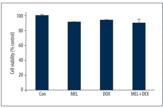
Figure 1. Cell viability across the groups. Con – control group; MEL – melatonin group; DOX – doxorubicin group; MEL+DOX – melatonin plus doxorubicin group. All data are expressed as mean ±SEM.
100 80 60 40 20 0
Con MEL DOX MEL+DEX
Cell viability (% contr
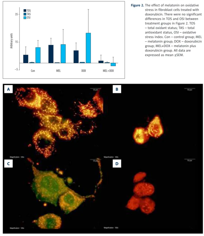
Figure 2. The effect of melatonin on oxidative stress in fibroblast cells treated with doxorubicin. There were no significant differences in TOS and OSI between treatment groups in Figure 2. TOS – total oxidant status; TAS – total antioxidant status, OSI – oxidative stress index. Con – control group; MEL – melatonin group; DOX – doxorubicin group; MEL+DOX – melatonin plus doxorubicin group. All data are expressed as mean ±SEM. 35 15 –5 Con TOS TAS OSI
MEL DOX MEL+DOX
Arbitrar y units
A
C
B
D
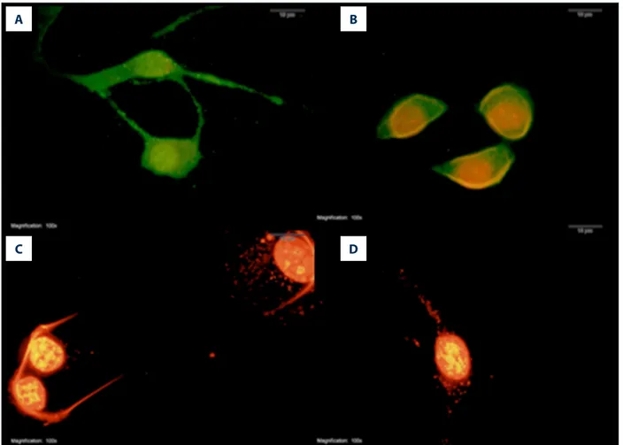
Figure 3. The effect of melatonin on mitochondrial membrane potential in fibroblast cells treated with doxorubicin. Red fluorescence represents healthy mitochondria having a high electrochemical potential and green fluorescence represents unhealthy mitochondria having a low electrochemical potential (100× magnification). Con – control group (A); MEL – melatonin group (B); DOX – doxorubicin group (C); MEL+DOX – melatonin plus doxorubicin group (D).
Results
Cell viabilityCell viability is expressed as the percentage of control cell vi-ability (taken to be 100%). We found no significant differenc-es between the groups (Figure 1).
The effect of melatonin on oxidative stress induced by doxorubicin
Figure 2 shows the effects of melatonin on oxidative stress in fibroblast cells across the groups. Even though it lowered TOS in the combination group (Mel+DOX) in respect to the DOX group (or other groups for that matter), the differences in TOS and OSI were not statistically significant. MEL groups of OSI, TOS, and TAS levels were not different from Con.
The effect of melatonin on doxorubicin-induced dissipation of mitochondrial membrane potential
The control and MEL group had normal healthy mitochondria, represented by red fluorescence (Figure 3A and B). Although DOX dissipated the MMP, resulting in green fluorescence (Figure 3C), MEL given with DOX restored the MMP (Figure 3D).
The effect of melatonin on doxorubicin-induced changes in protein levels
Cytochrome-c levels
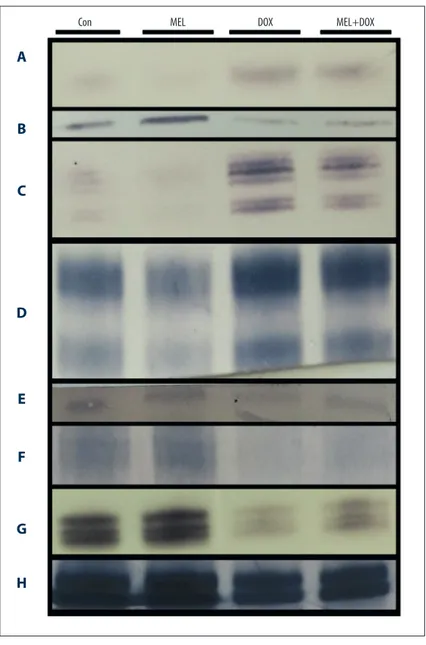
Cytochrome-c regulates the apoptosis-dependent mitochon-drial pathway. DOX increased its level but melatonin in the MEL+DOX group restored it to lower levels (Figure 4A).
Figure 4. The effect of melatonin on protein affected by oxidative stress in fibroblast cells treated with doxorubicin. Con – control group; MEL – melatonin group; DOX – doxorubicin group; MEL+DOX – melatonin plus doxorubicin group. (A) Cytochrome-c; (B) AIF – apoptosis inducing factor; (C) ERK1/2 – extracellular signal-regulated kinases; (D) SAPK/JNK – stress-activated kinases; (E) PPAR-g – peroxisome proliferator-activated receptor gamma; (F) AMPK – AMP-activated protein kinase; (G) PCNA – proliferating cell nuclear antigen; (H) b-actin.
Con MEL DOX MEL+DOX
A
C
E
G
B
D
F
H
Apoptosis-inducing factor levels
Apoptosis-inducing factor (AIF) plays a crucial role in caspase-independent apoptosis. It is located in the inner membrane of mitochondria. DOX caused its release into the cytosol (Figure 4), but MEL counteracted its release (Figure 4B).
Extracellular signal-regulated kinase expression
ERK 1 and 2 participate in the regulation of cell adhesion, cell migration, cell survival, and differentiation [27]. The DOX-treated cells showed a very low expression of both ERK 1 and 2. Co-treatment with MEL recovered their expressions (Figure 4C).
Stress-activated protein kinases levels
SAPK participates in cellular stress response, including differ-entiation and proliferation [28]. DOX lowered its level while co-treatment with MEL restored it (Figure 4D).
Peroxisome proliferator-activated receptor gamma protein levels
Peroxisome proliferator-activated receptor gamma (PPAR-g) plays a crucial role in fatty acid and glucose metabolism reg-ulation. DOX lowered PPAR-g levels, but MEL+DOX attenuat-ed its effect (Figure 4E).
AMP-activated protein kinase levels
AMPK maintains cell energy homeostasis by balancing ATP production when energy changes. DOX lowered the AMPK level, but it was improved in the MEL+DOX group (Figure 4F).
Proliferating cell nuclear antigen level
Proliferating cell nuclear antigen (PCNA) assists in DNA rep-lication and is a common marker of proliferation. As expect-ed, co-treatment with MEL countered the PCNA-lowering ef-fect of DOX (Figure 4G).
A
C
B
D
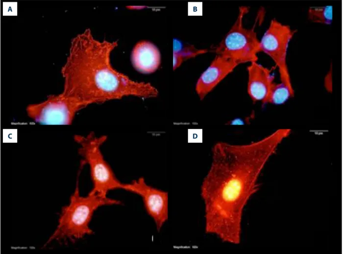
Figure 5. The effect of melatonin on F-actin distribution in fibroblasts treated with doxorubicin (100x magnification). Con – control group (A); MEL – melatonin group (B); DOX – doxorubicin group (C); MEL+DOX – melatonin plus doxorubicin group (D).
The effect of melatonin on doxorubicin-modulated distribution of F-actin
Figure 5 shows stained F-actin (red) and nucleus (blue) in NIH 3T3. DOX increased the formation of filopodia (Figure 5C), but co-treatment with MEL counteracted the stress, restoring F-actin to normal (Figure 5D). Only melatonin treatment also increased filopodia formation in the cells (Figure 5B).
The effect of melatonin on doxorubicin-induced apoptosis
DOX significantly increased the number of apoptotic cells to 123±37 vs. control (23±6), whereas co-treatment with MEL de-creased it to 30±6. Only MEL treated cells were not altered to apoptotic cells (22±5; Figures 6, 7).
Discussion
Our results have confirmed our hypothesis that melato-nin would counteract the adverse effects of doxorubicin: it
A
C
B
D
Figure 6. Apoptosis in fibroblasts treated with melatonin and doxorubicin (100× magnification). Green fluorescence represents healthier cells than red fluorescence (100× magnification). Con – control group (A); MEL – melatonin group (B); DOX – doxorubicin group (C); MEL+DOX – melatonin plus doxorubicin group (D).
Figure 7. Apoptotic cell counts in fibroblasts treated with melatonin and doxorubicin. Con – control group; MEL – melatonin group; DOX – doxorubicin group; MEL+DOX – melatonin plus doxorubicin group. a p<0.05
vs. Con, b p<0.05 vs. MEL, c p<0.05 vs. DOX. All data are
expressed as mean ±SEM. 150 100 50 0 Con MEL a,b c MEL+DOX DOX
References:
1. Coldwell K, Cutts SM, Ognibene TJ et al: Detection of adriamycin-DNA ad-ducts by accelerator mass spectrometry. Methods Mol Biol, 2010; 613: 103–18
2. Sauter KA, Magun EA, Iordanov MS, Magun BE: ZAK is required for doxo-rubicin, a novel ribotoxic stressor, to induce SAPK activation and apopto-sis in HaCaT cells. Cancer Biol Ther, 2010; 10(3): 258–66
3. Arif IS, Hooper CL, Greco F et al: Increasing doxorubicin activity against breast cancer cells using PPARgamma-ligands and by exploiting circadian rhythms. Br J Pharmacol, 2013; 169(5): 1178–88
4. Ta HQ, Thomas KS, Schrecengost RS, Bouton AH: A novel association be-tween p130Cas and resistance to the chemotherapeutic drug adriamycin in human breast cancer cells. Cancer Res, 2008; 68(21): 8796–804 5. Cao B, Li M, Zha W et al: Metabolomic approach to evaluating adriamycin
pharmacodynamics and resistance in breast cancer cells. Metabolomics, 2013; 9(5): 960–73
6. Dursun N, Taskin E, Yerer Aycan MB, Sahin L: Selenium-mediated cardio-protection against adriamycin-induced mitochondrial damage. Drug Chem Toxicol, 2011; 34(2): 199–207
7. Ozdogan K, Taskin E, Dursun N: Protective effect of carnosine on adriamy-cin-induced oxidative heart damage in rats. Anadolu Kardiyol Derg, 2011; 11(1): 3–10
8. Taskin E, Dursun N: The protection of selenium on adriamycin-induced mi-tochondrial damage in rat. Biol Trace Elem Res, 2012; 147(1–3): 165–71 9. Taskin E, Kindap EK, Ozdogan K et al: Acute adriamycin-induced
cardiotox-icity is exacerbated by angiotension II. Cytotechnology, 2016; 68(1): 33–43 10. Taskin E, Ozdogan K, Kunduz Kindap E, Dursun N: The restoration of kidney
mitochondria function by inhibition of angiotensin-II production in rats with acute adriamycin-induced nephrotoxicity. Ren Fail, 2014; 36(4): 606–12 11. Taskin E, Dursun N: Recovery of adriamycin induced mitochondrial
dysfunc-tion in liver by selenium. Cytotechnology, 2015; 67(6): 977–86 12. Yapislar H, Taskin E, Ozdas S et al: Counteraction of apoptotic and
inflam-matory effects of adriamycin in the liver cell culture by clinopitolite. Biol Trace Elem Res, 2015 [Epub ahead of print]
13. Quiles JL, Huertas JR, Battino M et al: Antioxidant nutrients and adriamy-cin toxicity. Toxicology, 2002; 180(1): 79–95
preserved mitochondrial membrane potential, and restored cytochrome-c and AIF levels as mechanisms of apoptosis and, therefore, decreased apoptotic cell count.
For the antioxidative action, our dosage was less efficient. An ear-lier study reported that oxidative stress induced by doxorubicin was higher in rats exposed to 24-h light than in rats kept in 24-h dark [29]. This finding suggests that melatonin could reverse oxi-dative stress induced by doxorubicin, even at physiological levels. However, our melatonin dose of 1 µM did not have a significant effect on OSI and has confirmed the findings by Santofimia-Castaño et al. [30]. Instead, it showed a dominant antiapoptotic effect. A previous study indicated that MEL cotreated with DOX in rats with hepatoma did not decrease the anticancer effect of DOX, and even decreased the effect of DOX on noncancerous tissue (e.g., heart) by decreasing apoptotic cell numbers [31]. In our study, melatonin restored MMP reduced by doxorubicin. This result is consistent with earlier reports [32,33]. Melatonin stimulates the biogenesis of mitochondria and upregulates PPAR-gamma. It has been reported to enhance mitochondri-al function and quantity [34]. In our study, doxorubicin low-ered the PPAR-gamma protein level, which plays an impor-tant role in regulating mitochondrial energy production [35]. Wang et al. [36] reported that one of the PPAR-gamma agonists attenuated apoptosis by inhibiting NF-kB and MAPK P38 sig-naling pathways. A similar antiapoptotic mechanism through PPAR-gamma was reported by Mahmoud-Awny et al. [37]. Currently, however, less is known about the modulation of apop-tosis by melatonin in fibroblasts exposed to doxorubicin. In our study, melatonin given with doxorubicin increased the PPAR-gamma and AMPK levels, which suggests that there is a link be-tween these 2 proteins [38]. Also, AMPK is related to circadian rythm [39]. To the best of our knowledge, our study is the first to suggest a trimodal relationship between AMPK-PPAR-gamma
and melatonin. Furthermore, we found a positive interaction between AMPK and ERK1/2. An earlier study reported that 1 µM of melatonin elevated ERK1/2 and SAP/JNK levels [30]. The observed changes could improve PCNA levels and cell prolif-eration of fibroblasts. All this suggests that melatonin coun-ters doxorubicin’s cytotoxicity by increasing cell proliferation and inhibiting apoptosis. The AMPK-PPARg pathway might be critical in the regulation of apoptosis induced by doxorubicin. Apoptosis is one of the most important pathways of doxoru-bicin toxicity. This is why we felt it was worth seeking the ex-act mechanism of apoptosis induced by doxorubicin, as finding this mechanism could lead to new cardioprotective chemo-therapy strategies.
However, our study has its limitations. First of all, the mela-tonin concentration we used is higher than the physiological level [34]. The other limitation is related to this study being
in vitro, as we cannot tell how melatonin will behave in living
organisms until in vivo studies are performed. Lastly, the pro-tein level of melatonin’s receptors was not evaluated. We hope that future studies will overcome these limitations and inves-tigate the relationship between melatonin and AMPK-PPARg in doxorubicin cytotoxicity even more closely.
Conclusions
Our study has provided evidence that melatonin has potential as an adjuvant in doxorubicin chemotherapy, which could at-tenuate its adverse effects without reducing its effectiveness in cancer cells. If proved efficient, this could result in new car-dioprotective strategies that may involve preserving the pa-tient’s circadian rhythm. Future in vivo studies will be necessary to test the therapeutic value of melatonin administration with doxorubicin to determine how it behaves in living organisms.
14. Chen Z, Chua CC, Gao J et al: Prevention of ischemia/reperfusion-induced cardiac apoptosis and injury by melatonin is independent of glutathione peroxdiase 1. J Pineal Res, 2009; 46(2): 235–41
15. Carbajo-Pescador S, Steinmetz C, Kashyap A et al: Melatonin induces tran-scriptional regulation of Bim by FoxO3a in HepG2 cells. Br J Cancer, 2013; 108(2): 442–49
16. Brzozowska I, Ptak-Belowska A, Pawlik M et al: Mucosal strengthening ac-tivity of central and peripheral melatonin in the mechanism of gastric de-fense. J Physiol Pharmacol, 2009; 60(Suppl.7): 47–56
17. Aziriova S, Repova Bednarova K, Krajcirovicova K et al: Doxorubicin-induced behavioral disturbances in rats: protective effect of melatonin and capto-pril. Pharmacol Biochem Behav, 2014; 124: 284–89
18. Govender J, Loos B, Engelbrecht AM: Melatonin: A protective role against doxorubicin-induced cardiotoxicity. Future Oncol, 2015; 11(14): 2003–6 19. Govender J, Loos B, Marais E, Engelbrecht AM: Mitochondrial catastrophe
during doxorubicin-induced cardiotoxicity: A review of the protective role of melatonin. J Pineal Res, 2014; 57(4): 367–80
20. Camelliti P, Borg TK, Kohl P: Structural and functional characterisation of cardiac fibroblasts. Cardiovasc Res, 2005; 65(1): 40–51
21. Souders CA, Bowers SL, Baudino TA: Cardiac fibroblast: the renaissance cell. Circ Res, 2009; 105(12): 1164–76
22. Jaworek J, Leja-Szpak A: Melatonin influences pancreatic cancerogenesis. Histol Histopathol, 2014; 29(4): 423–31
23. Kim TK, Kleszczynski K, Janjetovic Z et al: Metabolism of melatonin and bi-ological activity of intermediates of melatoninergic pathway in human skin cells. FASEB J, 2013; 27(7): 2742–55
24. Oz E, Erbas D, Surucu HS, Duzgun E: Prevention of doxorubicin-induced car-diotoxicity by melatonin. Mol Cell Biochem, 2006; 282(1–2): 31–37 25. Boucek RJ Jr., Miracle A, Anderson M et al: Persistent effects of
doxorubi-cin on cardiac gene expression. J Mol Cell Cardiol, 1999; 31(8): 1435–46 26. Nakamura T, Ueda Y, Juan Y et al: Fas-mediated apoptosis in
adriamycin-induced cardiomyopathy in rats: In vivo study. Circulation, 2000; 102(5): 572–78
27. Roskoski R Jr: ERK1/2 MAP kinases: structure, function, and regulation. Pharmacol Res, 2012; 66(2): 105–43
28. Paul A, Wilson S, Belham CM et al: Stress-activated protein kinases: Activation, regulation and function. Cell Signal, 1997; 9(6): 403–10 29. Escribano BM, Diaz-Moreno A, Tasset I, Tunez I: Impact of light/dark cycle
patterns on oxidative stress in an adriamycin-induced nephropathy mod-el in rats. PLoS One, 2014; 9(5): e97713
30. Santofimia-Castano P, Garcia-Sanchez L, Ruy DC et al: Melatonin induc-es calcium mobilization and influencinduc-es cell proliferation independently of MT1/MT2 receptor activation in rat pancreatic stellate cells. Cell Biol Toxicol, 2015; 31(2): 95–110
31. Dziegiel P, Podhorska-Okolow M, Zabel M: Melatonin: Adjuvant therapy of malignant tumors. Med Sci Monit, 2008; 14(5): RA64–70
32. Song N, Kim AJ, Kim HJ et al: Melatonin suppresses doxorubicin-induced premature senescence of A549 lung cancer cells by ameliorating mitochon-drial dysfunction. J Pineal Res, 2012; 53(4): 335–43
33. Teodoro BG, Baraldi FG, Sampaio IH et al: Melatonin prevents mitochon-drial dysfunction and insulin resistance in rat skeletal muscle. J Pineal Res, 2014; 57(2): 155–67
34. Kato H, Tanaka G, Masuda S et al: Melatonin promotes adipogenesis and mitochondrial biogenesis in 3T3-L1 preadipocytes. J Pineal Res, 2015; 59(2): 267–75
35. Guo J, Guo Q, Fang H et al: Cardioprotection against doxorubicin by metallo-thionein Is associated with preservation of mitochondrial biogenesis in-volving PGC-1alpha pathway. Eur J Pharmacol, 2014; 737: 117–24 36. Wang R, Yan Z, Wu X et al: Rosiglitazone attenuates renal injury caused by
hyperlipidemic pancreatitis. Int J Clin Exp Pathol, 2015; 8(5): 4332–43 37. Mahmoud-Awny M, Attia AS, Abd-Ellah MF, El-Abhar HS: Mangiferin
mit-igates gastric ulcer in ischemia/reperfused rats: Involvement of PPAR-gamma, NF-kappaB and Nrf2/HO-1 signaling pathways. PLoS One, 2015; 10(7): e0132497
38. Cheang WS, Tian XY, Wong WT et al: Metformin protects endothelial func-tion in diet-induced obese mice by inhibifunc-tion of endoplasmic reticulum stress through 5’ adenosine monophosphate-activated protein kinase-per-oxisome proliferator-activated receptor delta pathway. Arterioscler Thromb Vasc Biol, 2014; 34(4): 830–36
39. Menassol JB, Tautou C, Collet A et al: The effect of an intracerebroventric-ular injection of metformin or AICAR on the plasma concentrations of mel-atonin in the ewe: potential involvement of AMPK? BMC Neurosci, 2011; 12: 76