See discussions, stats, and author profiles for this publication at: https://www.researchgate.net/publication/301535768
Characterization of tyrosinase enzyme from native Bacillus megaterium SP.
STRAIN M36
Article in Journal of microbiology, biotechnology and food sciences · April 2016
DOI: 10.15414/jmbfs.2016.5.5.465-469 CITATIONS 0 READS 620 2 authors: Ebrahim Valipour Bülent Ecevit Üniversitesi 29PUBLICATIONS 36CITATIONS SEE PROFILE Burhan Arikan Cukurova University 40PUBLICATIONS 969CITATIONS SEE PROFILE
CHARACTERIZATION OF TYROSINASE ENZYME FROM NATIVE BACILLUS MEGATERIUM SP. STRAIN M36
Ebrahim Valipour, Burhan Arikan
Address(es): Ebrahim Valipour PhD.,
Molecular microbiology lab, Biotechnology Department, Institute of Basic and Applied Sciences, Cukuruva University, 01330, Adana, Turkey. Fax: 0090 3223386070, Tel No: 00905367304074.
*Corresponding author: [email protected] ABSTRACT
Keywords: Melanin, monophenolase, diphenolase, TLC
INTRODUCTION
Tyrosinase is a type 3 copper-containing enzyme that has been found widely distributed in microorganisms, plants and animals (Claus and Decker, 2006). Tyrosinase catalyses the hydroxylation of monophenol to o-diphenol (monophenolase or cresolase activity) and the oxidation of diphenol to o-quinones (diphenolase or catecholase activity). O-o-quinones are converted in to melanin by using nonenzymatic steps and molecular oxigene (Decker and
Tuczek, 2000). Howard et al., in 1948, elucidated the biosynthetic pathway for
melanin formation by tyrosinase enzyme. In mammals, tyrosinase catalyzes the biosynthesis of melanin pigments, which contributes to a fundamental part of the skin protection against UV radiation. It is also related to the browning reactions of fruit and vegetables (Seo et al., 2003). Tyrosinases have several biotechnological applications relying on the ability of the enzymes to oxidize both small phenolic molecules and protein-associated phenolic groups, i.e. the side chain of the amino acid tyrosine. Tyrosinase enzyme has very important role in bioremediation (Marino et al., 201128), production of L-DOPA, the preferred drug for treatment of Parkinson's disease and other antioxidants (having crucial application in medical field) (Xu et al., 2012), food industry (Allouche et al.,
2004), textile industry (Franciscon et al., 2012) and production of melanin (Kumar et al., 2011). Recently, because of increasing application of the
tyrosinase enzyme, the interest in the isolation of new tyrosinase enzyme has been increased. Up to present, several tyrosinase enzyme from microbial strains such as Bacillus thuringiensis (El-Shora and Metwally, 2008), Pseudomonas
putida F6 (McMahon et al., 2007), Ralstonia solanacearum
(Hernandez-Romero, 2005), Rhizobium etli (Pinero et al., 2007), Streptomyces antibioticus (Marino et al., 2010), Thermomicrobium roseum (Kong et al., 2000),
Streptomyces sp. REN-21(Ito and Inouye, 2005), Verrucomicrobium spinosum
(Fairhead and Thony-Meyer, 2010) have been isolated and characterized. Most
of the strains have multicatalytic functions such as peroxidase and laccases in addition to tyrosinase activity, these characteristics make more restrictions for the strains to be used in industrial and pharmaceutical applications (Dastager et al.,
2006), any way some strains which produce only tyrosinase enzyme has been
isolated from soil samples (Freddi et al., 2006). These strains are appropriate for industrial applications.
The commercial production of tyrosinase enzyme is mostly reported from the common mushroom Agaricus bisporus. Extensive research regarding this enzyme has been carried out using this mushroom tyrosinase. The mushroom’s tyrosinase enzyme exhibits relatively low pH and temperature stability and its purification is relatively hard, as compared to bacterial tyrosinases (Seo et al., 2003). To date,
this is the first time that isolation and characterization of a native tyrosinase enzyme from Bacillus megaterium strain was carried out.
MATERIAL AND METHODS
In this research all material for making medium were bought from sigma and merck. Also the substrate (l-tyrosine) was bought from sigma. According to its information wrote in sigma, L-tyrosine has the following properties; form: fine crystals and fragments, colour: white, molecular weight: 181.19 g/mol, water solubility: 0.479 g/l at 25 °c, formula: C9H11NO3
Production and partial purification of the m36 tyrosinase enzyme
Culture condition for tyrosinase enzyme production by the Bacillus megaterium sp. strain M36had been optimized previously and it was as follow: temperature (36 °C), pH (7.0), incubation time (16 hour), agitation (170rpm) , l-tyrosine (0.4mg/ml), yeast extract (0.05%), tryptone (0.423%), NaCl (3.4%) and CuSo4 (148.4µM). The native Bacillus sp.M36 was cultured at optimized culture condition and in order to enzyme extraction, to start with, the cell free extract was prepared then the extract was subjected to ammonium sulfate precipitation and dialysis.
For cell free extract preparation, the medium culture was centrifuged at 6000g for 10 min at 4°C when OD530 of medium culture was 1.3, Then the obtained supernatant was stored at +4°C and the pellets were washed twice in ice-cold 50mM potassium phosphate buffer, pH 7.0. After that the pellets were resuspended in 0.1M sodium phosphate pH 7.0 containing an inhibitory bacterial proteases cocktail (1: 4, µl: mg cell mass) and disrupted by sonication. The homogenate was centrifuged at 14000g for 15min. The supernatant achieved both by the previous centrifuge at 6000g and by centrifuge at 14000g were used as a cell free extract (Lopez-Serrano et al., 2002; McMahon et al., 2007; Michalik
et al., 1976). The cell free extract was subjected to precipitation with ammonium
sulfate (40, 50, 60, 70, 75, 80, 85 and 90% saturation) for 1 hour with gentle stirring. After fractionation with ammonium sulfate, the precipitated proteins are Tyrosinase is a type 3 copper-containing enzyme that catalyzes the conversion of l-tyrosine to L-DOPA and finally to melanin. In this study tyrosinase enzyme from native Bacillus megaterium sp. strain M36, was produces, characterized and used to produce L-DOPA. The M36 tyrosinase enzyme showed optimum monophenolase and diphenolase activity at pH 7.5 and conserved its maximum activity over than 95 % at pH ranging from 6.5 to 8.0. The M36 tyrosinase enzyme showed optimum monophenolase and diphenolase activity at 40 °C also, the enzyme conserved 100% of its original activity at 4-45 °C. The M36 tyrosinase enzyme was inhibited strongly by β-mercaptoethanol and about 90% by 5mmol of EDTA (a chelating agent). Although the enzyme was activated at the presence of 1mM SDS, it was strongly inhibited at high concentration of SDS (above 15mM). In TLC analysis, the transformation of tyrosine to L-DOPA was conspicuously detected.
ARTICLE INFO Received 10. 10. 2015 Revised 26. 11. 2015 Accepted 17. 12. 2015 Published 1. 4. 2016 Regular article doi: 10.15414/jmbfs.2016.5.5.465-469
J Microbiol Biotech Food Sci / Valipour and Arikan et al. 2016 : 5 (5) 465-469
recovered by centrifugation at 12000g for 30 minute and are dialyzed against 50mM sodium phosphate buffer, pH 6.8 with 0.02% sodium azide, 0.01mM CuSO4. The fractions were tested to tyrosinase activity and active fractions were stored at –20 ° C without loss of activity (El-Shora and Metwally, 2008). Protein contents of the samples were determined by Bradford method using bovine serum albumin (BSA) as the standard (Kohashi et al., 2004).
Enzyme assay
Tyrosinase activity is assayed by using L-tyrosine and L-DOPA as substrates. The appropriate concentration of the enzyme was determined before the enzyme activity was assayed and an aliquot of the enzyme solution is added to a 0.1M sodium phosphate buffer (pH 6.8) containing 1mM L-tyrosine and L-DOPA , and the formation of dopachrome is monitored by measuring the absorbance at 475 nm (Rao et al., 2013). The initial rate is used for the calculation of tyrosinase activity. One international unit (IU) of tyrosinase activity is defined as the amount of enzyme required to oxidize 1µmol of L-tyrosine to dopachrom per minute under the above conditions, which was calculated using the molar extinction coefficient of dopachrome (3600M-1 cm-1) by the following equation:
IU/ml ∼ µmol/ min /ml)
=absorption/ min · assay volume (ml) · dilution factor · 10 000 εnm(l · mol−1 cm−1) · 1 cm · enzyme volume (ml) Effect of pH and temperature on enzyme activity and stability
For this purpose, 200µl of enzyme solution (protein content, 0.05 mg/ml) was added to 1800µl buffer containing 1mM of L-dopa for diphenolase and 1mm of l-tyrosinae for monophenolase activity and incubated for 45min. The effect of pH on monophenolase activity was investigated by analyzing the activity at different pH values (pH 4, 5, 6, 7, 8, 9, 10, 11 and 12) and for diphenolase activity pH (4-7.5) were tested because L-DOPA spontaneously converted to dopachrome at pH values above 7.5. pH value in which the enzyme showed maximum relative activity was determined as optimum pH for the enzyme activity (Burhan et al.,
2003; McMahon et al., 2007).
Also, the enzyme activity was analyzed at a range of temperatures from 10 to 70°C (10, 20, 30, 40, 50, 60 and 70) and the temperature showing maximum relative activity was determined as an optimum temperature for the enzyme activity. In order to ascertain of the temperature stability, the enzyme solutions in different tubes are incubated at various temperatures in the range from 0°C to 70°C for 2 hour then residual activity is assayed in enzyme assay condition (Liu
et al., 2004).
Effect of detergents on enzyme activity
To examine the effects of sodium dodecyl sulphate (SDS), ethylene diamine tetraacetic acid (EDTA), Urea, Tween-80, TritonX-100, β-Mercaptoethanol and PMSF are analyzed by incubating enzyme in the presence of these detergents and substrate (Aygan et al., 2009; Caf et al., 2012).
Kinetic study of M36 tyrosinase enzyme
The initial rate of enzyme reaction for l-tyrosine and L-DOPA was determined at various concentrations. The resulting data was analyzed and the Km and Vmax values are calculated by Michaelis–Menten and Hill equation vi=Vkmax[S]
m+[s] and Lineweaver-burk equation v1 i= Km Vmax 1 [S]+ 1
Vmax (Mc Mahon et al., 2007; 45
Zanjani et al., 2009). After addition of 200µl of enzyme solution (protein
content, 0.05mg/ml) to potassium phosphat (50mM, pH, 7) containing various concentrations ranging from 0.02 to 0.8 for tyrosine and 0.06 to 2.0 for L-DOPA, the reaction medium with L-DOPA and with L-tyrosine was incubated at room temperature for 30min and 45 min, respectively. After that the reaction medium with L-tyrosine was diluted 5 times and reaction medium with L-DOPA was diluted 10 time and both of them was subjected to study of OD475 by spectrophotometer. The obtained data was used to calculation of velocity.
Electrophoretic study
The enzyme solution was loaded in several well of Non-denaturing PAGE (8% w/v) and after separating protein bands, a single lane of the gel was sliced out of the gel using a clean scalpel. The tyrosinase enzyme related band was stained by placing the gel slice in substrate solution (l-tyrosine (0.1mg/ml) and CuSO4 (50µM) in phosphate buffer (0.1M, pH 7)) for 60 min. The formation of a dark-brown band indicated the position of the tyrosinase enzyme. The remaining lanes of the gel were placed in 50mM phosphate buffer, pH 7.0. Using the activity stained lane as a guide to the location of tyrosinase, the corresponding band was sliced out of the unstained lanes. The gel slice was homogenized and resuspended in a 50mM phosphate buffer and left overnight at 4 ◦C. The gel suspension was centrifuged at 12000 g for 10 min to remove remaining gel fragments and the
obtained supernatant was subjected to SDS-PAGE (12%) analysis for determination of the tyrosinase enzyme molecular weight (Arikan, 2008).
Thin layer chromatography analysis of the reaction mixture
The conversion of L-tyrosine to L-DOPA by M36 tyrosinase enzyme was analyzed by thin layer chromatography. For this purpose, phenol-water system (75:25) (w/v) was used as a mobile phase and 3% ninhydrin in n-butanol as spray and staining reagent. Besides of TLC analysis, (Rani et al., 2007; Raval et al.,
2012).
Statistical analysis
All experiments were conducted in three replicates; data generated were subjected to statistical analysis using Microsoft Excel and presented as mean_SE.
RESULT AND DISCUSSION
Preparation of the M36 tyrosinase enzyme
The enzyme was precipitated by ammonium Sulfate 85% and centrifugation at 13000g and dialyzed against 50mM sodium phosphate buffer (pH 6.8 containing 0.02% sodium azide and 0.01mM CuSO4).
Effect of pH and temperature on enzyme activity and stability
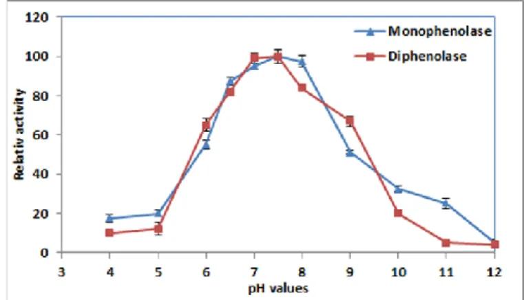
The result of this research showed that the M36 tyrosinase enzyme had maximum monophenolase and diphenolase activity at pH, 7.5 (Figure 1). This result was in accordance with tyrosinase enzyme originated from Streptomyces sp. REN-21 (pH 7.0) (Ito and Inouye, 2005), Rhizobium etli CFN42 (pH 7.5) (Pinero et al.
2007) and Pseudomonas putida F6 (pH 7.0) (McMahon et al., 2007)
notwithstanding, the tyrosinase enzyme from B. thuringiensis (Liu et al., 2004) and T. roseum (Kong et al., 2000) have shown to have maximum activity at 9.0 and 9.5, respectively. The M36 tyrosinase enzyme could conserve its maximum activity over than 95 % at pH (6.5-8.0). Before pH (6.5) and above pH (8.0) the activity and stability of the enzyme was dropped. These findings are similar to the finding of Shuster and Fishman (2009).
The tyrosinase enzymes have two cupper in its active site and each of the two metal atoms; CuA and CuB, of the active site are coordinated by three conserved histidines which are located in a ‘four α-helix bundle’ (Claus and Decker,
2006). The α-helix is structured by hydrogen bonds. Generally changing of pH
value (extremely basic or acidic) causes changes in the charge of H-bond donor and acceptor groups, it can rearrange the H-bonds and change the conformation/folding of the protein.
Figure 1 Effect of pH on activity and stability of the Bacillus megaterium M36
tyrosinase enzyme (monophenolase). The enzyme showed maximum activity (0.52 IU) at pH=7.5 and 97.5% of its maximum activity at pH=8.0. At pH lower than 6.5 and higher than 8.0 the activity of the enzyme was steeply decreased.
The M36 tyrosinase enzyme showed optimum monophenolase and diphenolase activity at 40 °C also, the enzyme conserved 100% of its original activity at 4-45 °C (Figure 2). The monophenolase and diphenolase activity of the enzyme was deeply decreased at temperature below 30 °C and above 55 °C, probably this result was related to that, the tyrosinase enzyme has mostly composed from α-helix, on the other hand α-helix is more flexible than the others structures. This result was more or less closed to other investigations.
The M36 tyrosinase enzyme showed up to 95% activity at temperature ranges from 35 °C-45°C, in contrast to this, the activity of tyrosinase enzyme from P.
putida F6 (McMahon et al., 2007) has been decreased dramatically at
temperature above 30°C and the enzyme of Streptomyces michiganensis DSM
(Philipp et al., 1991) has showed optimum activity at 33°C. Moreover there is
M36 tyrosinase enzyme was different with the tyrosinase from Rhizobium etli CFN42 (50°C) (Pinero et al., 2007), Bacillus (HR03) (55°C) (Dalfard et al.,
2006), Bacillus thuringiensis (75°C) (El-Shora, Metwally, 2008) and
Thermomicrobium roseum (70°C) (Kong et al., 2000). The M36 tyrosinase
enzyme conserved its original activity at 45°C, contrary to this Trichoderma
reesei (Cura et al., 2010) tyrosinase started to lose its activity relatively quickly
at temperature above 30°C.
Figure 2 Effect of temperature on a) monophenolase (L-tyrosine as a substrate) and b) diphenolase activity (L-DOPA as a substrate) of the
Bacillus megaterium M36 tyrosinase enzyme. The enzyme showed maximum monophenolase (0.56IU) and maximum diphenolase activity
(0.62IU) at 40°C. Both of the monophenolase and diphenolase activity were conserved 100% at temperature 0-45°C, after that the enzyme loosed its activity.
Effect of detergents on enzyme activity
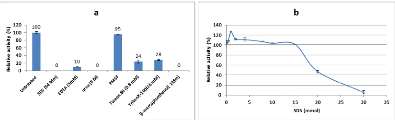
The M36 tyrosinase enzyme was studied in presence of various inhibitors (Figure 3a). The enzyme was inhibited strongly by mercaptoethanol. β-mercaptoethanol is a reducing agent which inhibit dopachrom and melanin synthesis by reducing qinones (an intermediate) to L-DOPA. Similar results were obtained for Bacillus megaterium tyrosinase (Shuster and Fishman, 2009), and
Thermomicrobium roseum tyrosinase (Kong et al., 2000) that was completely
inhibited by β-mercaptoethanol (1mmol). The M36 tyrosinase was inhibited about 90% by 5mM EDTA (a chelating agent). The agent can inhibit the enzyme by chelating of Cu from its active site. Similarly Bacills megaterium tyrosinase was inhibited up to 27% by 1mM EDTA (Shuster and Fishman, 2009) and
Bacillus (HR03) tyrosinase enzyme was partially inhibited by 1mM EDTA
(Dalfard et al., 2006). In contrast to the result of this research tyrosinase enzyme
from Bacillus thuringiensis (El-Shora and Metwally, 2008) was activated at high concentration EDTA from 200 to 400mM. Effect of different concentration of SDS (0.2- 30mM) on the M36 tyrosinase enzyme was studied. Although the enzyme was activated at the presence of 1mM SDS, it was strongly inhibited at high concentration of (above 15mM) SDS (Figure 5b). Previously, activating effect of SDS on tyrosinase enzyme from Xenopus laevis (Wittenberg and
Triplett, 1985), A. bisporus (Espin and Wichers, 1999), Bacillus sp. (Dalfard
et al., 2006) and Bacillus megaterium (Shuster and Fishman, 2009) has been
reported which was in agreement with our result. According to the paper published by Gandia-Herrero, although, active site of enzyme is not affected by SDS; a stepwise conformational change affected the enzyme activity by increasing accessibility of its active site to the substrate (Gandía-Herrero et al.,
2005).
Figure 3 a) Effect of inhibitors on Bacillus megaterium M36 tyrosinase activity. The activity of the sample containing the enzyme without
any of the additives was considered as control (100%). b) Effect of SDS concentration (w/v) on the Bacillus megaterium M36 tyrosinase activity. The enzyme showed maximum activity (126.6%) at the presence of 1mM of SDS and its activity was gradually decreased at the SDS concentration more than 1mM, so that it reached to 5.3% at concentration of 30mM. Sample having no SDS in reaction mixture was considered as a control (100%)
Production of L-DOPA from L-tyrosine
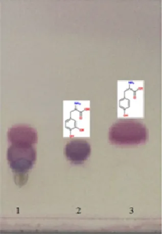
In TLC analysis, the transformation of L-tyrosine to L-DOPA was conspicuously detected. Ascorbic acid, used to prevent further oxidation of L-DOPA, did not give interfering spots (Figure 4).
Kinetic study of M36 tyrosinase enzyme
The M36 tyrosinase enzyme was shown to obey Michaelis-Menten kinetics when L-tyrosine and L-DOPA was used as a substrate. The Km value of M36 tyrosinase for l-tyrosine (0.15mM) was lower than L-DOPA (0.58mM). The obtained Vm was 1.7µM.min-1.ml-1for l-tyrosine and 6.2µM.min-1.ml-1 for L-DOPA. K
m value of M36 tyrosinase enzyme was similar to the previously reported Km values with the l-tyrosine, for example ; 0.2mM for Agaricus bisporus (Selinheimo et al.
2009) and 0.19mM for Rhizobium etli CFN42 (Cabrera-Valladares et al., 2006), also it is higher than the value (0.075mM) reported for Bacillus
megaterium (Shuster and Fishman, 2009) and it is less than the values
0.563mM, 1mM, 0.421mm reported for Bacillus huringiensis (El-Shora and
Metwally, 2008) , Streptomyces sp. REN-21(Ito and Inouye, 2005) and
Verrucomicrobium spinosum (Fairhead and Thony-Meyer, 2010), respectively.
The Km value of M36 tyrosinase enzyme for L-DOPA was higher than Km value of tyrosinase from Agaricus bisporus (0.17mM) (Selinheimo et al., 2009), P.
putida F6 (0.33) (McMahon et al., 2007) and Bacillus megaterium (0.35mM)
(Shuster and Fishman, 2009) for the same substrate, but it was lower than Km values of tyrosinase from Trichoderma reesei (7.5mM) (Selinheimo et al., 2009), Rhizobium etli CFN42 (2.44mM) (Cabrera-Valladares et al., 2006),
Streptomyces antibioticus (8.9mM) (Marino et al., 2011), Streptomyces castaneoglobisporus (8mM) (Kohashi et al., 2004) and Verrucomicrobium spinosum (7mM) (Fairhead and Thony-Meyer, 2010).
J Microbiol Biotech Food Sci / Valipour and Arikan et al. 2016 : 5 (5) 465-469
Figure 4 TLC analysis of L-tyrosine conversion to L-DOPA by Bacillus
megaterium M36 tyrosinase enzyme
Electrophoresis and enzymatic activities in gel
After dialysis, tyrosinase M36 was electrophoresed by using native polyacrylamide gel (8%), after specific staining, a distinct band was detected. by extracting of the tyrosinase enzyme from native gel using the method mentioned in material methods, and the enzyme was subjected to SDS-PAGE (12%) analysis. This analysis showed almost 34kDa bond of the enzyme (Figure 5). This result was similar to the result of Shuster and Fishman (2009) who have demonstrated the tyrosinase from Bacillus megaterium to be almost 35kDa.
Figure 5 Electrophoresis analysis of the Bacillus megaterium M36 tyrosinase
Enzyme. Lane (A) shows tyrosinase activity, lane (T) shows the enzyme molecular weight almost 34KDa, almost 15µg of protein was loaded, lane (M) shows protein marker.
Acknowledgments: This research was supported by the TUBITAK research
fund (No. 114Z065) and BAP research fund in CUKURUVA UNIVERSITY of Turkey (No. FEF2013D33).
REFERENCES
Ali, S., Shultz, J.L., Ikram, U.l.H. (2007). High performance microbiological transformation of L-tyrosine to L-dopa by Yarrowia lipolytica NRRL-143. BMC
Biotechnol 7:50. http://dx.doi.org/10.1186/1472-6750-7-50
Allouche, N., Damak, A., Ellouz, R., & Sayadi, S. (2004). Use of whole cells of
Pseudomonas aeruginosa for synthesis of the antioxidant hydroxytyrosol via
conversion of tyrosol. Applied and Environmental Microbiology, 70(4), 2105-2109. http://dx.doi.org/10.1128/aem.70.4.2105-2109.2004
Arikan, B. (2008). Highly thermostable, thermophilic, alkaline, SDS and chelator resistant amylase from a thermophilic Bacillus sp isolate A3-15. Bioresource
Technology, 99(8), 3071-3076. http://dx.doi.org/10.1016/j.biortech.2007.06.019 Aygan, A., & Arikan, B. (2009). Production and characterization of multifunctional endoxylanase by Bacillus sp X13. Turkish Journal of Biology, 33(3), 231-237.
Burhan, A., Nisa, U., Gokhan, C., Omer, C., Ashabil, A., & Osman, G. (2003). Enzymatic properties of a novel thermostable, thermophilic, alkaline and chelator resistant amylase from an alkaliphilic Bacillus sp isolate ANT-6. Process
Biochemistry, 38(10), 1397-1403.
http://dx.doi.org/10.1016/s0032-9592(03)00037-2
Cabrera-Valladares, N., Martinez, A., Pinero, S., Lagunas-Munoz, V. H., Tinoco, R., de Anda, R., Gosset, G. (2006). Expression of the melA gene from Rhizobium
etli CFN42 in Escherichia coli and characterization of the encoded tyrosinase.
Enzyme and Microbial Technology, 38(6), 772-779.
http://dx.doi.org/10.1016/j.enzmictec.2005.08.004
Caf, Yasemin, Maaşoğlu, Yelis, Valipour, Ebrahim, & Arikan, Burhan. Production and characterization of novel cold-active, pH tolerant and detergent-stable: α-amylase from a psychrotrophic bacterium from soil samples. New
Biotechnology, 29, Supplement, S82. http://dx.doi.org/10.1016/j.nbt.2012.08.227 Claus, H., & Decker, H. (2006). Bacterial tyrosinases. Systematic and Applied
Microbiology, 29(1), 3-14. http://dx.doi.org/10.1016/j.syapm.2005.07.012 Cura, D Ercili, Lille, M, Partanen, R, Kruus, K, Buchert, J, & Lantto, R. (2010). Effect of Trichoderma reesei tyrosinase on rheology and microstructure of acidified milk gels. International dairy journal, 20(12), 830-837.
http://dx.doi.org/10.1016/j.idairyj.2010.06.008
Dalfard, Arastoo Badoei, Khajeh, Khosro, Soudi, Mohammad Reza, Naderi-Manesh, Hossein, Ranjbar, Bijan, & Sajedi, Reza Hassan. (2006). Isolation and biochemical characterization of laccase and tyrosinase activities in a novel melanogenic soil bacterium. Enzyme and microbial technology, 39(7), 1409-1416. http://dx.doi.org/10.1016/j.enzmictec.2006.03.029
Dastager, S. G., Li, W. J., Dayanand, A., Tang, S. K., Tian, X. P., Zhi, X. Y., Jiang, C. L. (2006). Seperation, identification and analysis of pigment (melanin) production in Streptomyces. African Journal of Biotechnology, 5(11), 1131-1134. Decker, H., & Tuczek, F. (2000). Tyrosinase/catecholoxidase activity of hemocyanins: structural basis and molecular mechanism. Trends in Biochemical
Sciences, 25(8), 392-397. http://dx.doi.org/10.1016/s0968-0004(00)01602-9 El-Shora, Hamed M, & Metwally, M. (2008). Use of Tyrosinase Enzyme from
Bacillus thuringiensis for the Decontamination of Water Polluted with Phenols. Biotechnology, 7(2). http://dx.doi.org/10.3923/biotech.2008.305.310
Espín, Juan Carlos, van Leeuwen, Jeroen, & Wichers, Harry J. (1999). Kinetic study of the activation process of a latent mushroom (Agaricus bisporus) tyrosinase by serine proteases. Journal of agricultural and food chemistry, 47(9), 3509-3517. http://dx.doi.org/10.1021/jf9813539
Fairhead, M., & Thony-Meyer, L. (2010). Cross-linking and immobilisation of different proteins with recombinant Verrucomicrobium spinosum tyrosinase.
Journal of Biotechnology, 150(4), 546-551.
http://dx.doi.org/10.1016/j.jbiotec.2010.10.068
Franciscon, Elisangela, Grossman, Matthew James, Paschoal, Jonas Augusto Rizzato, Reyes, Felix Guillermo Reyes, & Durrant, Lucia Regina. (2012). Decolorization and biodegradation of reactive sulfonated azo dyes by a newly isolated Brevibacterium sp. strain VN-15. SpringerPlus, 1(1), 37.
http://dx.doi.org/10.1186/2193-1801-1-37
Freddi, G., Anghileri, A., Sampaio, S., Buchert, J., Monti, P., & Taddei, P. (2006). Tyrosinase-catalyzed modification of Bombyx mori silk fibroin: Grafting of chitosan under heterogeneous reaction conditions. Journal of Biotechnology,
125(2), 281-294. http://dx.doi.org/10.1016/j.jbiotec.2006.03.003
Gandía-Herrero, Fernando, Jiménez-Atiénzar, Mercedes, Cabanes, Juana, García-Carmona, Francisco, & Escribano, Josefa. (2005). Differential activation of a latent polyphenol oxidase mediated by sodium dodecyl sulfate. Journal of
agricultural and food chemistry, 53(17), 6825-6830.
http://dx.doi.org/10.1021/jf050505e
Hernandez-Romero, D., Solano, F., & Sanchez-Amat, A. (2005). Polyphenol oxidase activity expression in Ralstonia solanacearum. Applied and
Environmental Microbiology, 71(11), 6808-6815.
http://dx.doi.org/10.1128/aem.71.11.6808-6815.2005
Howard, Richard J, & Ferrari, Margaret A. (1989). Role of melanin in appressorium function. Experimental Mycology, 13(4), 403-418.
http://dx.doi.org/10.1016/0147-5975(89)90036-4
Ito, Masaaki, & Inouye, Kuniyo. (2005). Catalytic Properties of an Organic Solvent–Resistant Tyrosinase from Streptomyces sp. REN-21 and Its High-Level Production in E. coli. Journal of biochemistry, 138(4), 355-362.
http://dx.doi.org/10.1093/jb/mvi150
Kohashi, P. Y., Kumagai, T., Matoba, Y., Yamamoto, A., Maruyama, M., & Sugiyama, M. (2004). An efficient method for the overexpression and purification of active tyrosinase from Streptomyces castaneoglobisporus. Protein
Expression and Purification, 34(2), 202-207.
http://dx.doi.org/10.1016/j.pep.2003.11.015
Kong, K. H., Hong, M. P., Choi, S. S., Kim, Y. T., & Cho, S. H. (2000). Purification and characterization of a highly stable tyrosinase from
Thermomicrobium roseum. Biotechnology and Applied Biochemistry, 31,
113-118. http://dx.doi.org/10.1042/ba19990096
Kumar, C. G., Mongolla, P., Pombala, S., Kamle, A., & Joseph, J. (2011). Physicochemical characterization and antioxidant activity of melanin from a novel strain of Aspergillus bridgeri ICTF-201. Letters in Applied Microbiology,
469 Liu, N., Zhang, T., Wang, Y. J., Huang, Y. P., Ou, J. H., & Shen, P. (2004). A heat inducible tyrosinase with distinct properties from Bacillus thuringiensis.
Letters in Applied Microbiology, 39(5), 407-412.
http://dx.doi.org/10.1111/j.1472-765x.2004.01599.x
Lopez-Serrano, D., Sanchez-Amat, A., & Solano, F. (2002). Cloning and molecular characterization of a SDS-activated tyrosinase from Marinomonas
mediterranea. Pigment Cell Research, 15(2), 104-111.
http://dx.doi.org/10.1034/j.1600-0749.2002.1o068.x
Marino, S. M., Fogal, S., Bisaglia, M., Moro, S., Scartabelli, G., De Gioia, L. Bubacco, L. (2011). Investigation of Streptomyces antibioticus tyrosinase reactivity toward chlorophenols. Archives of Biochemistry and Biophysics,
505(1), 67-74. http://dx.doi.org/10.1016/j.abb.2010.09.019
McMahon, A. M., Doyle, E. M., Brooks, S., & O'Connor, K. E. (2007). Biochemical characterisation of the coexisting tyrosinase and laccase in the soil bacterium Pseudomonas putida F6. Enzyme and Microbial Technology, 40(5), 1435-1441. http://dx.doi.org/10.1016/j.enzmictec.2006.10.020
Michalik, J., Emilianowicz-Czerska, W., Switalski, L., & Raczynska-Bojanowska, K. (1975). Monophenol monooxygenase and lincomysin biosynthesis in Streptomyces lincolnensis. Antimicrob Agents Chemother, 8(5), 526-531. http://dx.doi.org/10.1128/aac.8.5.526
Philipp, Stephan, Held, Thomas, & Kutzner, Hans J. (1991). Purification and characterization of the tyrosinase of Streptomyces michiganensis DSM 40015.
Journal of basic microbiology, 31(4), 293-300.
http://dx.doi.org/10.1002/jobm.3620310412
Pinero, S., Rivera, J., Romero, D., Cevallos, M. A., Martinez, A., Bolivar, F., & Gosset, G. (2007). Tyrosinase from Rhizobium etli is involved in nodulation efficiency and symbiosis-associated stress resistance. Journal of Molecular
Microbiology and Biotechnology, 13(1-3), 35-44.
http://dx.doi.org/10.1159/000103595
Rani, Nisha, Joy, Beena, & Abraham, T Emilia. (2007). Cell suspension cultures of Portulaca grandiflora as potent catalysts for biotransformation of L-tyrosine into L-DOPA, an anti-Parkinson's drug. Pharmaceutical Biology, 45(1), 48-53.
http://dx.doi.org/10.1080/13880200601026341
Rao, K. R. S. S., Tripathy, N. K., Rao, D. S., & Prakasham, R. S. (2013). Production, Characterization, Catalytic and Inhibitory activities of Tyrosinase. Research Journal of Biotechnology, 8(1), 83-95.
Raval, Komal M, Vaswani, Pooja S, & Majumder, DR. (2012). Biotransformation of a single amino acid L tyrosine into a bioactive molecule L-DOPA. Int J Sci Res, 2, 2250-3153.
Selinheimo, Emilia, Gasparetti, Chiara, Mattinen, Maija-Liisa, Steffensen, Charlotte L, Buchert, Johanna, & Kruus, Kristiina. (2009). Comparison of substrate specificity of tyrosinases from Trichoderma reesei and Agaricus
bisporus. Enzyme and Microbial Technology, 44(1), 1-10.
http://dx.doi.org/10.1016/j.enzmictec.2008.09.013
Seo, S. Y., Sharma, V. K., & Sharma, N. (2003). Mushroom tyrosinase: Recent prospects. Journal of Agricultural and Food Chemistry, 51(10), 2837-2853.
http://dx.doi.org/10.1021/jf020826f
Surwase, S.N., Patil, S.A., Apine, O.A., Jadhav, J.P. (2012). Efficient microbial conversion of L-tyrosine to L-DOPA by Brevundimonas sp. SGJ. Appl Biochem
Biotechnol 167:1015-28. http://dx.doi.org/10.1007/s12010-012-9564-4 Shuster, V., & Fishman, A. (2009). Isolation, Cloning and Characterization of a Tyrosinase with Improved Activity in Organic Solvents from Bacillus
megaterium. Journal of Molecular Microbiology and Biotechnology, 17(4),
188-200. http://dx.doi.org/10.1159/000233506
Wittenberg, C, & Triplett, EL. (1985). A detergent-activated tyrosinase from Xenopus laevis. II. Detergent activation and binding. Journal of Biological Chemistry, 260(23), 12542-12546.
Xu, D. Y., Chen, J. Y., & Yang, Z. (2012). Use of cross-linked tyrosinase aggregates as catalyst for synthesis of L-DOPA. Biochemical Engineering Journal, 63, 88-94. http://dx.doi.org/10.1016/j.bej.2011.11.009
Zanjani Sorouri, R., Mir-Esmaili, S. M., Latifi, A.M, & Valipour, E. (2009). Isolation and identification of a type strain bacteria with the highest ability to produce organophosphorus acid anhidrase. Journal of Mazandaran University of Medical Sciences, 18(68).