73
Ankara Üniversitesi Tıp Fakültesi Mecmuası 2006; 59:73-76
Corresponding author
A. Ruchan Akar, MD, Department of Cardiovascular Surgery, Heart Center, University of Ankara School of Medicine, Dikimevi, 06340 Ankara, Turkey
Fax : +90 (312) 362 56 39 Tel : +90 (505) 527 96 80 E-mail adress : [email protected]
Received: 25.10.2005 • Accepted: 05.06.2006
CERRAHİ BİLİMLER / SURGICAL SCIENCES Derleme / Review
A
n abnormal connection between the aorta and cardiac chambers is a well recognised cardiac entity. Several types have been described, of which coronary-cameral fistulae or connections (congenital or acquired) are the most common (1). There have also been several reports of acquired aorta-cameral connections. Congenital aortico-aorta-cameral connections are much rarer and the majority are to the left ventricle (ALVT) (2;3). The aorticoright atrial tunnel (ARAT) is first described by Otero Coto and colleagues in 1980 (4). To date there have only been 13 cases reported of ARAT, most of which originated from the aortic left sinus of Valsalva whereas the tunnel in one case originated from the non-coronary sinus, in two cases from the right coronary sinus and in one case from the aortic isthmus (5-7). We report the third case of congenital ARAT arising from the right sinus of Valsalva.A 57-year-old man was admitted with unstable angina and a four month history of exertional chest pain. He was a smoker with a positive family history of ischaemic heart disease. His past medical history was otherwise unremarkable. The chest roent-genogram revealed mild cardiomegaly and the ECG showed sinus rhythm with evi-dence of T-wave inversion in leads 1, aVL, V2-V6. On admission his cardiac enzymes
were not elevated. He was managed conservatively and investigated.
Aortico-right atrial tunnel (ARAT) is a rare congenital entity. We report a case of 57-year-old man who presented with unstable angina and who had a continuous murmur at the right sternal edge. Coronary angiograpy revealed triple vessel coronary artery disease and a separate anoma-lous structure arising from the right sinus of Valsalva. Digital substraction angiography was nec-essary to define the distal end of this connection which was the roof of the right atrium. Our patient is unique in that the tunnel 5 cm in lenght originated from the right sinus of Valsalva and entered the right atrium anteriorly having traversed the right atrioventricular groove anterior, totally separate to the right coronary artery. The management of these congenital aorto-atrial connections is discussed.
Key words: Aortico-Cameral-Fistula, Aorta-Atrium, Sinus of Valsalva
Aortiko-sağ atriyal tünel nadir görülen konjenital anomalidir. Bu vaka takdiminde anstabil anjina-sı ve fizik muayenede sağ sternal bölgede devamlı üfürümü olan 57 yaşında erkek hasta sunul-maktadır. Koroner anjiyografide üç damar hastalığı ve sağ Valsalva sinüsünden ayrı olarak çıkan anormal bir yapı tespit edildi. Dijital substraksiyon anjiyografide bu yapının sağ atrium çatısıyla ilişkisi olduğu görüldü. Hastamızda, sağ koroner arterden tamamen ayrı olarak, sağ Valsalva sinü-sünden başlayıp sağ atriyoventriküler oluğun ön kısmında sonlanan, 5 cm uzunluğunda bir tünel tespit edildi.Yazımızda bu tip doğumsal aorto-atriyal bağlantıların tedavisi tartışıldı.
Anahtar sözcükler: Aortiko-kameral fistül, Aorta-atrium, Valsalva sinüsü
Aortico-cameral communication from right sinus of
valsalva to right atrium
Sağ valsalva sinüsü ile sağ atriyum arasında aertico-kameral komünikasyon
A. Rüçhan Akar, Mustafa Şırlak, Zeynep Eyileten, Refik Taşöz, Ümit Özyurda
Department of Cardiovascular Surgery, Heart Center, University of Ankara School of Medicine, Turkey
Ankara Üniversitesi Tıp Fakültesi Mecmuası 2006; 59(2)
74 Aortico-cameral communication from right sinus of valsalva to right atrium
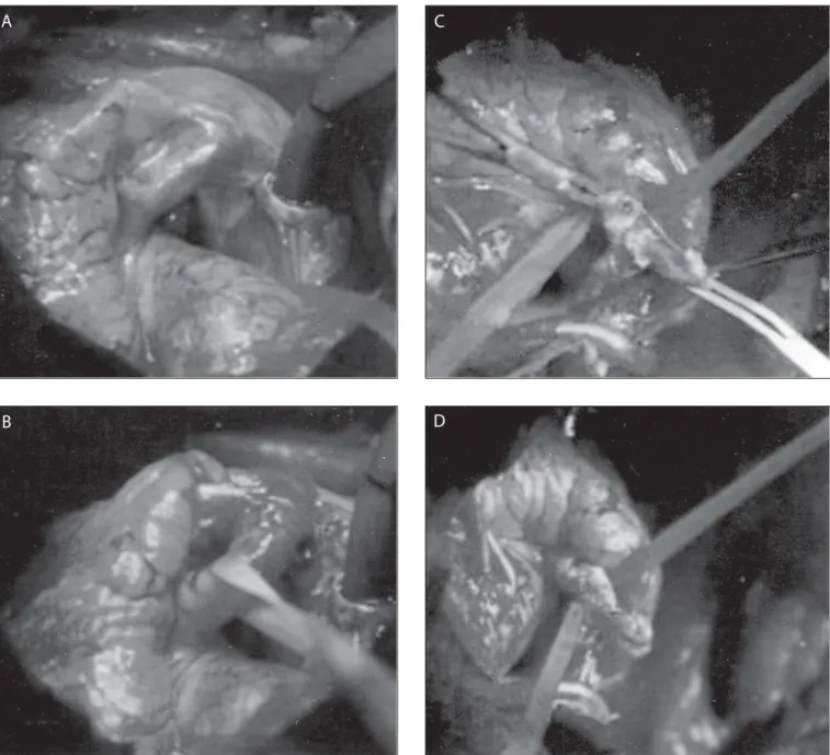
Selective coronary angiography revealed a normal left main stem with significant triple vessel coronary artery dis-ease and an anomalous tube like structure arising from the right sinus of Valsalva adjacent but totally separate from the right coronary artery and connecting the aorta to one of the cardiac chambers and gross macroscopic operative features confirmed these findings. (Fig.1). Transoesophageal echocar-diography confirmed these findings. Digital substraction an-giography (DSA) and CT scanning were, however, required to define the distal end of the connection as the roof of the right atrium. The ratio of pulmonary blood flow to systemic blood flow was measured as 1.9/1.
Figure 1. Intraoperative photograph showing aortico-right atrial tunnel (the patient’s head is on the lower end); (A) A systolic thrill is verified by palpation, (B) blunt dissection is used for posterior tunnel attachments, avoiding injury to right coronary artery, and the tunnel was taped, (C) Clamps have been
applied to the tunnel, with caution taken to monitor ECG changes before division, and completed division of the aortico-right atrial tunnel, (D) The ends of the tunnel are closed with 4-0 polypropylene suture. Ao=ascending aorta, RA=right atrium, RV=right ventricle.
The patient underwent operation via a median ster-notomy. A systolic thrill could be palpated over the base of aortico-right atrial connection. Cardiopulmonary bypass was established with moderate hypothermia (32 oC). After
coronary artery bypass grafting to left anterior descend-ing, intermediate, first obtuse marginal and right coronary arteries the anomalous structure was opened in the right atria-ventricular groove. The structure was described as 5-cm long fibrous tube anterior to the right coronary ar-tery in the right atrioventricular groove with an external diameter of 1.5-cm. The tube was divided between vas-cular clamps and the cut ends over sewn. The patient was
A
B
C
Journal of Ankara University Faculty of Medicine 2006; 59(2)
75
A.R. Akar, M. Şırlak, Z. Eyileten et al.
weaned off bypass without difficulty. Apart from a tempo-rary pacing requirement for nodal rhythm for 24 hours he made an uneventful recovery. Two years after the operation the patient remains asymptomatic.
Comment
Congenital aortico-cameral connections are extremely rare, but have been reported connecting the aorta to all four cardiac chambers. The majority of these are aortico-left ventricular connections which have been extensively reviewed and classified (3). Congenital ARAT is however, an extremely rare entity, and only 13 cases have been previ-ously reported in the English literature (Table 1). Mostly arise from the left sinus of Valsalva. This case is unique in that the origin of the aortico-right atrial connection was the right sinus of Valsalva, adjacent but separate to the right coronary ostium and the patient presented with un-stable angina. Furthermore, our case is the oldest patient in the literature presented with ARAT and surgically treated.
These connections can be difficult to define accurately as was experienced in this case, where conventional angiog-raphy and echocardiogangiog-raphy were inadequate. CT scanning and DSA did, however, illustrate the value of these imaging techniques in demonstrating abnormal cardiac structures.
Differential diagnoses included ruptured aneurysm of the sinus of Valsalva, coronary arteriovenous fistula, rup-ture of a dissecting aneurysm of the ascending aorta into the right atrium, and pseudoaneurysm of the right coro-nary artery followed by formation of a fistula between the aneurysm and the right atrium. There was no history of trauma in our case and no evidence of acquired aortic dis-ease. We conclude therefore, that this aortico-right atrial connection was a congenital structure. All previous con-genital connections of this type have been described in the pediatric population. Our patient is unique in having such a congenital structure diagnosed at the time of presenta-tion with a separate acquired cardiac condipresenta-tion. Indeed the histological findings confirm those described by Rosenberg et al. of a deficiency in smooth muscle, but abundance of elastic lamellae (8). The precise pathogenesis of this con-genital anomaly is however unknown; persistence of meso-cardial cysts or embryonic rests of the fifth aortic arch in the early stages of cardiogenesis, abnormal formation of supravalvular ridge producing aneurysmal dilatation of the primitive aorta, intrauterine rupture of a sinus of Valsalva aneurysm are possible explanations (4,8).
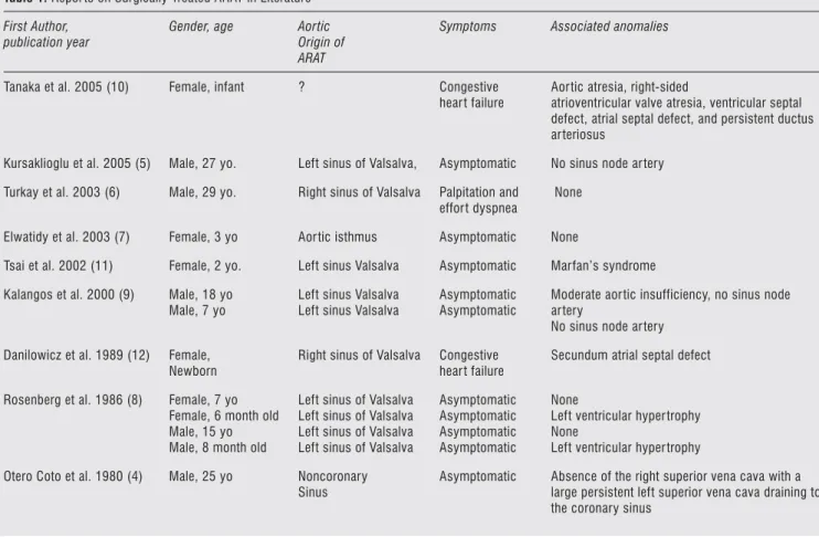
Table 1. Repor ts on Surgically Treated ARAT in Literature First Author,
publication year
Gender, age Aortic Origin of ARAT
Symptoms Associated anomalies
Tanaka et al. 2005 (10) Female, infant ? Congestive hear t failure
Aor tic atresia, right-sided
atrioventricular valve atresia, ventricular septal defect, atrial septal defect, and persistent ductus ar teriosus
Kursaklioglu et al. 2005 (5) Male, 27 yo. Left sinus of Valsalva, Asymptomatic No sinus node ar tery Turkay et al. 2003 (6) Male, 29 yo. Right sinus of Valsalva Palpitation and
effor t dyspnea
None Elwatidy et al. 2003 (7) Female, 3 yo Aor tic isthmus Asymptomatic None
Tsai et al. 2002 (11) Female, 2 yo. Left sinus Valsalva Asymptomatic Marfan’s syndrome Kalangos et al. 2000 (9) Male, 18 yo
Male, 7 yo
Left sinus Valsalva Left sinus Valsalva
Asymptomatic Asymptomatic
Moderate aor tic insufficiency, no sinus node ar tery
No sinus node ar tery Danilowicz et al. 1989 (12) Female,
Newborn
Right sinus of Valsalva Congestive hear t failure
Secundum atrial septal defect Rosenberg et al. 1986 (8) Female, 7 yo
Female, 6 month old Male, 15 yo Male, 8 month old
Left sinus of Valsalva Left sinus of Valsalva Left sinus of Valsalva Left sinus of Valsalva
Asymptomatic Asymptomatic Asymptomatic Asymptomatic
None
Left ventricular hyper trophy None
Left ventricular hyper trophy Otero Coto et al. 1980 (4) Male, 25 yo Noncoronary
Sinus
Asymptomatic Absence of the right superior vena cava with a large persistent left superior vena cava draining to the coronary sinus
Ankara Üniversitesi Tıp Fakültesi Mecmuası 2006; 59(2)
76 Aortico-cameral communication from right sinus of valsalva to right atrium
Previous reports have suggested that patients with aor-tico-right atrial tunnel may present with continuous heart murmur, mild cardiomegaly or clinical evidence of ven-tricular overload due to left-to-right shunt. Angina and myocardial infarction in contrast have not been reported secondary to aortico-right atrial communications. The ischemic symptoms in our patient could theoretically have been due to coronary artery steal, but in reality probably was due to the proximal stenosis of left anterior descend-ing artery.
Of the congenital aortico-cameral connections, at least 37 cases have been reported of aortico-left ventricular tunnel (ALVT) (3). Surgery is clearly indicated in this condition to prevent progressive aortic incompetence and cardiac failure. Little, is however, known of the natural history of untreated
References
1. Fernandes ED, Kadivar H, Hallman GL et al. Congenital malformations of the coronary arteries: the Texas Heart Institute experience. Ann.Thorac.Surg. 1992; 54:732-740.
2. Akalin H, Erol C, Oral D et al. Aortico-left ventricular tunnel: successful diagnostic and surgical approach to the oldest patient in the literature. J.Thorac.Cardiovasc.Surg. 1989; 97:804-805. 3. Hovaguimian H, Cobanoglu A, Starr A. Aortico-left ventricular
tunnel: a clinical review and new surgical classification. Ann. Thorac.Surg. 1988; 45:106-112.
4. Coto EO, Caffarena JM, Such M et al. Aorta--right atrial communication. Report of an unusual case. J.Thorac.Cardiovasc. Surg. 1980; 80:941-944.
5. Kursaklioglu H, Iyisoy A, Celik T et al. Aortico-right atrial tunnel in an adult patient. Int.J.Cardiovasc.Imaging 2005; 21:383-385. 6. Turkay C, Golbasi I, Belgi A et al. Aorta-right atrial tunnel.
J.Thorac.Cardiovasc.Surg. 2003; 125:1058-1060.
aortico-right atrial connections. Congenital ARAT is also uncommon except for complications with a coronary artery fistula. We, however, assume that this communication was not an equivalent of a coronary artery fistula, since the coro-nary arteries in our case had normal origin, distribution and no dilatation. In general, surgical correction is recommend-ed to avoid the long term effects of left-to-right shunting thus preventing left ventricular impairment and improving life expectancy. Furthermore because of possible complica-tions such as bacterial endocarditis, aneurysm formation, or spontaneous rupture, the closure of ARAT is recommended (9). However, it should be kept in mind during surgery that the “tunnel” can also give origin to coronary arteries. Defini-tive surgical correction has been shown to be effecDefini-tive and safe, with good long term results.
7. Elwatidy AF, Galal AN, Rhydderch D et al. Aorto-right atrial fistula. Ann.Thorac.Surg. 2003; 76:929-931.
8. Rosenberg H, Williams WG, Trusler GA et al. Congenital aortico-right atrial communications. The dilemma of differentiation from coronary-cameral fistula. J.Thorac.Cardiovasc.Surg. 1986; 91:841-847. 9. Kalangos A, Beghetti M, Vala D et al. Aorticoright atrial tunnel.
Ann.Thorac.Surg. 2000; 69:635-637.
10. Tanaka T, Tomita H, Watanabe K et al. A case of aorto-right atrial tunnel associated with aortic and tricuspid atresia. Pediatr.Int. 2005; 47:466-468.
11. Tsai YC, Wang JN, Yang YJ et al. Aortico-cameral communication from left sinus Valsalva aneurysm to right atrium via a tortuous tunnel with aneurysmal dilatation. Pediatr.Cardiol. 2002; 23:108-109.
12. Danilowicz D, Presti S, Colvin S et al. Congenital fistulous tract between aorta and right atrium presenting as heart failure in a newborn. Pediatr.Cardiol. 1989; 10:93-97.