MELD-XI score predicts in-hospital mortality
independent of simplified pulmonary
embolism severity index among patients with
intermediate-to-high risk acute pulmonary
thromboembolism
doi • 10.5578/tt.68614 Tuberk Toraks 2019;67(3):169-178
Geliş Tarihi/Received: 28.05.2019 • Kabul Ediliş Tarihi/Accepted: 14.08.2019
KLİNİK Ç
ALIŞMA
RESEARCH
AR
TICLE
Orçun ÇİFTCİ1 Çaşıt Olgun ÇELİK1 Güldeniz UZAR1 Elif KÜPELİ2 İbrahim HaldunMÜDERRİSOĞLU1
1Department of Cardiology, Baskent University Hospital, Ankara, Turkey 1Başkent Üniversitesi Ankara Hastanesi, Kardiyoloji Anabilim Dalı,
Ankara, Türkiye
2Department of Chest Diseases, Baskent University Hospital, Ankara, Turkey 2Başkent Üniversitesi Ankara Hastanesi, Göğüs Hastalıkları Anabilim Dalı,
Ankara, Türkiye
ABSTRACT
MELD-XI score predicts in-hospital mortality independent of simplified pulmonary embolism severity index among patients with intermediate-to-high risk acute pulmonary thromboembolism
Introduction: Acute pulmonary thromboembolism (PTE) is a highly morbid
and fatal condition. Although several risk stratification models exist for prediction of mortality risk in PTE, no study has yet focused on the effect of impaired vital organ function, such as renal or hepatic impairment, on mortality in PTE. MELD-XI (Model for end-stage liver disease excluding INR) score predicts mortality among patients with end-stage hepatic and cardiovascular disorders. Herein, we aimed to test MELD-XI score for predicting in-hospital prognosis of patients with intermediate-to-high risk acute PTE.
Materials and Methods: We reviewed the medical records patients older than
18 years hospitalized with intermediate-to-high risk PTE between 01.06.2011 and 01.01.2019. Simplified pulmonary embolism severity index (sPESI) score and MELD-XI score were calculated, and in-hospital mortality determined. MELD-XI score was compared between patients with and without in-hospital mortality and was correlated to sPESI score. The predictive power of MELD-XI score for in-hospital mortality was sought and an in-hospital survival analysis with Kaplan Meier curve and log-rank test was done for MELD-XI score.
Results: A total of 104 patients [mean age of 70.8 ± 15.9 years; 68 (65.4%)
females]. Fourteen (13.5%) patients died at hospital. MELD-XI and sPESI
Dr. Orçun ÇİFTCİ
Başkent Üniversitesi Ankara Hastanesi, Kardiyoloji Anabilim Dalı, ANKARA - TÜRKİYE e-mail: [email protected]
Yazışma Adresi (Address for Correspondence)
Cite this arcticle as: Çiftci O, Çelik ÇO, Uzar G, Küpeli E, Müderrisoğlu İH. MELD-XI score predicts in-hospital mortality independent of simplified pulmonary embolism severity index among patients with intermediate-to-high risk acute pulmonary thromboembolism. Tuberk Toraks 2019;67(3):169-78.
©Copyright 2019 by Tuberculosis and Thorax. Available on-line at www.tuberktoraks.org.com
INTRODUCTION
Acute pulmonary thromboembolism (PTE) is a highly morbid and fatal condition, being responsible for most vascular deaths after myocardial infarction and stroke (1,2). There are several risk stratification sys-tems and scores are available for risk and mortality prediction in PTE. The most commonly used risk stratification scheme is the ESC risk stratification sys-tem, which mostly concentrates on cardiac involve-ment by the increased right ventricular afterload imposed by pulmonary embolus, and thus requires cardiac evaluaiton via electrocardiography, echocar-diography, and cardiac biomarker evaluation (1). Other risk stratification systems are also available, such as pulmonary embolism severity index (PESI) and its simplified form, simplified PESI (SPESI), which mostly concentrate on demographic, clinical and
vital sign characteristics (3,4). However, no study has yet focused on the effect of impaired vital organ func-tion, such as of the kidneys and the liver, on mortali-ty in PTE, which may occur due to the hemodynamic burden of PTE and the underlying comorbidities. MELD-XI (Model for end-stage liver disease excluding INR) score is a modification of the original MELD (Model for end-stage liver disease) score, which is a novel score derived for mortality prediction among patients with end-stage liver disorders and incorpo-rates the logarithmic conversions of serum total biliru-bin, creatinine concentrations, and INR level (5,6). MELD-XI score was primarily developed for patients with end-stage liver disease using anticoagulants and does not use INR level (7). Recently, MELD-XI score has also been shown to be able to predict prognosis in critically ill patients (7). Likewise, it could predict
scores were significantly correlated to each other and were higher in deceased patients than the survivors [17.3 (IQR 14.3) vs. 10.12 (IQR 2.99); p< 0.05 and 2 (IQR 1) vs. 1 (IQR 1); p< 0.05, respectively]. MELD-XI score and sPESI score were significant predictor of in-hospital mortality in multivariate analysis. A MELD-XI score ≥ 10.25 had a sensitivity of 78.6% and a specificity of 70.0% for in-hospital mortality. A survival analysis revealed that a high MELD-XI category (MELD-XI score ≥ 10.2) significantly worsened in-hospital survival (p< 0.01; log rank test).
Conclusion: MELD-XI score performs well for mortality prediction among patients with intermediate-to-high risk PTE. This subject
needs to be further studied by large, randomized controlled studies.
Key words: Acute pulmonary thromboembolism; MELD-XI score; mortality; intermediate-to-high risk ÖZ
MELD-XI skoru orta-yüksek riskli pulmoner tromboemboli hastalarında basitleştirilmiş pulmoner emboli ciddiyet indeksinden bağımsız olarak hastane içi mortaliteyi öngörmektedir
Giriş: Akut pulmoner tromboemboli (PTE) yüksek oranda morbid ve fatal bir durumdur. PTE’de mortalite riskinin öngörülmesi için
çeşitli risk sınıflama modelleri olsa da, henüz hiçbir çalışma PTE’de karaciğer ve böbrek fonksiyon bozukluğu gibi vital organların bozulmuş fonksiyonunun mortalite üzerine etkisine yoğunlaşmamıştır. MELD-XI (INR çıkarılmış son dönem karaciğer hastalığı mode-li), son dönem karaciğer ve kardiyovasküler hastalıklarda mortaliteyi öngörmektedir. Bu çalışmada MELD-XI skorunun orta-yüksek riskli PTE hastalarında hastane içi ölümü öngörücü rolünü belirlemeyi amaçladık.
Materyal ve Metod: 01.06.2011 ve 01.01.2019 tarihleri arasında orta-yüksek PTE ile yatırılan hastaların tıbbi kayıtları tarandı.
Basitleştirilmiş pulmoner emboli ciddiyet indeksi (sPESI) skoru ve XI skoru hesaplandı ve hastane içi mortalite belirlendi. MELD-XI skoru hastane içinde ölen ve ölmeyen hastalar arasında karşılaştırıldı ve sPESI skoru ile korele edildi. MELD-MELD-XI skorunun hastane içi ölüm içi öngördürücü değeri test edildi ve Kaplan Meier eğrisi ve log rank testi kullanılarak MELD-XI skoru için sağkalım analizi yapıl-dı.
Bulgular: Çalışmaya toplam 104 hasta [ortalama yaş 70.8 ± 15.9 yıl; 68 (%65.4) kadın] alındı. On dört (%13.5) hasta hastanede
öldü. MELD-XI ve sPESI skorları anlamlı şekilde birbirleriyle koreleydi ve ölenlerde yaşayanlara göre anlamlı şekilde daha yüksekti [17.3 (sırasıyla ÇAA 14.3)’e karşılık 10.12 (ÇAA 2.99); p< 0.05 ve 2 (ÇAA 1)’ye karşılık 1 (ÇAA 1); p< 0.05]. MELD-XI skoru ve sPESI skoru anlamlı şekilde korele idiler (r= 0.232; p< 0.05). MELD-XI skoru ve sPESI skoru çok değişkenli analizde hastane içi ölümün anlamlı öngördürücüsüydüler. MELD-XI skorunun ≥ 10.25 olması hastane içi mortaliteyi %78.6 duyarlılık ve %70.0 özgüllükle öngö-rebiliyordu. Sağkalım analizinde yüksek MELD-XI kategorisi (MELD-XI skoru ≥ 10.2) hastane içi sağkalımı anlamlı şekilde kötüleştir-mekteydi (p< 0.01; log rank testi).
Sonuç: MELD-XI skoru orta-yüksek riskli PTE hastalarında mortalitenin öngörülmesinde iyi performansı göstermektedir. Bu konunun
geniş, randomize kontrollü çalışmalarla ileri araştırılması gerekir.
patient outcomes in a number of cardiac disorders, namely heart failure, left ventricular assist device implanted patients, heart transplantation, infective endocarditis, mitral valve repair surgery, among oth-ers (8-12). The suggested mechanism underlying the ability of MELD-XI score to predict mortality among cardiac, along with other critically ill, patients is renal and/or hepatic hypoperfusion and/or congestion resulting from forward and/or backward heart failure, resulting in deranged end-organ function (13-15). Although it has been shown to predict outcomes in various cardiovascular disorders, MELD-XI score has never been studied among patients with PTE. Hence, in this study we aimed to determine the role of MELD-XI score for predicting in-hospital prognosis of outpa-tients presenting with intermediate-to-high risk acute PTE and compare it with the sPESI score, a score with already proven role in predicting mortality in PTE. We used MELD-XI score instead of the original MELD score as the patients with PTE are almost always treat-ed with anticoagulants.
MATERIALS and METHODS
This study was approved by the Local Ethics Committee and supported by the Institutional Research Fund (No: KA19/172). The medical records of patients aged older than 18 years who presented to our hospital’s emer-gency department as outpatients with acute PTE and were admitted to hospital for intermediate-to-high risk PTE between 01.01.2013 and 01.01.2019 were retro-spectively reviewed and recorded from written medi-cal reports and hospital’s data automation system. We recorded and analyzed demographic variables includ-ing age, gender; clinical variables includinclud-ing presentinclud-ing symptom (chest pain, dyspnea, syncope, or altered consciousness and poor overall condition), comorbid-ities (cardiac, pulmonary, or systemic), presumed, definable cause of PTE such as cancer or recent trau-ma or surgery, administered anticoagulant and/or fibrinolytic therapy; vital signs including body tem-perature, respiratory rate, heart rate, and admission systolic blood pressure; echocardiographic variables including right ventricular apical four chamber basal size, right ventricular tricuspid annular plane systolic excursion (TAPSE), systolic pulmonary artery pressure (sPAP), grade of tricuspid insufficiency (1-4), and left ventricular ejection fraction (LVEF); electrocardio-graphic variables including sinus tachycardia, S1Q3T3 pattern, pathological Q waves in inferior leads, right axis deviation, right ventricular hypertrophy,
incom-plete or comincom-plete right bundle branch block, anteroseptal T inversion, anteroseptal ST depression, and profound bradycardia (< 40/min); biochemical variables including blood urea nitrogne (BUN), creati-nine, total bilirubin, INR level, white blood cell count (WBC), hemoglobin concentration, serum C-reactive protein (CRP) level, maximal cardiac troponin I level at the time of emergency department admission, D-dimer level; and arterial blood gas variables includ-ing pH, PaO2, SO2, PCO2, lactate level, and HCO3 level.
This study only included intermediate and high risk PTE patients determined according to the ESC 2014 Pulmonary Embolism Guidelines. As such, high-risk PTE was defined as acute PTE with persistent hypoten-sion [systolic blood pressure < 90 mmHg or > 40 mmHg drop for at least 15 minutes or needing inotro-pic support, not due to a cause other than PE, such as arrhythmia, hypovolemia, sepsis, or left ventricular (LV) dysfunction], pulselessness, or persistent profound bradycardia (heart rate < 40 bpm with signs or symp-toms of shock). Intermediate-risk PTE was defined as acute PTE without systemic hypotension (systolic blood pressure > 90 mmHg) but with either RV dys-function or myocardial necrosis. RV dysdys-function was considered positive when end-diastolic right ventricu-lar (RV) to left ventricuventricu-lar (LV) diameter ratio was greater than 0.9 in apical four chamber view and/or there occurred RV systolic dysfunction evidenced by reduced tricuspid annular plane excursion (TAPSE) on echocardiography; or when there were newly devel-oped electrocardiographic changes (new complete or incomplete right bundle-branch block, anteroseptal ST elevation or depression, or anteroseptal T-wave inver-sion). Myocardial necrosis was defined by elevated admission or follow-up troponin I level (> 0.4 ng/mL) (1).
Simplified PESI (pulmonary embolism severity index) (sPESI) score was calculated for all patients. In this score, 1 point is given to each of the following charac-teristics: age > 80 years; male gender; chronic obstruc-tive pulmonary disease or chronic heart failure; pulse rate ≥ 110 bpm; systolic blood pressure < 100 mmHg; and arterial SO2 < 90% (4,16).
MELD-XI score was calculated by the following formu-la: 5.11 × ln (serum bilirubin in mg/dL) + 11.76 × ln (serum creatinine in mg/dL) + 9.44. Serum creatinine and bilirubin values below 1.0 mg/dL were rounded
up to 1.0 and serum creatinine values above 4 were rounded to 4.0. Serum creatinine was rounded to 4 in patients on hemodialysis therapy (17).
The exclusion criteria included patients younger than 18 years, patients with low-risk thromboembolism, patients with missing medical records, and patients that were treated as outpatients. In-hospital all-cause mortality was defined as death by any cause during the time of hospital stay.
Statistical Analysis
All statistical analyses were performed using SPSS v 20 software package (IBM SPSS statistics). The nor-mality of the distribution of continuous variables was tested using the Kolmogorov-Smirnov test. The nor-mally distributed continuous variables were expressed as mean ± standard deviation; the non-normally dis-tributed ones as median and interquartile range-(IQR); and categoric variables as number and per-centage. Normally distributed continuous variables were compared with the independent samples t-test; non-normally distributed continuous variables with the Mann-Whitney U test; and the categoric variables with the Chi-square test or Fisher’s exact test. MELD-XI score was compared between patients with and without in-hospital mortality. Correlation between the scoring systems was tested using Spearman bivar-iate correlation analysis. The significant predictors of in-hospital mortality were initially tested with a variate analysis using all available variables. All uni-variate predictors of mortality with p value ≤ 0.2 were then used in a binary logistic regression model to determine the independent predictors of in-hospi-tal morin-hospi-tality. The predictive power of MELD-XI score for in-hospital mortality was sought using ROC (Receiver operating characteristics) analysis to deter-mine the area under the curve, sensitivity, specificity, and positive and negative predictive values. A surviv-al ansurviv-alysis with Kaplan Meier curve and log-rank test was used to determine the effect high vs low MELD-XI score category on in-hospital survival. A p value of less than 0.05 was considered statistically significant for all statistical tests.
RESULTS
A total of 104 patients with a mean age of 70.8 ± 15.9 (range 24-96 years) were included, of which 68 (65.4%) were female. General clinical and demo-graphic characteristics of the study population were
shown on Table 1. Based on the ESC 2014 PTE gide-line, a total of 9 (8.7%) patients had high-risk PTE and 95 (91.3%) had intermediate-risk PTE. The mean sPESI score of the patients was 1.31 ± 1.02 and the mean MELD-XI score was 11.44 ± 3.78. A total of 14 (13.5%) of the patients died at hospital. The com-parison between the deceased and surviving patients with respect to clinical, echocardiographic, electro-cardiographic, and biochemical parameters were shown on Table 2. MELD-XI score was significantly higher in deceased patients than the survivors [17.3 (IQR 14.3) vs. 10.12 (IQR 2.99); p< 0.05] as was the median SPESI score [2 (1) vs. 1 (1); p< 0.05] and serum CRP level [110.0 (IQR 68.0) vs 27.2 (IQR 49.2); p< 0.05] whereas the deceased ones had a significantly lower hemoglobin count than the survi-vors [9.7 (IQR 2.1) vs. 13.4 (IQR 2.3); p< 0.05]. Other parameters were similar between the two groups.
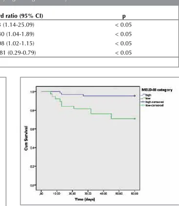
In correlation analysis MELD-XI score and sPESI score were significantly correlated (r= 0.232; p< 0.05). Univariate predictors of death were MELD-XI score, sPESI score, pulmonary artery systolic pres-sure, serum CRP, and hemoglobin count, whereas clinical PTE severity was not predictive of mortality in univariate analysis. A multivariate analysis using binary logistic regresion analysis with forward Wald method showed that MELD-XI score was an indepen-dent predictor of in-hospital death along with sPESI, systolic pulmonary artery pressure, and hemoglobin count (Table 3). ROC analyses showed that sPESI score (AUC = 0.744 95%CI 0.631-0.856; p= 0.001) and MELD-XI score (AUC = 0.765 95%CI 0.618-0.912; p= 0.001) were significantly predictive of in-hospital death (Figure 1, 2). A MELD-XI score of ≥ 10.25 had a sensitivity of 78.6% and a specificity of 70.0%, while a sPESI score of ≥ 1.5 had a sensitivity of 78.6%, a specificity of 64.6%. A survival analysis based on a MELD-XI cut-off point of 10.2 revealed that patients in the high MELD-XI category (MELD-XI score ≥ 10.2) had a significantly worse in-hospital survival than those in the low MELD-XI category (MELD-XI score < 10.2) (p< 0.01; log rank test), so that 95.5% of patients with a MELD-XI score of < 10.2 survived by 60 days after hospitalization where-as only 71.1% of those with a MELD-XI score of ≥ 10.2 survived. (Figure 3).
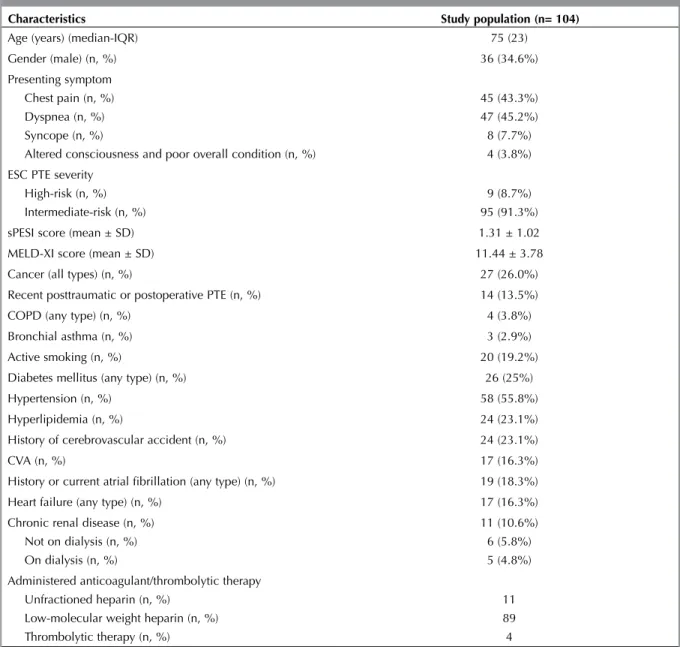
Table 1. General clinical and demographic characteristics of the study population
Characteristics Study population (n= 104)
Age (years) (median-IQR) 75 (23)
Gender (male) (n, %) 36 (34.6%)
Presenting symptom Chest pain (n, %) Dyspnea (n, %) Syncope (n, %)
Altered consciousness and poor overall condition (n, %)
45 (43.3%) 47 (45.2%) 8 (7.7%) 4 (3.8%) ESC PTE severity
High-risk (n, %) Intermediate-risk (n, %)
9 (8.7%) 95 (91.3%)
sPESI score (mean ± SD) 1.31 ± 1.02
MELD-XI score (mean ± SD) 11.44 ± 3.78
Cancer (all types) (n, %) 27 (26.0%)
Recent posttraumatic or postoperative PTE (n, %) 14 (13.5%)
COPD (any type) (n, %) 4 (3.8%)
Bronchial asthma (n, %) 3 (2.9%)
Active smoking (n, %) 20 (19.2%)
Diabetes mellitus (any type) (n, %) 26 (25%)
Hypertension (n, %) 58 (55.8%)
Hyperlipidemia (n, %) 24 (23.1%)
History of cerebrovascular accident (n, %) 24 (23.1%)
CVA (n, %) 17 (16.3%)
History or current atrial fibrillation (any type) (n, %) 19 (18.3%)
Heart failure (any type) (n, %) 17 (16.3%)
Chronic renal disease (n, %) Not on dialysis (n, %) On dialysis (n, %)
11 (10.6%) 6 (5.8%) 5 (4.8%) Administered anticoagulant/thrombolytic therapy
Unfractioned heparin (n, %) Low-molecular weight heparin (n, %) Thrombolytic therapy (n, %)
11 89 4
IQR: Interquartile range, ESC: European Society of Cardiology, PTE: Pulmonary thromboembolism, sPESI: Simplified pulmonary embolism severity index, COPD: Chronic obstructive pulmonary disease.
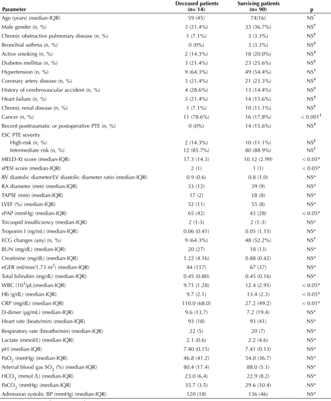
Table 2. Comparison deceased and surviving patients with respect to clinical, echocardiographic, electrocardiographic, and biochemical parameters
Parameter Deceased patients (n= 14) Surviving patients (n= 90) p
Age (years) (median-IQR) 59 (45) 74(16) NS*
Male gender (n, %) 3 (21.4%) 33 (36.7%) NS†
Chronic obstructive pulmonary disease (n, %) 1 (7.1%) 3 (3.3%) NS‡
Bronchial asthma (n, %) 0 (0%) 3 (3.3%) NS‡
Active smoking (n, %) 2 (14.3%) 18 (20.0%) NS‡
Diabetes mellitus (n, %) 3 (21.4%) 23 (25.6%) NS‡
Hypertension (n, %) 9 (64.3%) 49 (54.4%) NS†
Coronary artery disease (n, %) 3 (21.4%) 21 (23.3%) NS‡
History of cerebrovascular accident (n, %) 4 (28.6%) 13 (14.4%) NS‡
Heart failure (n, %) 3 (21.4%) 14 (15.6%) NS‡
Chronic renal disease (n, %) 1 (7.1%) 10 (11.1%) NS‡
Cancer (n, %) 11 (78.6%) 16 (17.8%) < 0.001†
Recent posttraumatic or postoperative PTE (n, %) 0 (0%) 14 (15.6%) NS‡
ESC PTE severity High-risk (n, %) Intermediate risk (n, %) 2 (14.3%) 12 (85.7%) 10 (11.1%) 80 (88.9%) NS‡ NS†
MELD-XI score (median-IQR) 17.3 (14.3) 10.12 (2.99) < 0.05*
sPESI score (median-IQR) 2 (1) 1 (1) < 0.05*
RV diastolic diameter/LV diastolic diameter ratio (median-IQR) 0.9 (0.6) 0.8 (1.0) NS*
RA diameter (mm) (median-IQR) 33 (12) 39 (9) NS*
TAPSE (mm) (median-IQR) 17 (2) 18 (8) NS*
LVEF (%) (median-IQR) 52 (11) 55 (8) NS*
sPAP (mmHg) (median-IQR) 65 (42) 43 (28) < 0.05*
Tricuspid insufficiency (median-IQR) 2 (1-3) 2 (1-3) NS*
Troponin I (ng/mL) (median-IQR) 0.06 (0.41) 0.05 (1.15) NS*
ECG changes (any) (n, %) 9 (64.3%) 48 (52.2%) NS†
BUN (mg/dL) (median-IQR) 20 (27) 18 (13) NS*
Creatinine (mg/dL) (median-IQR) 1.22 (4.16) 0.88 (0.42) NS*
eGFR (ml/min/1.73 m2) (median-IQR) 44 (117) 67 (37) NS*
Total bilirubin (mg/dL) (median-IQR) 0.45 (0.80) 0.45 (0.16) NS*
WBC (103/µL)(median-IQR) 9.71 (1.28) 12.4 (2.95) < 0.05*
Hb (g/dL) (median-IQR) 9.7 (2.1) 13.4 (2.3) < 0.05*
CRP (mg/dL) (median-IQR) 110.0 (68.0) 27.2 (49.2) < 0.01*
D-dimer (µg/mL) (median-IQR) 9.6 (13.7) 7.2 (19.4) NS*
Heart rate (beats/min) (median-IQR) 93 (18) 93 (41) NS*
Respiratory rate (breaths/min) (median-IQR) 22 (5) 20 (7) NS*
Lactate (mmol/L) (median-IQR) 2.1 (0.6) 2.2 (4.6) NS*
pH (median-IQR) 7.40 (0.15) 7.41 (0.13) NS*
PaO2 (mmHg) (median-IQR) 46.8 (41.2) 54.0 (36.7) NS*
Arterial blood gas SO2 (%) (median-IQR) 80.4 (17.4) 88.0 (5.1) NS*
HCO3 (mmol /L) (median-IQR) 23.0 (6.4) 22.9 (8.2) NS*
PaCO2 (mmHg) (median-IQR) 35.7 (3.5) 29.6 (10.4) NS*
Admission systolic BP (mmHg) (median-IQR) 120 (18) 136 (46) NS*
* Mann-Whitney U test; † Chi-square test; ‡ Fisher’s exact test.
ESC: European Society of Cardiology, PTE: Pulmonary thromboembolism, RV: Right ventricle, RA: Right atrium, TAPSE: Tricuspid annular plane systolic excursion, sPAP: Systolic pulmonary artery disease, BUN: Blood urea nitrogen, eGFR: Estimated glomerular filtration rate, WBC: White blood cell count, Hb: Hemoglobin, CRP: C-reactive protein, PaO2: Partial oxygen pressure, SO2: Oxygen saturation, PaCO2: Partial carbondioxide pressure, BP: Blood pressure.
DISCUSSION
The results of the present study revealed that, among hospital admitted intermediate-to-high risk PTE patients, MELD-XI score was significantly higher in deceased versus survivors. Furthermore, MELD-XI score was an independent predictor of in-hospital mortality and had an identical sensitivity and a better specificity than sPESI score for the same outcome. Finally, a higher MELD-XI score category (MELD-XI≥ 10.2) was associated with a significantly lower sur-vival after hospitalization. These results collectively indicate that MELD-XI score is a useful tool for mor-tality prediction among hospitalized intermedi-ate-to-high risk PTE patients.
PTE is a condition associated with a fairly high mor-bidity and mortality risk, being the third most com-mon vascular disease, only after myocardial infarc-tion and stroke and having an increasing incidence in parallel to cancer, immobility, and obesity (1,18-22). There are some risk stratification schemes and scores for mortality prediction among PTE patients, the most commonly used of which is the ESC risk stratification Table 3. Independent predictors of in-hospital death in binary logistic regression analysis
Variable Hazard ratio (95% CI) p
sPESI score 5.3 (1.14-25.09) < 0.05
MELD-XI score 1.40 (1.04-1.89) < 0.05
Systolic pulmonary artery pressure 1.08 (1.02-1.15) < 0.05
Hemoglobin count 0.481 (0.29-0.79) < 0.05
Figure 2. ROC curve for sPESI score.
Figure 3. Kaplan Meier in-hospital survival curve for MELD-XI score.
model with a proved role for mortality prediction (1,23). According to that model, pulmonary embo-lism severity is classified as high-risk, intermedi-ate-risk, and low-risk, and is based on cardiovascu-lar hemodynamics (syncope, persistent hypotension, profound bradycardia), right ventricular dilatation (increased right-to-left ventricular diameter ratio), systolic dysfunction (reduced right ventricular systol-ic function and/or wall hypo/akinesis), or myocardial injury (increased cardiac enzymes or B-natriuretic peptide level). There are other risk stratification models and scores, of which the most commonly utilized ones are PESI score and its simplified form, sPESI score, which are mainly based on demograph-ic and clindemograph-ical variables such as demographdemograph-ic factors (age, sex) comorbidities [cancer, heart failure (HF) or chronic obstructive pulmonary disease (COPD)] and vital signs (heart rate, systolic blood pressure, body temperature, respiratory rate, and consciousness level). Both scores have been linked to increase mor-tality risk in PTE (24-29). However, the afore-men-tioned scores primarily incorporate cardiopulmo-nary variables but not other systems, and it is unclear how impaired end-organ function, such as renal and hepatic functions, affects mortality. Herein we aimed to use MELD-XI to predict mortality in PTE. MELD-XI score was significantly higher in deceased patients and was an independent predictor of mortality apart from sPESI score. Furthermore, its predictive power for mortality was fairly good. Our positive results for MELD-XI score may indicate three possibilities about its ability to predict mortality in PTE. First, MELD-XI score may reflect a mortality increase as a result of impaired end-organ function and multiorgan failure secondary to cardiovascular derangement and result-ing low-output/congestive state due to PTE (30,31). Second, although being statistically non-significant, our results showed that PaO2 and SO2 levels in arte-rial blood gas analysis were lower in the deceased patients than the survivors. This may reflect that tis-sue hypoxia may also have increased mortality by impairing renal and/or hepatic function and thus increasing MELD-XI score. So, far studies have shown that tissue hypoxia may impair renal and hepatic function and/or cause reversible or irrevers-ible injury (32,33). As PTE may create severe hypox-ia and depress tissue oxygenization, this mechanism may be a link between mortality from PTE and an increased MELD-XI score. As the third and the last possibility, MELD-XI score may in fact predit
mortal-ity increase due to underlying disorders impairing renal and hepatic function, such as poor overall status, comorbidities or underlying disorders like cancer, COPD, HF, CAD, and recent surgery, among others. However, irrespective of which possibility is more important, these both of the two possibilities ultimately lead to the same conclusion that sicker patients with impaired renal and/or hepatic function of any reason are more likely to die. Therefore, MELD-XI score may be in fact may be assessing a common final pathway during the events culminat-ing into death in PTE. Hence, although it is a rela-tively simpler score incorporating serum levels of only two serum levels, it may be one, if not the only one, of the ultimate risk stratifiers in PTE, as in other criticall ilnesses. Besides, as it is an easy score requiring only two parameters readily available in serum biochemistry, it may readily calculated and give an immediate idea about the outlook of a patient. Therefore, our results seem very relevant with the goal of predicting mortality among these patients, perhaps not in the form of guiding deci-sions regarding thrombolytic therapy, but being aware of the more grave prognosis to prompt admin-istering more aggressive treatments to correct end-or-gan perfusion and/or congestion and to address causative or comorbid conditions.
LIMITATIONS
This study had some limitations. First of all, this was a retrosepctive study with a relatively small sample size and a low number of deceased patients. Second, we did not use B-type natriuretic peptide level for determining cardiac injury and based the latter sole-ly on troponin I measurement. Third, we assessed only in-hospital mortality but not long-term mortali-ty. Lastly, we only included hospitalized patients and we did not evaluate the role of MELD-XI for mortal-ity prediction for PTE patients managed on an outpa-tient basis.
CONCLUSION
The present study implies that MELD-XI score per-forms well and independently of sPESI score for mortality prediction among in-patients with interme-diate-to-high risk PTE. This subject must be further studied by large, randomized controlled studies. CONFLICT of INTEREST
AUTHORSHIP CONTRIBUTIONS Concept/Design: OÇ, ÇOÇ
Analysis/Interpretation: OÇ, ÇOÇ, GU Data Acquisition: OÇ, GU
Writting: Oç
Critical Revision: OÇ, EK Final Approval: OÇ, EK, İHM REFERENCES
1. Konstandinides SV, Torbicki A, Agnelli G, Danchin N, Fitzmaurice D, Galie N, et al. 2014 ESC Guidelines on the diagnosis and management of acute pulmonary embo-lism. Eur Heart J 2014;35:3033-69.
2. Goldhaber SZ, Visani L, De Rosa M. Acute pulmonary embolism: clinical outcome in the International Cooperative Pulmonary Embolism Registry. Lancet 1999;353:1386-9. 3. Donzé J, Le Gal G, Fine MJ, Roy PM, Sanchez O,
Verschuren F, et al. Prospective validation of the Pulmonary Embolism Severity Index. A clinical prognostic model for pulmonary embolism. Thromb Haemost 2008;100:943-8. 4. Jiménez D, Aujesky D, Moores L, Gómez V, Lobo
JL, Uresandi F, et al.; RIETE Investigators. Simplification of the pulmonary embolism severity index for prognostica-tion in patients with acute symptomatic pulmonary embo-lism. Arch Intern Med 2010;170:1383-9.
5. Kamath PS, Kim WR; Advanced Liver Disease Study Group. The model for end-stage liver disease (MELD). Hepatology 2007;45:797-805.
6. Cholongitas E, Marelli L, Shusang V, Senzolo M, Rolles K, Patch D, et al. A systematic review of the performance of the model for end-stage liver disease (MELD) in the setting of liver transplantation. Liver Transpl 2006;12:1049-61. 7. Wernly B, Lichtenauer M, Franz M, Kabisch B, Muessig J,
Masyuk M, et al. Model for End-stage Liver Disease excluding INR (MELD-XI) score in critically ill patients: Easily available and of prognostic relevance. PLoS One 2017;12:e0170987.
8. Abe S, Yoshihisa A, Takiguchi M, Shimizu T, Nakamura Y, Yamauchi H. Liver dysfunction assessed by model for end-stage liver disease excluding INR (MELD-XI) scoring sys-tem predicts adverse prognosis in heart failure. PLoS One 2014;9:e100618.
9. Critsinelis A, Kurihara C, Volkovicher N, Kawabori M, Sugiura T, Manon M 2nd, et al. Model of end-stage liver
disease-excluding international normalized ratio (MELD-XI) scoring system to predict outcomes in patients who undergo left ventricular assist device implantation. Ann Thorac Surg 2018;106:513-9.
10. Assenza GE, Graham DA, Landzberg MJ, Valente AM, Singh MN, Bashir A. MELD-XI score and cardiac mortality or transplantation in patients after Fontan surgery. Heart 2013;99:491-6.
11. He PC, Wei XB, Luo SN, Chen XL, Ke ZH, Yu DQ, et al. Risk prediction in infective endocarditis by modified MELD-XI score. Eur J Clin Microbiol Infect Dis 2018;37:1243-50. 12. Spieker M, Hellhammer K, Wiora J, Klose S, Zeus T, Jung C,
et al. Prognostic value of impaired hepato-renal function assessed by MELD-XI score in patientsundergoing percuta-neous mitral valve repair. Catheter Cardiovasc Interv 2018.
13. Inohara T, Kohsaka S, Shiraishi Y, Goda A, Sawano M, Yagawa M, et al. Prognostic impact of renal and hepatic dysfunction based on MELD-XI score in patients with acute heart failure. Int J Cardiol 2014;176:571-3. 14. Ronco C, McCullough P, Anker SD, Anand I, Aspromonte
N, Bagshaw SM, et al. Cardio-renal syndromes: report from the consensus conference of the acute dialysis quality ini-tiative. Eur Heart J 2010;31:703-11.
15. Nikolaou M, Parissis J, Yilmaz MB, Seronde MF, Kivikko M, Laribi S, et al. Liver function abnormalities, clinical profile, and outcome in acute decompensated heart fail-ure. Eur Heart J 2014;34:742-9.
16. Erol S, Gürün Kaya A, Arslan Ciftçi F, Çiledağ A, Şen E, Kaya A, et al. Is oxygen saturation variable of simplified pulmonary embolism severity index reliable for identifica-tion of patients, suitable for outpatient treatment. Clin Respir J 2018;12:762-6.
17. Heuman DM, Mihas AA, Habib A, Gilles HS, Stravitz RT, Sanyal AJ, et al. MELD-XI: a rational approach to “sick-est first” liver transplantation in cirrhotic patients requiring anticoagulant therapy. Liver Transpl 2007;13:30-7. 18. Condliffe R, Kiely DG, Gibbs JS, Corris PA, Peacock
AJ, Jenkins DP, et al. Prognostic and aetiological factors in chronic thromboembolic pulmonary hypertension. Eur Respir J 2009;33:332-8.
19. Fanikos J, Piazza G, Zayaruzny M, Goldhaber SZ. Long-term complications of medical patients with hospital-acquired venous thromboembolism. Thromb Haemost 2009;102:688-93.
20. Klok FA, van Kralingen KW, van Dijk AP, Heyning FH, Vliegen HW, Kaptein AA, et al. Quality of life in long-term survivors of acute pulmonary embolism. Chest 2010;138:1432-40.
21. Cohen AT, Agnelli G, Anderson FA, Arcelus JI, Bergqvist D, Brecht JG, et al; VTE Impact Assessment Group in Europe (VITAE). Venous thromboembolism (VTE) in Europe. The number of VTE events and associated mor-bidity and mortality. Thromb Haemost 2007;98:756-64. 22. Heit JA. The epidemiology of venous thromboembolism in
the community. Arterioscler Thromb Vasc Biol 2008;28:370-2.
23. Becattini C, Agnelli G, Lankeit M, Masotti L, Pruszczyk P, Casazza F, et al. Acute pulmonary embolism: mortality prediction by the 2014 European Society of Cardiology risk stratification model. Eur Respir J 2016;48:780-6.
24. Aujesky D, Obrosky DS, Stone RA, Auble TE, Perrier A, Cornuz J, et al. Derivation and validation of a prognostic model for pulmonary embolism. Am J Respir Crit Care Med 2005;172:1041-6.
25. Righini M, Roy PM, Meyer G, Verschuren F, Aujesky D, Le Gal G. The Simplified Pulmonary Embolism Severity Index (PESI): validation of a clinical prognostic model for pul-monary embolism. J Thromb Haemost 2011;9:2115-7. 26. Piovella F, Iosub DI. Acute pulmonary embolism: risk
assessment, risk stratification and treatment options. Clin Respir J 2016;10:545-54.
27. Meyer G, Vicaut E, Danays T, Agnelli G, Becattini C, Beyer-Westerndorf J, et al. Fibrinolysis for patients with interme-diate-risk pulmonary embolism. N Engl J Med 2014;370:1402-11.
28. Hobohm L, Hellenkamp K, Hasenfub G, Meunzel T, Konstantinides S, Lankeit M. Comparison of risk assess-ment strategies for not-high-risk pulmonary embolism. Eur Respir J 2016;47:1170-8.
29. Elias A, Mallett S, Daoud-Elias M, Poggi JN, Clarke M. Prognostic models in acute pulmonary embolism: a sys-tematic review and meta-analysis. BMJ Open 2016;6:e010324.
30. Gonsalez SR, Cortês AL, Silva RCD, Lowe J, Prieto MC, Silva Lara LD. Acute kidney injury overview: from basic findings to new prevention and therapy strategies. Pharmacol Ther 2019;200:1-12.
31. Seeto RK, Fenn B, Rockey DC. Ischemic hepatitis: clinical presentation and pathogenesis. Am J Med 2000;109:109-13.
32. Tögel F, Westenfelder C. Recent advances in the under-standing of acute kidney injury. F1000Prime Rep 2014;6:83.
33. Fuhrmann V, Kneidinger N, Herkner H, Heinz G, Nikfardjam M, Bojic A, et al. Hypoxic hepatitis: underlying conditions and risk factors for mortality in critically ill patients. Intensive Care Med 2009;35:1397-405.