O
riginal paperT
he pOTenTial prOgnOsTic rOle Of periTumOraleOsinOphils wiThin whOle TumOr
-
assOciaTedinflammaTOry cells and sTrOmal hisTOlOgical
characTerisTics in cOlOrecTal cancer
Saime Ramadan1, BuRcu Saka2, enveR YaRikkaYa3, ahmet Bilici4, muStafa Oncel5
1Department of Pathology, Baskent University Istanbul Hospital, Istanbul, Turkey 2Department of Pathology, Istanbul Medipol University Hospital, Istanbul, Turkey 3Department of Pathology, Istanbul Training and Research Hospital, Istanbul, Turkey 4Department of Medical Oncology, Istanbul Medipol University Hospital, Istanbul, Turkey 5Department of General Surgery, Istanbul Medipol University Hospital, Istanbul, Turkey
We aimed to determine the prognostic role of whole tumor-associated inflamma-tory cells, especially eosinophils, and stromal histological characteristics in relation to other prognostic parameters in patients with colorectal carcinoma (CRC). A to-tal of 122 patients who underwent an operation for CRC were included in this retrospective study. Conventional (tumor grade, TNM stage and venous invasion [VI]) and other histopathological (intratumoral/peritumoral budding [ITB/PTB], desmoplasia) tumor parameters were recorded and classified by density, as were the tumor-associated inflammatory parameters (intratumoral/peritumoral lympho-cytes [ITL/PTL], eosinophils [IE/PTE], overall inflammation [ITI/PTI], Crohn-like inflammation [CLI]). Cancer-specific survival data were analyzed with respect to all tumor parameters. High ITB and PTB were significantly correlated with a higher rate of pT4, VI and desmoplasia (p < 0.05). An association of moderate ITL and extensive PTL with lesser likelihood of VI and metastasis; an association of ex-tensive CLI with a significantly lower rate of metastasis and TNM stage IV; and minimal PTE with a significantly higher rate of pT4 stage, metastasis and ITB were detected (p < 0.05 for each). Our findings revealed that low score tumoral budding and an increase in tumor-related inflammation were associated with lesser likelihood of poor prognostic tumor parameters. Nonetheless, given the associa-tion of an increase in PTE with lesser likelihood of ITB, pT4, metastasis, and with non-significantly for better survival rates, our findings emphasize the potential role of peritumoral eosinophils as an additional prognostic parameter in CRC.
Key words: colorectal cancer, survival, tumor-related inflammation, tumor budding.
Introduction
Colorectal cancer (CRC) is one of the most common cancers worldwide and the third leading cause of can-cer-related mortality, despite better understanding
of its pathogenesis and advances in therapy. Colorectal cancer incidence has been increasing steadily, espe-cially in developing countries. Obesity, sedentary life style, red meat consumption, alcohol and tobacco are the major risk factors behind the growth of CRC [1].
Colon cancer and rectal cancer have similar histo-pathologic risk factors and similar survival rates [1, 2].
Colorectal cancer comprises a group of diseases driven by several mutations and mutagens. Heredi-tary CRCs account for 7-10% of all cases and include Lynch syndrome, adenomatous and hamartomatous syndromes [3]. Up to 30% of CRC patients have a family history of the neoplasm, meaning there are probably predisposing germ-line mutations. To date, several nuclear DNA variants have been shown to be associated with increased risk of CRC. However, there are conflicting results about the role of inherit-ed mitochondrial DNA (mtDNA) mutations in CRC carcinogenesis. Previous studies suggested that cer-tain polymorphic mtDNA positions were associated with increased risk of CRC in different nations [4, 5] but a British study [6] and a recent Polish [7] study did not support the hypothesis that mtDNA variants contribute to inherited predisposition of CRC.
Currently, post-surgical staging according to the American Joint Committee on Cancer (AJCC)/Union for International Cancer Control (UICC), tumor node metastasis (TNM) system is a standard reference for prognosis and a guide for therapeutic decision mak-ing in CRC [8]. However, TNM classification reflects an anatomical extent rather than biological tumor characteristics [9]. Alongside considerable variation in the clinical course of disease among patients with an identical stage [8, 10], unexpected progression during the follow-up of early stage, lymph node neg-ative patients causes difficulties to decide the ther-apeutic modalities [9, 10, 11, 12]. Tumor budding seems important in this regard, as a strong adverse prognostic factor to select patients for adjuvant ther-apy particularly in those with early stage, lymph node negative disease [12, 13].
However, given the heterogeneity of tumor biol-ogy within each stage, there is a need for additional stage-independent prognostic histopathological pa-rameters to identify patients with more aggressive clinical behavior. This will guide clinical decisions in terms of follow-up scheduling and provision of ad-juvant therapy tailored to individualized risk of pro-gression [8, 10, 12].
In accordance with the increasingly apparent rec-ognition of CRC outcome to be dependent not only on tumor characteristics but also on the interactions between host immune/inflammatory response and tumor [14], assessment of tumor-related local in-flammation has been proposed as a novel prognostic parameter [10, 14].
Several studies to date have confirmed the prog-nostic value of the tumor-related inflammatory or immune cell infiltrate especially the adaptive immune response (i.e. infiltration of T cells) in patients with
CRC [14, 15, 16]. For example, Galon et al. found
a positive correlation between the presence of
mark-ers for Th1 polarization of cytotoxic and memory T cells and a low incidence of tumor recurrence [15]. An international concortitum suggested the Immu-noscore assay to assess the prognostic value of total tumor-infiltrating T-cell counts and cytotoxic tu-mor-infiltrating T-cell counts in patients with stage I-III colon cancer. They found that the Immunoscore provides a reliable estimate of the risk of recurrence in patients with colon cancer [16]. There are a few studies that have focused on the innate immune cells (i.e. eosinophils) with a standardized assessment re-garding type, density and location of inflammatory infiltrate [17, 18, 19]. A simplified histopatholo-gy-based scoring method described by Klintrup et al. [18] is important in this regard, since it represents standard criteria for the prognostic value of tumor- related inflammatory infiltrate assessments.
This study was therefore designed to investigate the prognostic role of tumor-associated inflammatory parameters and tumor budding in relation to other prognostic tumor parameters in patients operated on CRC. Tumor-associated inflammatory infiltrates were assessed based on standardized criteria being strati-fied by location (intratumoral, peritumoral), inflam-matory cell subtype (lymphocyte, eosinophil, overall inflammation) and density.
Material and methods
Study population
A total of 122 patients operated on for colorectal cancer (mean (SD) age: 59.9 (12.4) years, 59.0% were men) were included in this retrospective study that was conducted at a tertiary care center between 2013 and 2014 (Table I). Rare histologic subtypes such as medullary and squamous cell carcinoma were exclud-ed from the study and patients with insufficient dis-ease information were excluded from data analysis.
The study was conducted in full accordance with local good clinical practice guidelines and current legislations, while permission was obtained from our institutional ethics committee for the use of pa-tient data for publication purposes. The need for con-sent from the patients was waived because of the ret-rospective nature of this study.
Study parameters
Data on patient demographics (age, gender), tumor grade and pT and pN stages, and neoadjuvant thera-py history were retrospectively obtained from patient charts. Metastases were recorded both at the time of diagnosis and during follow-up. Intratumoral (ITB), peritumoral budding (PTB), venous invasion and desmoplasia were evaluated in addition to tumor- associated inflammatory parameters (intratumoral
lymphocyte [ITL], peritumoral lymphocyte [PTL], Crohn-like inflammation [CLI], intratumoral eosino-phils [IE], peritumoral eosinoeosino-phils [PTE], intratumor-al overintratumor-all inflammation [ITI], peritumorintratumor-al overintratumor-all in-flammation [PTI]). Survival data were obtained from patients’ medical records or via phone call interviews.
The prognostic role of tumor-associated inflamma-tory parameters and desmoplasia in relation to conven-tional prognostic parameters were evaluated, along with 5-year cancer-specific survival (CSS) rate and mean CSS time. The relation of tumor budding with conventional prognostic parameters, tumor-related inflammatory pa-rameters and desmoplasia was also analyzed along with the concordance between ITB and PTB.
Histopathology
For the classification of TNM, AJCC/UICC 2017 criteria were used [20], while histological tumor type and differentiation (grade) were assessed based on the 2010 edition of the World Health Organization classification [21].
We analyzed venous invasion on hematoxylin and eo-sin (HE) slides with attention to the orphan arteriole and protruding tongue signs. An orphan arteriole sign is described as a circumscribed tumor nodule adjacent to a muscularized artery without an obvious accompa-nying vein. The protruding tongue sign is described as a smooth bordered protrusion of a tumor into pericolic fat adjacent to an artery [22]. Besides these morpho-logic findings, many studies have demonstrated the su-periority of elastin staining compared to (HE) alone in the detection of VI [22, 23]. Since determination of the true prevalence of VI is difficult based on HE, es-pecially when the muscular wall of the vein is obliterat-ed, elastin stain was also performed on all tumor blocks. Venous invasion was considered positive when a tumor was observed in an endothelium-lined space [22].
Both intratumoral and peritumoral budding were assessed on HE slides as described previously by
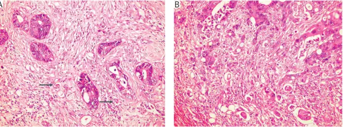
Gra-ham et al. [24]. The intensity of budding was scored accordingly as no budding, low (1-9 budding foci), and high (≥ 10 foci) (Figs. 1, 2). Desmoplasia was scored semi-quantitatively as minimal, moderate and severe.
Table I. Demographic and clinical characteristics of the study group age, years (mean, range) 60 28-87 n % Gender Male 72 59 Female 50 41 Grade Low 107 87.7 High 15 12.3 T 1-2 23 18.8 3 70 53.4 4 29 27.8 N 0 72 59.0 1 33 26.9 2 17 14.1 Metastasis No 97 79.5 Yes 25 20.5 TNM Stage I 21 17.2 II 44 36.1 III 35 28.7 IV 22 18.0
Fig. 1. Low (shown by arrows) and high score intratumoral budding (HE, magnification 200×)
Tumor-associated inflammation
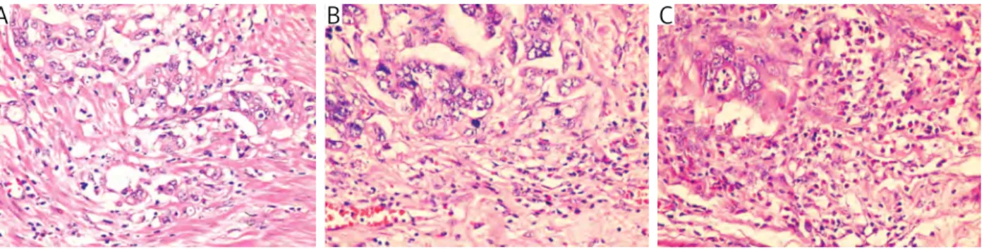
The intensity of overall inflammatory cell reaction was assessed separately within the tumor center (in-tratumoral) and in the stroma at the invasive tumor margin (peritumoral) with a modified method de-scribed by Klintrup et al. [18] and scored from 1 to 3 based on a mild or patchy increase (score 1), a prom-inent inflammatory reaction (score 2), and a florid “cuplike” inflammatory infiltrate (score 3; Fig. 3).
The numbers of intratumoral and peritumoral eosinophils were evaluated separately on HE stained slides, using a 40× objective lens in a high power
field (HPF) measuring 0,24 mm2 (Olympus BX45).
IE and PTE were categorized according to modified Fernandez-Acenero criteria, considering of < 10 cells/
0.24 mm2 area as minimal, 10-50 cells/0.24 mm2
area as moderate and > 50 cells/0.24 mm2 area as
extensive infiltration [25] (Fig. 4).
Crohn-like response was scored as none or mild-to-moderate and marked according to College of Amer-ican Pathologists (CAP) criteria [26]. Intratumoral lymphocytes were evaluated based on CAP criteria and graded as none, mild-to-moderate (1-2 per HPF) and marked (≥ 3 per HPF), (Fig. 5).
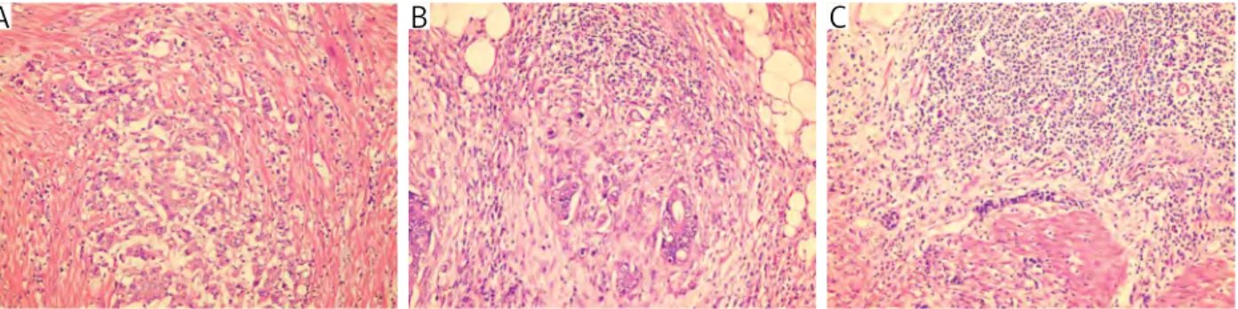
Peritumoral lymphocytes were evaluated according to modified Huh et al. [27] criteria on HE stained slides and categorized as minimal, moderate and severe (Fig. 6).
Fig. 2. Low (shown by arrows) and high score peritumoral budding (HE, magnification 200×)
Fig. 3. The intensity of overall inflammatory cell reaction in order to minimal, moderate and severe (HE, magnification 200×)
Fig. 4. The intensity of eosinophil infiltration in order to minimal, moderate and severe (HE, magnification 400×)
A
B
C
A
B
Statistical analysis
Statistical analysis was performed using IBM SPSS Statistics for Windows, version 22.0 (IBM Corp., Ar-monk, NY). The χ2 test was used for the comparison
of categorical data, while the one way ANOVA test was used for the parametric variables. The κ concor-dance index was used to determine the concorconcor-dance between intratumoral and peritumoral budding. Cancer-specific survival was assessed using the Ka-plan-Meier method and the significance among pa-tient groups was calculated using the log-rank test. Cox proportional hazards regression model was used to determine hazard ratios and data were expressed as “mean (standard deviation; SD)”, minimum-max-imum, 95% confidence interval (CI) and percent (%) where appropriate. P < 0.05 was considered statisti-cally significant.
Results
Tumor-associated inflammatory parameters and tumor budding according to grade, TNM stage and venous invasion
Tumor-associated inflammatory parameters and tumor budding according to TNM stage are shown in Table II.
High ITB as compared with the absence of ITB and low ITB was associated with significantly higher rate of pT4 (42.0% vs. 6.3% and 12.5%, respective-ly, p = 0.002) and venous invasion (54.0% vs. 18.7% and 33.9%, respectively, p = 0.019).
High PTB as compared with the absence of PTB and low PTB was associated with significantly higher rate of pT4 (41.5% vs. 6.3% and 11.3%, respective-ly, p = 0.001) and TNM stage IV (28.3% vs. 12.5% and 9.4%, respectively, p = 0.043) alongside higher likelihood of venous invasion (52.8% vs. 18.8% and 34.0%, respectively, p = 0.024).
Most of our cases were low grade (87.7%). We only found a statistically significant positive correla-tion with grade and ITL. Extensive ITL was strong-ly associated with a higher likelihood of high grade tumor rather than mild-to-moderate ITL (26.9% vs. 7.2%, p = 0.033). However extensive ITL was in-versely correlated with venous invasion (p = 0.048).
The presence of metastasis was more likely to occur in cases of minimal (28.1%) and moderate (17.9%) PTL in comparison to extensive (0.0%) PTL (p = 0.025).
Marked CLI as compared to the absence of CLI and mild-to-moderate CLI was associated with a sig-nificantly lower rate of metastasis and TNM stage IV (p = 0.005).
Fig. 5. Crohn-like response: minimal and marked (HE, magnification 40×)
Fig. 6. Peritumoral lymphocyte infiltration in order to minimal, moderate and severe (HE, magnification 200×)
A
B
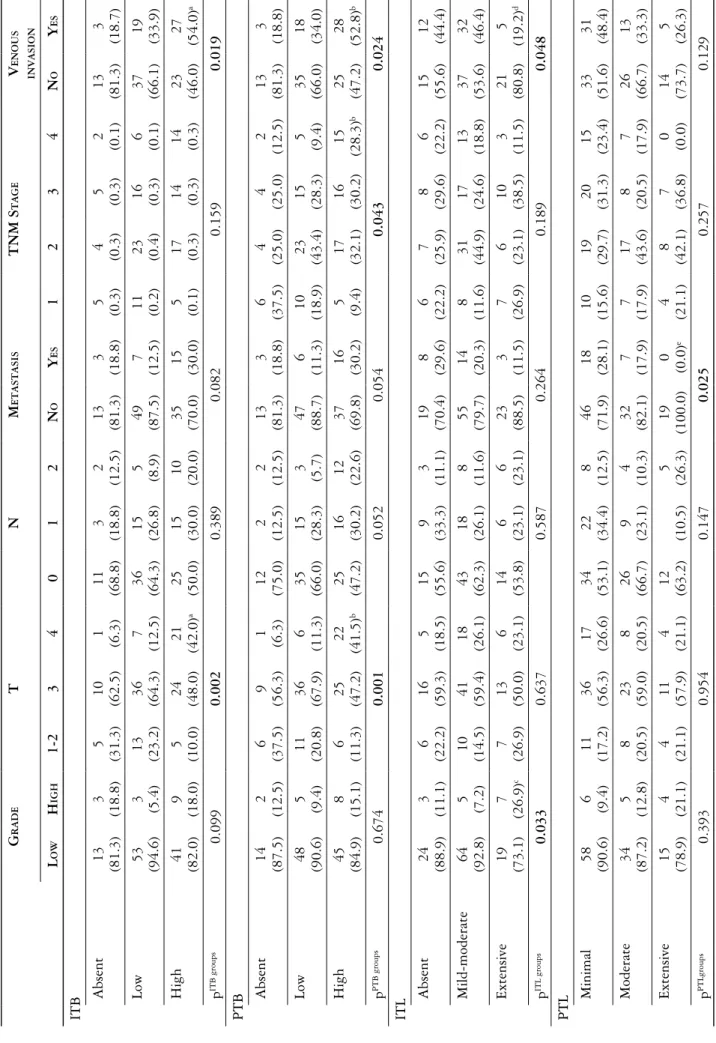
Table II.
T
umor budding and tumor
-associated inflammatory parameters with respect to tumor grade, TNM stage and venous invasion
g rade T n m eT as T asis T nm s T age V en O us in V asi O n l O w h igh 1-2 3 4 0 1 2 n O y es 1 2 3 4 n O y es ITB Absent 13 (81.3) 3 (18.8) 5 (31.3) 10 (62.5) 1 (6.3) 11 (68.8) 3 (18.8) 2 (12.5) 13 (81.3) 3 (18.8) 5 (0.3) 4 (0.3) 5 (0.3) 2 (0.1) 13 (81.3) 3 (18.7) Low 53 (94.6) 3 (5.4) 13 (23.2) 36 (64.3) 7 (12.5) 36 (64.3) 15 (26.8) 5 (8.9) 49 (87.5) 7 (12.5) 11 (0.2) 23 (0.4) 16 (0.3) 6 (0.1) 37 (66.1) 19 (33.9) High 41 (82.0) 9 (18.0) 5 (10.0) 24 (48.0) 21 (42.0) a 25 (50.0) 15 (30.0) 10 (20.0) 35 (70.0) 15 (30.0) 5 (0.1) 17 (0.3) 14 (0.3) 14 (0.3) 23 (46.0) 27 (54.0) a p ITB groups 0.099 0.002 0.389 0.082 0.159 0.019 PTB Absent 14 (87.5) 2 (12.5) 6 (37.5) 9 (56.3) 1 (6.3) 12 (75.0) 2 (12.5) 2 (12.5) 13 (81.3) 3 (18.8) 6 (37.5) 4 (25.0) 4 (25.0) 2 (12.5) 13 (81.3) 3 (18.8) Low 48 (90.6) 5 (9.4) 11 (20.8) 36 (67.9) 6 (11.3) 35 (66.0) 15 (28.3) 3 (5.7) 47 (88.7) 6 (11.3) 10 (18.9) 23 (43.4) 15 (28.3) 5 (9.4) 35 (66.0) 18 (34.0) High 45 (84.9) 8 (15.1) 6 (11.3) 25 (47.2) 22 (41.5) b 25 (47.2) 16 (30.2) 12 (22.6) 37 (69.8) 16 (30.2) 5 (9.4) 17 (32.1) 16 (30.2) 15 (28.3) b 25 (47.2) 28 (52.8) b p PTB groups 0.674 0.001 0.052 0.054 0.043 0.024 ITL Absent 24 (88.9) 3 (11.1) 6 (22.2) 16 (59.3) 5 (18.5) 15 (55.6) 9 (33.3) 3 (11.1) 19 (70.4) 8 (29.6) 6 (22.2) 7 (25.9) 8 (29.6) 6 (22.2) 15 (55.6) 12 (44.4) Mild-moderate 64 (92.8) 5 (7.2) 10 (14.5) 41 (59.4) 18 (26.1) 43 (62.3) 18 (26.1) 8 (11.6) 55 (79.7) 14 (20.3) 8 (11.6) 31 (44.9) 17 (24.6) 13 (18.8) 37 (53.6) 32 (46.4) Extensive 19 (73.1) 7 (26.9) c 7 (26.9) 13 (50.0) 6 (23.1) 14 (53.8) 6 (23.1) 6 (23.1) 23 (88.5) 3 (11.5) 7 (26.9) 6 (23.1) 10 (38.5) 3 (11.5) 21 (80.8) 5 (19.2) d p ITL groups 0.033 0.637 0.587 0.264 0.189 0.048 PTL Minimal 58 (90.6) 6 (9.4) 11 (17.2) 36 (56.3) 17 (26.6) 34 (53.1) 22 (34.4) 8 (12.5) 46 (71.9) 18 (28.1) 10 (15.6) 19 (29.7) 20 (31.3) 15 (23.4) 33 (51.6) 31 (48.4) Moderate 34 (87.2) 5 (12.8) 8 (20.5) 23 (59.0) 8 (20.5) 26 (66.7) 9 (23.1) 4 (10.3) 32 (82.1) 7 (17.9) 7 (17.9) 17 (43.6) 8 (20.5) 7 (17.9) 26 (66.7) 13 (33.3) Extensive 15 (78.9) 4 (21.1) 4 (21.1) 11 (57.9) 4 (21.1) 12 (63.2) (10.5) 5 (26.3) 19 (100.0) 0 (0.0) e 4 (21.1) 8 (42.1) 7 (36.8) 0 (0.0) 14 (73.7) 5 (26.3) p PTLgroups 0.393 0.954 0.147 0.025 0.257 0.129
g rade T n m eT as T asis T nm s T age V en O us in V asi O n l O w h igh 1-2 3 4 0 1 2 n O y es 1 2 3 4 n O y es CLI Absent 43 (93.5) 3 (6.5) 13 (28.3) 26 (56.5) 7 (15.2) 29 (63) 12 (26.1) 5 (10.9) 32 (69.6) 14 (30.4) 13 (28.3) 10 (21.7) 11 (23.9) 12 (26.1) 29 (63) 17 (37.0) Mild-moderate 34 (87.2) 5 (12.8) 5 (12.8) 21 (53.8) 13 (33.3) 21 (53.8) 11 (28.2) 7 (17.9) 29 (74.4) 10 (25.6) 4 (10.3) 16 (41.0) 10 (25.6) 9 (23.1) 19 (48.7) 20 (51.3) Marked 30 (81.1) 7 (18.9) 5 (13.5) 23 (62.2) 9 (24.3) 22 (59.5) 10 (27) 5 (13.5) 36 (97.3) 1 (2.7) f 4 (10.8) 18 (48.6) 14 (37.8) 1 (2.7) f 25 (67.6) 12 (32.4) p CLI groups 0.230 0.157 0.893 0.005 0.005 0.210 IE Minimal 71 (85.5) 12 (14.5) 13 (15.7) 49 (59.0) 21 (25.3) 45 (54.2) 26 (31.3) 12 (14.5) 63 (75.9) 20 (24.1) 12 (14.5) 28 (33.7) 26 (31.3) 17 (20.5) 45 (54.2) 38 (45.8) Moderate 20 (100.0) 0 (0.0) 4 (20.0) 12 (60.0) 4 (20.0) 15 (75.0) 3 (15.0) 2 (10.0) 17 (85.0) 3 (15.0) 3 (15.0) 11 (55.0) 3 (15.0) 3 (15.0) 15 (75.0) 5 (25.0) Extensive 16 (84.2) 3 (15.8) 6 (31.6) 9 (47.4) 4 (21.1) 12 (63.2) 4 (21.1) 3 (15.8) 17 (89.5) 2 (10.5) 6 (31.6) 5 (26.3) 6 (31.6) 2 (10.5) 13 (68.4) 6 (31.6) p IE groups 0.185 0.600 0.491 0.334 0.249 0.166 PTE Minimal 53 (82.8) 11 (17.2) 10 (15.6) 34 (53.1) 20 (31.3) 32 (50.0) 21 (32.8) 11 (17.2) 45 (70.3) 19 (29.7) 9 (14.1) 18 (28.1) 21 (32.8) 16 (25.0) 33 (51.6) 31 (48.4) Moderate 29 (93.5) 2 (6.5) 4 (12.9) 25 (80.6) 2 (6.5) g 21 (67.7) 8 (25.8) 2 (6.5) 28 (90.3) 3 (9.7) h 4 (12.9) 16 (51.6) 8 (25.8) 3 (9.7) 21 (67.7) 10 (32.3) Extensive 25 (92.6) 2 (7.4) 9 (33.3) 11 (40.7) 7 (25.9) 19 (70.4) 4 (14.8) 4 (14.8) 24 (88.9) 3 (11.1) 8 (29.6) 10 (37.0) 6 (22.2) 3 (11.1) 19 (70.4) 8 (29.6) p PTE groups 0.223 0.008 0.206 0.030 0.094 0.144 ITI Minimal 57 (86.4) 9 (13.6) 12 (18.2) 38 (57.6) 16 (24.2) 39 (59.1) 18 (27.3) 9 (13.6) 48 (72.7) 18 (27.3) 11 (16.7) 22 (33.3) 18 (27.3) 15 (22.7) 36 (54.5) 30 (45.5) Moderate 29 (87.9) 4 (12.1) 4 (12.1) 22 (66.7) 7 (21.2) 19 (57.6) 10 (30.3) 4 (12.1) 28 (84.8) 5 (15.2) 4 (12.1) 14 (42.4) 10 (30.3) 5 (15.2) 20 (60.6) 13 (39.4) Extensive 21 (91.3) 2 (8.7) 7 (30.4) 10 (43.5) 6 (26.1) 14 (60.9) 5 (21.7) 4 (17.4) 21 (91.3) 2 (8.7) 6 (26.1) 8 (34.8) 7 (30.4) 2 (8.7) 17 (73.9) 6 (26.1) p ITI groups 0.824 0.424 0.954 0.111 0.643 0.263 Table II. Cont.
g rade T n m eT as T asis T nm s T age V en O us in V asi O n l O w h igh 1-2 3 4 0 1 2 n O y es 1 2 3 4 n O y es PTI Minimal 57 (87.7) 8 (12.3) 11 (16.9) 37 (56.9) 17 (26.2) 36 (55.4) 20 (30.8) 9 (13.8) 48 (73.8) 17 (26.2) 10 (15.4) 20 (30.8) 20 (30.8) 15 (23.1) 37 (56.9) 28 (43.1) Moderate 28 (82.4) 6 (17.6) 7 (20.6) 21 (61.8) 6 (17.6) 21 (61.8) 10 (29.4) 3 (8.8) 28 (82.4) 6 (17.6) 6 (17.6) 14 (41.2) 9 (26.5) 5 (14.7) 20 (58.8) 14 (41.2) Extensive 22 (95.7) 1 (4.3) 5 (21.7) 12 (52.2) 6 (26.1) 15 (65.2) 3 (13.0) 5 (21.7) 21 (91.3) 2 (8.7) 5 (21.7) 10 (43.5) 6 (26.1) 2 (8.7) 16 (69.6) 7 (30.4) p PTI groups 0.325 0.869 0.399 0.182 0.683 0.563 Desmoplasia Minimal 53 (85.5) 9 (14.5) 13 (21.0) 39 (62.9) 10 (16.1) 39 (62.9) 14 (22.6) 9 (14.5) 51 (82.3) 11 (17.7) 12 (19.4) 21 (33.9) 20 (32.3) 9 (14.5) 39 (62.9) 23 (37.1) Moderate 31 (91.2) 3 (8.8) 6 (17.6) 18 (52.9) 10 (29.4) 21 (61.8) 10 (29.4) 3 (8.8) 23 (67.6) 11 (32.4) 5 (14.7) 15 (44.1) 4 (11.8) 10 (29.4) 19 (55.9) 15 (44.1) Extensive 23 (88.5) 3 (11.5) 4 (15.4) 13 (50.0) 9 (34.6) 12 (46.2) 9 (34.6) 5 (19.2) 23 (88.5) 3 (11.5) 4 (15.4) 8 (30.8) 11 (42.3) 3 (11.5) 15 (57.7) 11 (42.3) p desmopl. groups 0.713 0.367 0.527 0.105 0.122 0.774 Neoadj tx. Absent 83 (88.3) 11 (11.7) 11 (11.7) 58 (61.7) 25 (26.6) 55 (58.5) 23 (24.5) 16 (17.0) 76 (80.9) 18 (19.1) 10 (10.6) 39 (41.5) 27 (28.7) 18 (19.1) 53 (56.4) 4 (43.6) Present 24 (85.7) 4 (14.3) 12 (42.9) 12 (42.9) 4 (14.3) i 17 (60.7) 10 (35.7) 1 (3.6) 21 (75.0) 7 (25.0) 11 (39.3) i 5 (17.9) i 8 (28.6) 4 (14.3) 20 (71.4) 8 (28.6) p tx groups 0.746 0.001 0.148 0.501 0.003 0.154
ITB – intratumoral budding; PTB – peritumoral budding; ITL – intratumoral lymphocyte; P
TL – peritumoral lymphocyte; CLI – Crohn-like inflammation; IE – intratumoral eosinophils; PTE – peritumoral eosinophils; ITI –
intratumoral
overall inflammation; PTI – peritumoral overall inflammation p <
0.05;compared to
alow and absent ITB groups, blow and absent PTB groups, cminimal ITL group, d absent and minimal ITL groups, e minimal and moderate PTL groups, fabsent and mild-moderate CLI groups, gminimal and extensive PTE
groups,
hminimal PTE group and iabsence of neoadjuvant therapy ( χ 2 test) Table II. Cont.
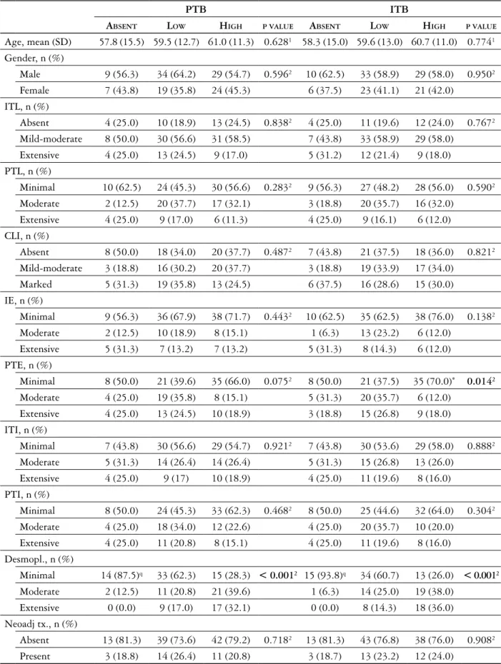
Table III. Concordance between ITB and PTB and association of tumor budding with study parameters
pTB iTB
aBsenT lOw high pValue aBsenT lOw high pValue
Age, mean (SD) 57.8 (15.5) 59.5 (12.7) 61.0 (11.3) 0.6281 58.3 (15.0) 59.6 (13.0) 60.7 (11.0) 0.7741 Gender, n (%) Male 9 (56.3) 34 (64.2) 29 (54.7) 0.5962 10 (62.5) 33 (58.9) 29 (58.0) 0.9502 Female 7 (43.8) 19 (35.8) 24 (45.3) 6 (37.5) 23 (41.1) 21 (42.0) ITL, n (%) Absent 4 (25.0) 10 (18.9) 13 (24.5) 0.8382 4 (25.0) 11 (19.6) 12 (24.0) 0.7672 Mild-moderate 8 (50.0) 30 (56.6) 31 (58.5) 7 (43.8) 33 (58.9) 29 (58.0) Extensive 4 (25.0) 13 (24.5) 9 (17.0) 5 (31.2) 12 (21.4) 9 (18.0) PTL, n (%) Minimal 10 (62.5) 24 (45.3) 30 (56.6) 0.2832 9 (56.3) 27 (48.2) 28 (56.0) 0.5902 Moderate 2 (12.5) 20 (37.7) 17 (32.1) 3 (18.8) 20 (35.7) 16 (32.0) Extensive 4 (25.0) 9 (17.0) 6 (11.3) 4 (25.0) 9 (16.1) 6 (12.0) CLI, n (%) Absent 8 (50.0) 18 (34.0) 20 (37.7) 0.4872 7 (43.8) 21 (37.5) 18 (36.0) 0.8212 Mild-moderate 3 (18.8) 16 (30.2) 20 (37.7) 3 (18.8) 19 (33.9) 17 (34.0) Marked 5 (31.3) 19 (35.8) 13 (24.5) 6 (37.5) 16 (28.6) 15 (30.0) IE, n (%) Minimal 9 (56.3) 36 (67.9) 38 (71.7) 0.4432 10 (62.5) 35 (62.5) 38 (76.0) 0.1382 Moderate 2 (12.5) 10 (18.9) 8 (15.1) 1 (6.3) 13 (23.2) 6 (12.0) Extensive 5 (31.3) 7 (13.2) 7 (13.2) 5 (31.3) 8 (14.3) 6 (12.0) PTE, n (%) Minimal 8 (50.0) 21 (39.6) 35 (66.0) 0.0752 8 (50.0) 21 (37.5) 35 (70.0)* 0.0142 Moderate 4 (25.0) 19 (35.8) 8 (15.1) 5 (31.3) 20 (35.7) 6 (12.0) Extensive 4 (25.0) 13 (24.5) 10 (18.9) 3 (18.8) 15 (26.8) 9 (18.0) ITI, n (%) Minimal 7 (43.8) 30 (56.6) 29 (54.7) 0.9212 7 (43.8) 30 (53.6) 29 (58.0) 0.8882 Moderate 5 (31.3) 14 (26.4) 14 (26.4) 5 (31.3) 15 (26.8) 13 (26.0) Extensive 4 (25.0) 9 (17) 10 (18.9) 4 (25.0) 11 (19.6) 8 (16.0) PTI, n (%) Minimal 8 (50.0) 24 (45.3) 33 (62.3) 0.4682 8 (50.0) 25 (44.6) 32 (64.0) 0.3042 Moderate 4 (25.0) 18 (34.0) 12 (22.6) 4 (25.0) 20 (35.7) 10 (20.0) Extensive 4 (25.0) 11 (20.8) 8 (15.1) 4 (25.0) 11 (19.6) 8 (16.0) Desmopl., n (%) Minimal 14 (87.5)q 33 (62.3) 15 (28.3) < 0.0012 15 (93.8)q 34 (60.7) 13 (26.0) < 0.0012 Moderate 2 (12.5) 11 (20.8) 21 (39.6) 1 (6.3) 14 (25.0) 19 (38.0) Extensive 0 (0.0) 9 (17.0) 17 (32.1) 0 (0.0) 8 (14.3) 18 (36.0) Neoadj tx., n (%) Absent 13 (81.3) 39 (73.6) 42 (79.2) 0.7182 13 (81.3) 43 (76.8) 38 (76.0) 0.9082 Present 3 (18.8) 14 (26.4) 11 (20.8) 3 (18.7) 13 (23.2) 12 (24.0)
ITB – intratumoral budding; PTB – peritumoral budding; ITL – intratumoral lymphocyte; PTL – peritumoral lymphocyte; CLI – Crohn-like inflammation; IE – intratumoral eosinophils; PTE – peritumoral eosinophils; ITI – intratumoral overall inflammation; PTI – peritumoral overall inflammation; Desmopl. – desmoplasia; tx – therapy
1 Oneway ANOVA test, χ2 test
Minimal PTE was associated with a significantly higher rate of metastasis and pT4 when compared with moderate and extensive PTE (p = 0.03 and 0.008, respectively).
Concordance between ITB and PTB and their association with study parameters
ITB and PTB were positive totally in 86.9% of pa-tients with high concordance between ITB and PTB (92.8%, p = 0.000). The absence of ITB (87.5% vs. 12.5% and 0.0%, respectively, p < 0.001) and PTB (93.8% vs. 6.3% and 0.0%, respectively, p < 0.001) were both associated with having a higher probabili-ty of minimal desmoplasia. The presence of high ITB was associated with a higher rate of minimal PTE (70.0% vs. 12.0 and 18.0%, respectively, p = 0.014). No other significant association of PTB or ITB was noted within the study parameters other than those mentioned above (Table III).
Survival data
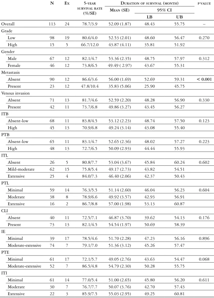
Five-year CSS rate was 78.7%, mean (SE) dura-tion of survival was 52.09 (1.87) months. The ab-sence of metastasis when compared to the preab-sence of metastasis was associated with significantly higher 5-year survival rate (86.6% vs. 47.8%) and a longer duration of survival (mean [SE] 56.0 [1.69] vs. 35.83 [5.06] months) (p < 0.001). TNM stage, tumor grade, venous invasion, desmoplasia, neoadjuvant therapy, budding or other tumor-related
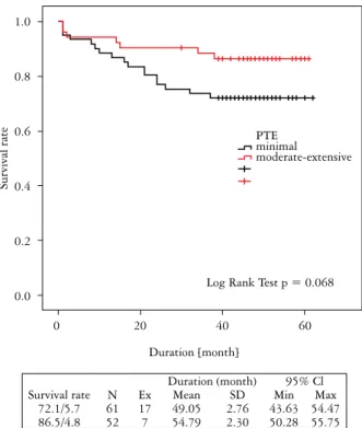
inflamma-tory parameters had no significant impact on CSS. Albeit not statistically significant, there was a higher rate of 5-year CSS (86.5% vs. 72.15%) and a lon-ger duration of survival mean [SE] 54.79 [2.30] vs. 49.05 [2.76] months) in the presence of a moder-ate-extensive rather than a minimal PTE (p = 0.068) (Table IV, Fig. 7).
Cox proportional hazards regression model assessing the prognostic significance of study parameters
In Cox proportional hazards regression analyses, an increasing age (hazard ratio 1.076, 95% CI: 1.025-1.130, p = 0.003) and the presence of metastasis (haz-ard ratio: 4.591, 95% CI: 1.814-11.619, p = 0.001) were independently associated with poor CSS.
Discussion
Colorectal cancer is classically seen in aged people and more common in males [1]. In accordance with this, the mean age of our study group was 60 and the majority (59%) were male.
Our findings revealed that there was an associa-tion of an increase in tumor-related inflammatory pa-rameters with lesser likelihood of certain convention-al poor prognostic tumor parameters such as pT4, TNM stage IV, venous invasion and metastasis, and that of higher PTE with lower ITB. This seems in agreement with the consistently reported association of a high-grade peritumoral or intratumoral local inflammatory response with an effective anti-tumor host immune responses and an improved prognosis following a potentially curative resection for CRC [14, 15, 16, 17].
Our findings emphasize the potential role of PTE in prognostic outcome in operated CRC patients giv-en the association of an increase in PTE with a less-er likelihood of ITB and metastasis as well as with a non-significantly for higher 5-year CSS rate and longer duration of CSS.
High IE and PTE counts were shown to be cor-related with improved survival in operable CRC in previous studies [10, 14, 25, 28, 29], being inde-pendent of AJCC/UICC stage [14, 25, 28] and more common for tumors lacking lymph node or distant metastases [30, 31]. However, consistent with our findings non-significantly better survival rather than a significant relationship was also reported for PTE in patients with CRC [18].
In a recent study by Harbaum et al. [10] on retro-spective analysis of peri- and intratumoral eosinophil counts in 381 CRC patients, the presence or an in-creasing number of eosinophils at the tumor margin was reported to be strongly associated with a favor-able tumor phenotype in terms of TNM stage, tu-mor grade, vascular invasion, and tutu-mor budding as
Fig. 7. Survival rate in minimal vs. moderate-extensive peri-tumoral eosinophils 1.0 0.8 0.6 0.4 0.2 0.0 Survival rate PTE minimal moderate-extensive
Log Rank Test p = 0.068
Duration [month]
0 20 40 60
Duration (month) 95% Cl
Survival rate N Ex Mean SD Min Max
72.1/5.7 61 17 49.05 2.76 43.63 54.47
Table IV. Survival data in relation to study parameters
n ex 5-year
surViValraTe
(%/se)
duraTiOnOfsurViVal (mOnTh) pValue
mean (se) 95% ci lB uB Overall 113 24 78.7/3.9 52.09 (1.87) 48.43 55.75 – Grade Low 98 19 80.6/4.0 52.53 (2.01) 48.60 56.47 0.270 High 15 5 66.7/12.0 43.87 (4.11) 35.81 51.92 Gender Male 67 12 82.1/4.7 53.36 (2.35) 48.75 57.97 0.312 Female 46 12 73.8/6.5 49.49 ( 2.97) 43.67 55.31 Metastasis Absent 90 12 86.6/3.6 56.00 (1.69) 52.69 59.31 < 0.001 Present 23 12 47.8/10.4 35.83 (5.06) 25.90 45.75 Venous invasion Absent 71 13 81.7/4.6 52.59 (2.20) 48.28 56.90 0.330 Present 42 11 73.7/6.8 49.86 (3.27) 43.45 56.27 ITB Absent-low 68 11 83.8/4.5 53.12 (2.23) 48.74 57.50 0.123 High 45 13 70.9/6.8 49.24 (3.14) 43.08 55.40 PTB Absent-low 65 11 83.1/4.7 52.65 (2.36) 48.02 57.27 0.223 High 48 13 72.7/6.5 50.09 (2.93) 44.44 55.93 ITL Absent 26 5 80.8/7.7 53.04 (3.67) 45.84 60.24 0.602 Mild-moderate 62 15 75.8/5.4 49.17 (2.73) 43.82 54.51 Extensive 25 4 84.0/7.3 46.40 (2.06) 42.37 50.43 PTL Minimal 59 14 76.3/5.5 51.14 (2.60) 46.04 56.23 0.604 Moderate 38 8 78.9/6.6 49.92 (3.57) 42.93 56.91 Extensive 16 2 86.7/8.8 57.00 (1.98) 53.13 60.87 CLI Absent 40 11 72.5/7.1 46.87 (3.70) 39.62 54.13 0.176 Present 73 13 82.1/4.5 54.54 (1.97) 50.69 58.39 IE Minimal 39 17 78.5/4.6 51.70 (2.28) 47.23 56.16 0.896 Moderate-extensive 74 7 79.1/7.0 51.36 (3.12) 45.26 57.47 PTE Minimal 61 17 72.1/5.7 49.05 (2.76) 43.63 54.47 0.068 Moderate-extensive 52 7 86.5/4.8 54.79 (2.30) 50.28 55.75 ITI Minimal 61 14 77.0/5.4 51.00 (2.65) 45.80 56.20 0.611 Moderate 30 7 76.7/7.7 50.07 (3.76) 42.70 57.43 Extensive 22 3 85.9/7.5 55.03 (2.95) 49.25 60.81
well as with improved survival [10]. The authors also noted that although the PTE count correlated with the intensity of the overall inflammatory cell reac-tion, it was independently associated with the out-come [10]. In our study, although we found that high PTE was strongly associated with better survival rates, probably due to the relatively small sample size, we did not obtain statistically significant p val-ues (p = 0.068). Similarly, in our cohort, although extensive CLI was also associated with a decreased rate of metastasis and TNM stage 4, only PTE was associated significantly with ITB and potential sur-vival. This seems to support the potential role of eo-sinophil infiltration as an antitumoral mechanism in-dependent of overall host inflammatory cell reaction in CRCs [10, 32].
In a recent study by Briede et al. on retrospective analysis of 553 CRC cases, high grade peritumoral in-flammation was associated with beneficial morpholog-ic CRC features, including less frequent manifestations of invasion [19]. In our study, we found no associ-ation of ITI and PTI with any of the tumor prog-nostic parameters. This may be due to high density of neutrophil leukocytes in ITI and PTI, since there are controversial findings on the prognostic value of neutrophils in CRC [33, 34]. These controversies may be explained by the duality of neutrophils com-prising both a tumor suppressive N1 population and tumor supportive N2 neutrophils [35].
Nonetheless, while the evaluation of PTE seems to be a promising tool with potential to improve risk stratification in CRC patients [10], inconsistency re-garding its relationship on overall inflammation or staging of the disease [10, 17, 28, 29] is considered
to challenge its incorporation in the routine prognos-tic assessment in clinical pracprognos-tice [17].
In a study by Nagtegaal et al. [28] in 160 CRC pa-tients, an increasing peritumoral, but not intratumoral eosinophil count was reported to be associated with better CSS and lower rates of recurrence. This effect was shown to be dependent on both TNM stage and overall inflammatory cell reaction [28]. In another study carried out by Fisher et al. [29] higher numbers of eosinophils were found to be associated with better overall survival, when dependent at the tumor stage but not on the overall inflammatory cell reaction [29].
In a meta-analysis of 30 studies on 2988 patients on the impact of tumor-infiltrating inflammation on survival outcomes in terms of generalized tumor inflammatory infiltrate (n = 12) and T lymphocyte subsets (n = 18), the authors concluded the associ-ation of high density of generalized tumor inflam-matory infiltrate to be a good prognostic marker for CRC. This was due to its significant association with improved survival, and emphasized a need for further prospective studies on subsets of T lymphocytes due to significant heterogeneity and an insufficient num-ber of studies [36].
In addition, mechanisms of antitumoral activi-ty exhibited by eosinophils also remain not eluci-dated, while emerging evidence indicates that they may exert their anti-tumor effect not only through their cytotoxicity (such as TNF-α, granzyme, cationic proteins and IL-8), but also via immunomodulatory mechanisms including secretion of T-cell cytokines, activation of dendritic cells or through antigen pre-sentation to T-cells [10, 17, 37, 38]. Additionally, a plethora of factors produced by cancer and immune
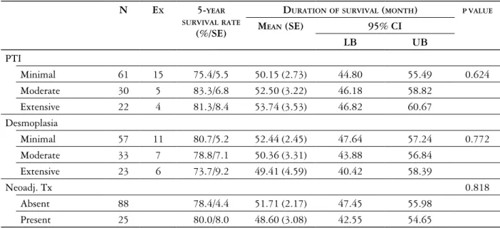
n ex 5-year
surViValraTe
(%/se)
duraTiOnOfsurViVal (mOnTh) pValue
mean (se) 95% ci lB uB PTI Minimal 61 15 75.4/5.5 50.15 (2.73) 44.80 55.49 0.624 Moderate 30 5 83.3/6.8 52.50 (3.22) 46.18 58.82 Extensive 22 4 81.3/8.4 53.74 (3.53) 46.82 60.67 Desmoplasia Minimal 57 11 80.7/5.2 52.44 (2.45) 47.64 57.24 0.772 Moderate 33 7 78.8/7.1 50.36 (3.31) 43.88 56.84 Extensive 23 6 73.7/9.2 49.41 (4.59) 40.42 58.39 Neoadj. Tx 0.818 Absent 88 78.4/4.4 51.71 (2.17) 47.45 55.98 Present 25 80.0/8.0 48.60 (3.08) 42.55 54.65
ITB – intratumoral budding; PTB – peritumoral budding; ITL – intratumoral lymphocyte; PTL – peritumoral lymphocyte; CLI – Crohn-like inflammation; IE – Intratumoral eosinophils; PTE – peritumoral eosinophils; ITI – intratumoral overall inflammation; PTI – peritumoral overall inflammation; tx – therapy; CI – confidence interval; LB – lower bound; UB – upper bound; SE – standard error. Log Rank Test
cells can attract and/or activate eosinophils in the tu-mor microenvironment [38].
An association of high-grade tumor infiltrating lymphocytes with a higher likelihood of deriving a survival benefit from adjuvant chemotherapy in patients undergoing curative resection of CRC was reported [17, 39]. Therefore, the increase in ITL with higher tumor grade and lower likelihood of venous invasion in our cohort seems notable. This seems to also emphasize the likelihood of the presence of a dis-tinct lymphocytic infiltrative pattern rather than spe-cific tumor infiltrating lymphocytes to be the most important determinant of survival [14, 40].
In a retrospective analysis of 120 patients with AJCC/UICC stage II colorectal cancer by Betge et al., high score tumor budding was reported to be sig-nificantly associated with tumor grade and lympho-vascular invasion and thought to be an independent predictor of disease progression and cancer-related death [41]. Our findings revealed a high concor-dance between ITB and PTB, in parallel to their similar pathogenesis as morphologic manifestations
of epithelial-mesenchymal transition [42]. Both high
ITB and high PTB were associated with higher rates of pT4 stage, venous invasion and
moderate-to-ex-tensive desmoplasia. This indicates that tumor
bud-ding is frequently found in tumors with an invasive growth pattern and desmoplastic stromal response. Neither ITB nor PTB was associated with a metasta-sis rate in our cohort despite the reported association of tumor budding with an increased probability for metastatic dissemination to lymph nodes and dis-tant organs. Our findings were thus compatible with the literature [43].
The presence of higher ITB in patients with mini-mal as compared to moderate-to-extensive PTE in our cohort seems to support the potential role of eosino-phil infiltration as an antitumoral mechanism [10]. It also seems notable given the infrequently encoun-tered tumor budding in patients with a strong per-itumoral infiltration caused by CD8+ cytotoxic T cells, indicating the likelihood of an interaction between tumor budding cells and the host immune system as two opposing sides of an attacker-defender model [13, 43].
Our findings emphasized the potential role of ad-ditional prognostic parameters related to tumor mi-croenvironment alongside the conventional morpho-logical prognostic parameters in the identification of high-risk pathological features and in guiding pro-vision of adjuvant therapy in CRCs [10, 14, 15].
The retrospective and single center nature of our study was an important limitation and might have influenced our findings in establishing the temporal-ity between cause and effect as well as generalizing our findings to an overall CRC population. The other limitation of this study was the relatively small sample
size that might have prevented from obtaining statis-tical significance concerning prognostic role of tumor inflammatory parameters. The inclusion of patients with and without neoadjuvant therapy together was another limitation of the study. Nevertheless, de-spite these limitations, given the paucity of the solid information available in this area, our findings rep-resent a valuable contribution to the literature by providing data on assessment of the prognostic role of tumor-associated inflammatory infiltrates based on standardized criteria and being stratified by lo-cation (intratumoral, peritumoral) and inflammatory cell subset of both the adaptive and innate immune system (lymphocytes, eosinophils).
In conclusion, our findings revealed that low score tumoral budding and an increase in tumor-related in-flammatory parameters were associated with a lesser likelihood of conventional poor prognostic tumor pa-rameters such as pT4, TNM stage IV, venous invasion and metastasis. Nonetheless, given the association of an increase in peritumoral eosinophil infiltrates with a lower probability of intratumoral budding, pT4, me-tastasis and for better survival rates, our findings em-phasize the potential role of peritumoral eosinophils independent of the overall inflammatory reaction. This can be considered as an additional prognostic parameter related to the tumor microenvironment in better stratification of progression and guiding provi-sion of adjuvant therapy tailored to individualized risk.
The authors declare no conflict of interest. References
1. Rawla P, Sunkara T, Barsouk A. Epidemiology of colorectal cancer: incidence, mortality, survival and risk factors. Gastro-enterology Rev 2019; 14: 89-103.
2. Lee CH, Tseng PL, Tung HY, et al. Comparison of risk factors between colon cancer and rectum cancer in a single medical center hospital, Taiwan. Arch Med Sci 2020; 16: 102-111. 3. Malyarchuk BA, Grzybowski T, Derenko MV, et al.
Mitochon-drial DNA variability in Poles and Russians. Ann Hum Genet 2002; 66: 261-283.
4. Mohammed F, Rezaee Khorasany AR, Mosaileby E, et al. Mi-tochondrial A12308G alteration in tRNA(Leu (CUN)) in colo- rectal cancer samples. Diagn Pathol 2015; 10: 115.
5. Kumar B, Bhat ZI, Bansal S, et al. Asscociation of mitochon-drial copy number variation and T16189C polymorphism with colorectal cancer in North Indian population. Tumour Biol 2017; 39: 1010428317740296.
6. Webb E, Broderick P, Chandler I, et al. Comprehensive analysis of common mitochondrial DNA variants and colorectal cancer risk. Br J Cancer 2008; 99: 2088-2093.
7. Skonieczna K, Jawien A, Marszalek A, et al. Mitogenome germ- line mutations and colorectal cancer risk in Polish population. Arch Med Sci 2020; 16: 366-373.
8. Compton CC. Optimal pathologic staging: defining stage II disease. Clin Cancer Res 2007; 13: 6862-6870.
9. Barresi V, Reggiani Bonetti L, Ieni A, et al. Poorly differenti-ated clusters: clinical impact in colorectal cancer. Clin Colo- rectal Cancer 2017; 16: 9-15.
10. Harbaum L, Pollheimer MJ, Kornprat P, et al. Peritumoral eo-sinophils predict recurrence in colorectal cancer. Mod Pathol 2015; 28: 403-413.
11. O’Connell JB, Maggard MA, Ko JY. Colon cancer survival rates with the new American Joint Committee on Cancer sixth edition staging. J Natl Cancer Inst 2004; 96: 1420-1425. 12. Wang LM, Sheahan K. Prognostic markers in colorectal
pathol-ogy: is morphology enough? Diag Pathol 2011; 17: 386-394. 13. Lugli A, Karamitopoulou E, Panayiotides I, et al. CD8+
lym-phocytes/tumour-budding index: an independent prognostic factor representing a ‘pro-/anti-tumour’ approach to tumour host interaction in colorectal cancer. Br J Cancer 2009; 101: 1382-1392.
14. Roxburgh CS, McMillan DC. The role of the in situ local in-flammatory response in predicting recurrence and survival in patients with primary operable colorectal cancer. Cancer Treat Rev 2012; 38: 451-466.
15. Galon J, Costes A, Sanchez-Cabo F, et al. Type, density and lo-cation of immune cells within human colorectal tumors predict clinical outcome. Science 2006; 313: 1960-1964.
16. Pagès F, Mlenic B, Marliot F, et al. International validation of the consensus Immunoscore for the classification of colon cancer: a prognostic and accuracy study. Lancet 2018; 391: 2128- 2139.
17. Prizment AE, Vierkant RA, Smyrk TC, et al. Tumor eosinophil infiltration and improved survival of colorectal cancer patients: Iowa Women’s Health Study. Mod Pathol 2016; 29: 516-527. 18. Klintrup K, Makinen JM, Kauppila S, et al. Inflammation and prognosis in colorectal cancer. Eur J Cancer 2005; 41: 2645-2654.
19. Briede I, Strumfa I, Vanags A, et al. The association between inflammation, epithelial mesenchymal transition and stemness in colorectal carcinoma. J Inflamm Res 2020; 13: 15-34. 20. Amin MB, Edge SB, Greene FL, et al. AJCC Cancer Staging
Manual, 8th ed. Springer, New York 2017.
21. Hamilton SR, Bosman FT, Boffetta P. Carcinoma of the colon and rectum. Bosman FT, Carneiro F, Hruban RH (eds.). World Health Organization classification of tumours of the digestive system. IARC, Lyon 2010; 131-181.
22. Dawson H, Kirsch R, Driman DK, et al. Optimizing the de-tection of venous invasion in colorectal cancer: the Ontario, Canada, experience and beyond. Front Oncol 2015; 4: 354. 23. Kirch R, Messenger D, Riddell R, et al. Venous invasion in
colorectal cancer: Impact of an elastin stain on detection and interobserver agreement among gastrointestinal and nongastrointestinal pathologists. Am J Surg Pathol 2013; 37: 200-210.
24. Graham RP, Vierkant RA, Tillmans LS, et al. Tumor budding in colorectal carcinoma: confirmation of prognostic signifi-cance and histologic cutoff in a population-based cohort. Am J Surg Pathol 2015; 39: 1340-1346.
25. Fernández-Aceñero MJ, Galindo-Gallego M, Sanz J, Aljama A. Prognostic influence of tumor-associated eosinophilic infiltrate in colorectal carcinoma. Cancer 2000; 88: 1544-1548. 26. College of American Pathologists protocol for the examination
of specimens from patients with primary carcinoma of the co-lon and rectum. Based on AJCC/UICC TNM, 8th edition Pro-tocol web posting date: February 2020 Available from: https:// documents.cap.org/protocols/cp-gilower-colonrectum-biop-sy-20-4100.pdf.
27. Huh JW, Lee JH, Kim HR. Prognostic significance of tu-mor-infiltrating lymphocytes for patients with colorectal can-cer. Arch Surg 2012; 147: 366-371.
28. Nagtegaal ID, Marijnen CA, Kranenbarg EK, et al. Local and distant recurrences in rectal cancer patients are predicted by the nonspecific immune response; specific immune response has only a systemic effect – a histopathological and immuno-histochemical study. BMC Cancer 2001; 1: 7.
29. Fisher ER, Paik SM, Rockette H, et al. Prognostic significance of eosinophils and mast cells in rectal cancer: findings from the National Surgical Adjuvant Breast and Bowel Project (pro-tocol R-01). Hum Pathol 1989; 20: 159-163.
30. McGinnis MC Jr., Bradley EL, Pretlow TP, et al. Correlation of stromal cells by morphometric analysis with metastatic behavior of human colonic carcinoma. Cancer Res 1989; 49: 5989-5993.
31. Pretlow TP, Boohaker EA, Pitts AM, et al. Heterogeneity and subcompartmentalization in the distribution of eosinophils in human colonic carcinomas. Am J Pathol 1984; 116: 207-213. 32. Richards CH, Flegg KM, Roxburgh CS, et al. The relationships
between cellular components of the peritumoural inflammato-ry response, clinicopathological characteristics and survival in patients with primary operable colorectal cancer. Br J Cancer 2012; 106: 2010-2015.
33. Rao H, Chen J, Li M, et al. Increased intratumoral neutro-phil in colorectal carcinomas correlates closely with malignant phenotype and predicts patients’ adverse prognosis. PLoS One 2012; 7: e30806.
34. Akishima-Fukasawa Y, Ishikawa Y, Akasaka Y, et al. Histo-pathological predictors of regional lymph node metastasis at the invasive front in early colorectal cancer. Histopathology 2011; 59: 470-481.
35. Mizuno R, Kawada K, Itatani Y, et al. The role of tumor-asscoci-ated neutrophils in colorectal cancer. Int J Mol Sci 2019; 20: 529. 36. Mei Z, Liu Y, Liu C, et al. Tumour-infiltrating inflammation
and prognosis in colorectal cancer: systematic review and me-ta-analysis. Br J Cancer 2014; 110: 1595-1605.
37. Kita H. Eosinophils: Multifaceted biological properties and roles in health and disease. Immunol Rev 2011; 242: 161-177. 38. Varricchi G, Galdiero MR, Loffredo S, et al. Eosinophils:the un-sung heroes in cancer? Oncoimmunology 2018; 7: e139134. 39. Morris M, Platell C, Iacopetta B. Tumor-infiltrating
lympho-cytes and perforation in colon cancer predict positive response to 5-fluorouracil chemotherapy. Clin Cancer Res 2008; 14: 1413-1417.
40. Ogino S, Nosho K, Irahara N, et al. Lymphocytic reaction to colorectal cancer is associated with longer survival, indepen-dent of lymph node count, microsatellite instability, and CpG island methylator phenotype. Clin Cancer Res 2009; 15: 6412-6420.
41. Betge J, Kornprat P, Pollheimer MJ, et al. Tumor budding is an independent predictor of outcome in AJCC/UICC stage II colorectal cancer. Ann Surg Oncol 2012; 19: 3706-3712. 42. Lugli A, Kirsch R, Ajioka Y, et al. Recommendations for
re-porting tumor budding in colorectal cancer based on the In-ternational Tumor Budding Consesnsus Conference (ITBCC) 2016. Mod Pathol 2017; 30: 1299-1311.
43. Koelzer VH, Zlobec I, Lugli A. Tumor budding in colorectal cancer – ready for diagnostic practice? Hum Pathol 2016; 47: 4-19.
Address for correspondence
Saime Ramadan
Department of Pathology,
Baskent University Istanbul Hospital
Altunizade Mah. 7. Kisikli cad. Oymacı Sk. 34662 Uskudar/Istanbul, Turkey
tel. +905335255563 fax +902164743149