Research Article
Benign Solitary Pulmonary Necrotic Nodules: How Effectively
Does Pathological Examination Explain the Cause?
Halide Nur Urer ,
1Mehmet Zeki Gunluoglu,
2Nurcan Unver,
1Sezer Toprak,
3and Mediha Gonenc Ortakoylu
41University of Health Sciences Turkey, Yedikule Chest Diseases and Thoracic Surgery Training and Research Hospital,
Department of Pathology, Istanbul, Turkey
2Medipol University School of Medicine, Thoracic Surgery, Istanbul, Turkey
3University of Health Sciences Turkey, Yedikule Chest Diseases and Thoracic Surgery Training and Research Hospital,
Department of Microbiology, Istanbul, Turkey
4University of Health Sciences Turkey, Yedikule Chest Diseases and Thoracic Surgery Training and Research Hospital,
Department of Pulmonary Medicine, Istanbul, Turkey
Correspondence should be addressed to Halide Nur Urer; [email protected]
Received 19 April 2020; Revised 7 June 2020; Accepted 15 June 2020; Published 6 July 2020 Academic Editor: Akiteru Goto
Copyright © 2020 Halide Nur Urer et al. This is an open access article distributed under the Creative Commons Attribution License, which permits unrestricted use, distribution, and reproduction in any medium, provided the original work is properly cited.
Aims. We investigated the histopathological features of solitary pulmonary necrotic nodules (NNs) of undetermined cause. We combined our findings with those obtained using other methods to determine how well the etiological factors were explained. Methods. We screened patients who underwent surgery to treat solitary pulmonary granulomatous and nongranulomatous NNs of undetermined cause. The NN sizes and features of both the NNs and adjacent parenchyma were evaluated. Histochemical analyses included Ehrlich–Ziehl–Neelsen (EZN), Grocott, and Gram staining. Polymerase chain reaction (PCR) was used to detect tuberculous and nontuberculous mycobacteria, panfungal DNA, Nocardia, Francisella tularensis types A and B, and actino-mycetes. Results. The NNs were granulomatous in 78.9% and nongranulomatous in 21% of the 114 patients included. EZN staining or PCR was positive for Mycobacterium in 53.5% of all NNs: 62.2% of granulomatous and 20.8% of nongranulomatous NNs. We found a weak but significant correlation between granulomatous NNs and Bacillus positivity and a significant correlation between granulomas surrounding the NNs and the presence of multiple necroses. The NN etiology was determined via histopathological, histochemical, and PCR analyses in 57% of patients but remained undetermined in 42.9%. Conclusion. The causes of both granulomatous and nongranulomatous NNs can be determined by pathological examination. Granulomatous necrosis and granulomas in the adjacent parenchyma are important for differential diagnosis. When both features are present, they strongly support a diagnosis of tuberculosis, even in the absence of bacilli.
1. Introduction
Pulmonary necrotic nodules (NNs) are space-occupying lung lesions. The principal etiologies include infection, collagen tissue disease, thromboembolism, vasculitis, and aspiration. NNs are often difficult to distinguish from ma-lignancies on clinical examination and radiological imaging. The underlying cause of pulmonary NNs can sometimes be
determined based on morphology. Identification of an in-fectious agent facilitates diagnosis. However, the causes of some benign pulmonary nodules cannot be determined.
Here, we explored how often the etiological factors can be identified. The diagnostic and histopathological charac-teristics of the lesions were defined and evaluated in com-bination with histochemical and polymerase chain reaction (PCR) analyses.
Volume 2020, Article ID 7850750, 6 pages https://doi.org/10.1155/2020/7850750
2. Materials and Methods
Cases of benign pulmonary NNs and granulomatous NNs treated between January 1, 2015, and December 31, 2017, in the Pathology Department of Yedikule Pulmonary Diseases and Thoracic Surgery Training and Research Hospital were screened. Archived slides and pathology reports were ana-lyzed. Patients who received specific diagnoses based on microbiological and serological tests performed during clinical evaluation were excluded. Patients in whom the etiology was identified by routine histopathological evalu-ation, those with synchronous tumors, and those who re-ceived neoadjuvant therapy were also excluded. Age, sex, and type of surgical resection were recorded in all cases. A total of four sections of 3 μm thickness were obtained from one or two paraffin blocks. We performed routine hema-toxylin-and-eosin, Ehrlich–Ziehl–Neelsen (EZN), Gram, and Grocott staining. All slides were examined under a light microscope by two pathologists (H.N.U. and N.U.). Both our hospital institutional review board and our local ethics committee approved the study.
2.1. Histopathological Evaluation. The NNs were divided
into two groups: granulomatous NNs (those exhibiting epithelioid histiocytes in the periphery) and non-granulomatous NNs (all others). The nature and size of each NN and the surrounding tissue features were evaluated. Necrosis within an NN was recorded in terms of size (≤1 or >1 cm), border type (smooth or irregular), shape (round, triangular, or amorphous), necrotic quality (caseified, infarctoid, apoptotic, or “dirty” with many polymorpho-nuclear leukocytes), number (single or multiple), and lo-cation. The parenchyma surrounding each NN was evaluated in terms of granuloma status, vasculitis (granu-lomatous, lymphoplasmacytic, necrosis, and leukocyto-clastic), parenchymal changes, and foreign body and thromboembolism statuses. NNs positive on PCR and/or EZN staining were considered to contain Mycobacterium
tuberculosis (MT).
2.2. Histochemical Evaluation. Each slide was stained with
EZN, Grocott, and Gram stains. The entire sample area was observed under a light microscope at 40× magnification. When required, 100× magnification was used for closer examination. Acid-fast bacilli (AFB) were assessed by EZN staining, fungal spores and hyphae by Grocott staining, and Gram-positive and -negative bacteria by the Gram staining.
2.3. PCR Procedures. Necrotic areas were marked in one or
two paraffin blocks. Microdissection was performed under sterile conditions. The samples were transferred to coded, sterile Eppendorf tubes. After deparaffinization, DNA was isolated using the InstaGene Matrix kit (Bio-Rad, USA). MT and nontuberculous mycobacteria were detected using a commercial kit (GenMark, Turkey). Each tube contained 10 μL Real-time PCR Master Mix with UDG, 5 μL Primer Probe Mix, and purified DNA (5 μL). PCR was performed
using the CFX96 Real-time PCR device (Bio-Rad). The following primer sets specific to panfungal, Nocardia,
Francisella tularensis types A and B, and actinomycetes were
used (5′ to 3′): GCATCGATGAAGAACGCAGC and TCCTCCGCTTATTGATATGC, 300 nM; ACCGACCA-CAAGGGG and GGTTGTAACCTCTTCGA, 300 nM; GAGACATCAATTAAAAGAAG and CCAAGAGTACT-ATTTCCGGTTGGT, 400 nM; CTTGTACTTTTATTTG-GCTACTGAGAAACT and CTTGCTTGGTTTGTAAA-TATAGTGGAA, 300 nM; and GGATGAGCCCGCG-GCCTA and CCAGCCCCACCTTCGAC, 400 nM, respec-tively. Each tube contained 10 μL Real-time PCR Master Mix with EVAGREEN (GenMark, Turkey), 2 μL primer mix, 3 μL double-distilled water, and 5 μL sample DNA. PCR was performed as described above. The specificities of the PCR products were assessed by melting curve analysis.
2.4. Statistical Analysis. All data were evaluated statistically.
Correlations of the morphological characteristics with the histochemical and PCR data were analyzed. The chi-squared or Fisher’s exact test was used to compare the frequencies of categorical variables, and Spearman’s correlation test was used to determine the correlations between variables. A
pvalue < 0.05 was considered statistically significant.
3. Results
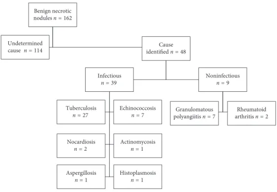
We examined 162 patients with benign NNs. Of these, 48 were excluded because the NN etiology was determined based on clinical examination, radiological findings, mi-crobiological or serological tests, or routine histopatholog-ical evaluation (Figure 1). The remaining 114 patients (60 males and 54 females) with NNs of unknown cause were analyzed. The mean (range) and median patient ages were 48.3 (20–79) and 50 years, respectively. The surgery per-formed was wedge resection in 97 (85%) and anatomical resection in 17 (15%).
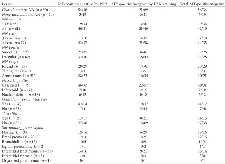
NNs were classified as granulomatous in 90 patients (78.9%) and nongranulomatous in 24 (21%) (Figure 2). AFB were detected by EZN staining (Figure 3). PCR was used for MT and panfungal detection. The histochemical and PCR results are shown in Table 1.
Vasculitis was found in 29 cases. Lymphoplasmacytic vasculitis was detected in 89.6% (26). Granulomatous vas-culitis was just in 10.3% (3) cases. There was no leukocy-toclastic vasculitis and necrotic vasculitis.
Two patients exhibited foreign bodies and two throm-boembolisms around the NNs. All four were negative for bacilli. Both patients with foreign bodies and one with a thromboembolism had granulomatous NNs.
MT was detected by PCR and/or EZN in 53.5% of all NNs: 62.2% of granulomatous NNs and 20.8% of non-granulomatous NNs. Both methods yielded positive results in 22 patients and negative results in 53 patients. Two pa-tients were negative by PCR but positive by EZN staining, whereas 37 were positive by PCR but negative by EZN staining. We found a weak (Spearman’s coefficient, r � 0.4) but highly significant (p � 0.0001) correlation between the
PCR and EZN staining results. Granulomatous NNs were significantly associated with other histopathological pa-rameters and Bacillus positivity (Table 2).
We found a weak but significant correlation between granulomatous NN and Bacillus positivity, which was
correlated significantly with the presence of a granuloma around the NN and the presence of multiple necrotic foci (p < 0.0001, r � 0.45; p < 0.0001, r � 0.28, respectively).
Ba-cillus positivity was not correlated significantly with NN size,
border type, shape, necrotic quality, vasculitis, or foreign body or thromboembolic status (p � 0.49, p � 0.57,
p �0.61, p � 0.33, and p � 0.66, respectively).
Gram and Grocott staining did not identify a specific infectious cause in any case. Panfungal PCR was positive in two cases. PCR did not detect nontuberculous mycobacteria,
Nocardia, F. tularensis type A or B, or actinomycetes in any
case. Thus, the etiologies of benign pulmonary NNs were determined in 65 patients (57%) but not in 49 (42.9%) (Table 3).
4. Discussion
The leading causes of benign pulmonary NNs are infections (bacterial, fungal, and parasitic), vasculitis, pulmonary in-farction, aspiration, rheumatoid arthritis, and necrotizing sarcoidosis [1–3]. NNs can be granulomatous or non-granulomatous. Necrosis commonly develops in patients with pulmonary granulomatous diseases and is caused by infection in nearly 50% of cases [4]. The coexistence of necrosis and a granuloma limits the number of etiological factors to consider during diagnosis. Indeed, diagnosis fa-cilitates identification of the specific infectious agent [4]. Our results corroborate this, as the most common specific agent identified was MT. Pathological findings of granulomatous necrosis and a granuloma around the nodule are associated with a higher likelihood of tuberculosis. Lesions should be exhaustively tested for bacilli.
The diagnosis of pulmonary NN requires an algorithmic approach. Re-evaluation of new slides after histochemical Benign necrotic nodules n = 162 Undetermined cause n = 114 Cause identified n = 48 Infectious n = 39 Tuberculosis n = 27 Echinococcosis n = 7 Nocardiosis n = 2 Actinomycosisn = 1 Aspergillosis n = 1 Histoplasmosis n = 1 Noninfectious n = 9 Granulomatous polyangiitis n = 7 Rheumatoid arthritis n = 2
Figure 1: The study population.
Figure 2: Nongranulomatous necrotic nodule, H&E, ×40.
Table 1: Characteristics of the histopathological lesions.
Lesion MT-positive/negative by PCR AFB-positive/negative by EZN staining Total MT-positive/negative
Granulomatous NN (n � 90) 54/36 21/69 56/34 Nongranulomatous NN (n � 24) 5/19 3/21 5/19 NN number 1 (n � 53) 19/34 3/50 19/34 >1 (n � 61) 40/21 21/40 42/19 NN size ≤1 cm (n � 35) 17/18 3/32 17/18 >1 cm (n � 79) 42/37 21/58 44/35 NN border Smooth (n � 52) 27/25 6/46 27/26 Irregular (n � 62) 32/30 18/44 34/28 NN shape Round (n � 57) 28/29 7/50 28/29 Triangular (n � 6) 3/3 1/5 3/3 Amorphous (n � 51) 28/23 16/35 30/21 Necrotic quality Caseified (n � 79) 46/33 22/57 48/31 Infarctoid (n � 17) 7/10 2/15 7/10 Nuclear debris (n � 18) 6/12 0/18 6/12
Granuloma around the NN
Yes (n � 56) 42/14 19/37 44/12 No (n � 58) 17/41 5/53 17/41 Vasculitis Yes (n � 29) 12/17 8/21 14/15 No (n � 85) 47/38 16/69 47/38 Surrounding parenchyma Normal (n � 35) 19/16 6/29 19/16 Emphysema (n � 28) 12/16 3/25 12/16 Bronchiolitis (n � 15) 10/5 6/9 10/5 Lipoid pneumonia (n � 2) 1/1 0/2 1/1 Interstitial pneumonia (n � 30) 14/16 9/21 16/14 Interstitial fibrosis (n � 3) 3/0 0/3 3/0 Organized pneumonia (n � 1) 0/1 0/1 0/1
MT: Mycobacterium tuberculosis; EZN: Ehrlich–Ziehl–Neelsen stain; NN: necrotic nodule.
Table 2: Features of granulomatous necrotic nodules.
Chi-squared p value Correlation coefficient Correlation p value
Bacillus positivity 0.001 0.34 0.0001 NN number 0.001 0.31 0.001 NN size 1 −0.01 0.94 NN border 0.41 0.14 0.15 NN shape 0.65 0.06 0.53 Necrotic quality 0.24 −0.13 0.18
Presence of granuloma around the NN 0.0001 0.37 0.0001
Vasculitis 0.40 0.10 0.27
NN: necrotic nodule.
Table 3: Pathological diagnosis of necrotic nodules.
Granulomatous necrotic nodules Nongranulomatous necrotic nodules
MT 56 5 MT + panfungal 1 — Fungal 1 — Infarct 1 1 Foreign body 2 — Indeterminable 31 18
staining, microbiological culture, fungal serological tests, PCR analysis, and clinical correlations all support a diagnosis. The specific agent can be determined retrospectively. Mukhopadya et al. reported that diagnosis was assisted by histological findings in 60% of cases, by microbiological culture in 47%, by fungal serology in 14%, and by clinical radiological confir-mation in 8% [5]. PCR of NNs allows accurate detection of tuberculosis bacilli in the paraffin block [6]. Moreover, the importance of confirmation using other methods such as EZN staining has been emphasised [7–9]. Such staining is of un-deniable diagnostic utility, being cost effective, rapid, and easy to perform [9]. Any step-by-step pathological evaluation should aim to detect tuberculosis bacilli.
In our study, vasculitis was detected in 25% of NNs. We observed no correlation between the granuloma status and
Bacillus positivity. Thus, vasculitis is not specific in terms of
NN differentiation. Perivascular and intramural inflam-mation may develop in patients with pulmonary tubercu-losis and infarctoid necrosis [10]. In particular, the morphological lesions of granulomatous polyangiitis may not differ from lesions that develop secondary to infection [11]. Thus, the presence of vasculitis neither rules out nor compromises a preliminary diagnosis of tuberculosis.
Infectious granulomatous necrosis may mimic a thromboembolic infarction because the necrosis may be of a coagulative or dirty nature in the former condition [12]. Moreover, contrary to general belief, the borders of an infarct can be either irregular or round. Vascular inflammation may be evident. Activated epithelioid histiocytes at the border of an infarctoid necrosis render a definitive diagnosis difficult [13]. We found that granulomatous necroses containing bacilli can feature an infarctoid or even dirty necrosis.
Our study had certain limitations, of which the first is its retrospective design. Clinical data were obtained from files. However, in patients with conditions such as rheumatoid arthritis and vasculitis, serological tests may be negative during certain periods. Another limitation was that all pa-tients underwent resection because malignancy was sus-pected; therefore, pre- or postoperative microbiological culture was not performed in all cases.
In conclusion, undetermined diagnoses of pulmonary NN need to be minimized. Causative agents may be detected via pathological evaluation of both granulomatous and nongranulomatous NNs. In particular, the presence of both a necrotizing granuloma and a granuloma in the adjacent parenchyma is particularly important. Even if bacilli cannot be detected, tuberculosis must be considered. Integration of pathological and microbiological methods is essential for diagnosis of pulmonary NNs.
Data Availability
The data used to support the findings of this study are available from the corresponding author upon request.
Ethical Approval
The study has been approved by the local ethical committee (University of Health Sciences Istanbul Research and
Training Hospital Ethics Committee Approval. The study protocol number and date are 1407/31.08.2018).
Disclosure
None of the authors has a financial relationship with a commercial entity that has an interest in the subject of this manuscript.
Conflicts of Interest
The authors declare that they have no conflicts of interest.
Authors’ Contributions
Halide Nur Urer constructed an idea or hypothesis for research and manuscript and designed and planned the method to achieve the results; Halide Nur Urer, Mehmet Zeki Gunluoglu, and Sezer Toprak were responsible for the evaluation and conclusion of findings; Halide Nur Urer, Nurcan Unver, Mehmet Zeki Gunluoglu, and Mediha Gonenc Ortakoylu were responsible for the writing of the entire work or its noticable parts; Halide Nur Urer and Mediha Gonenc Ortakoylu re-evaluated the study in the scientific sense, regardless of language and literary correc-tions, prior to the delivery of the manuscript.
References
[1] S. Ohshimo, J. Guzman, U. Costabel, and F. Bonella, “Dif-ferential diagnosis of granulomatous lung disease: clues and pitfalls,” European Respiratory Review, vol. 26, no. 145, pp. 170012–170016, 2017.
[2] K. K. Shah, B. S. Pritt, and M. P. Alexander, “Histopathologic review of granulomatous inflammation,” Journal of Clinical Tuberculosis and Other Mycobacterial Diseases, vol. 7, pp. 1– 12, 2017.
[3] A. C. Roden and A. N. Schuetz, “Histopathology of fungal diseases of the lung,” Seminars in Diagnostic Pathology, vol. 34, no. 6, pp. 530–549, 2017.
[4] S. Mukhopadhyay, C. F. Farver, L. T. Vaszar et al., “Causes of pulmonary granulomas: a retrospective study of 500 cases from seven countries,” Journal of Clinical Pathology, vol. 65, no. 1, pp. 51–57, 2012.
[5] S. Mukhopadhyay, B. E. Wilcox, J. L. Myers et al., “Pulmonary necrotizing granulomas of unknown cause,” Chest, vol. 144, no. 3, pp. 813–824, 2013.
[6] C. Nopvichai, A. Sanpavat, R. Sawatdee et al., “PCR detection of Mycobacterium tuberculosis in necrotising non-granulo-matous lymphadenitis using formalin-fixed paraffin-embed-ded tissue: a study in Thai patients,” Journal of Clinical Pathology, vol. 62, no. 9, pp. 812–815, 2009.
[7] P. Mahaisavariya, A. Chaiprasert, J. Manonukul, S. Khemngern, and N. Tingtoy, “Detection and identification of Mycobacterium species by polymerase chain reaction (PCR) from paraffin-embedded tissue compare to AFB staining in pathological sections,” Journal of the Medical Association of Thailand � Chotmaihet Thangphaet, vol. 88, no. 1, pp. 108–113, 2005.
[8] K. Miller, S. M. Harrington, and G. W. Procop, “Acid-fast smear and histopathology results provide guidance for the appropriate use of broad-range polymerase chain reaction and
sequencing for mycobacteria,” Archives of Pathology & Lab-oratory Medicine, vol. 139, no. 8, pp. 1020–1023, 2015. [9] A. Karadaǧ, E. Usta, K. Bilgin, A. K. G¨uney, C. Eroǧlu, and
M. G¨unaydin, “Comparison of culture, real-time DNA am-plification assay and Ehrlich-Ziehl-Neelsen for detection of Mycobacterium tuberculosis,” Balkan Medical Journal, vol. 30, no. 1, pp. 13–15, 2013.
[10] D. Jain, S. Ghosh, L. Teixeira, and S. Mukhopadhyay, “Pa-thology of pulmonary tuberculosis and non-tuberculous mycobacterial lung disease: facts, misconceptions, and prac-tical tips for pathologists,” Seminars in Diagnostic Pathology, vol. 34, no. 6, pp. 518–529, 2017.
[11] J. Ramsey, M. Amari, and S. P. Kantrow, “Pulmonary vas-culitis: clinical presentation, differential diagnosis, and management,” Current Rheumatology Reports, vol. 12, no. 6, pp. 420–428, 2010.
[12] M.-C. Aubry, “Necrotizing granulomatous inflammation: what does it mean if your special stains are negative?” Modern Pathology, vol. 25, no. S1, pp. S31–S38, 2012.
[13] S. A. Yousem, “The surgical pathology of pulmonary infarcts: diagnostic confusion with granulomatous disease, vasculitis, and neoplasia,” Modern Pathology, vol. 22, no. 5, pp. 679–685, 2009.