Revista de cercetare [i interven]ie social\
ISSN: 1583-3410 (print), ISSN: 1584-5397 (electronic) Selected by coverage in Social Sciences Citation Index, ISI databasesDEAF ATHLETE: IS THERE ANY DIFFERENCE BEYOND
THE HEARING LOSS?
Ebru AKGUL ERCAN, Alparslan KILIÇ, Seyfi SAVAS, Mahmut ACAK, Zeynep BIYIKLI, Hasan Fehmi TORE
Revista de cercetare [i interven]ie social\, 2016, vol. 52, pp. 241-251 The online version of this article can be found at:
www.rcis.ro, www.doaj.org and www.scopus.com
Published by:
Expert Projects Publishing House
On behalf of:
„Alexandru Ioan Cuza” University, Department of Sociology and Social Work
and
Holt Romania Foundation
REVISTA DE CERCETARE SI INTERVENTIE SOCIALA is indexed by ISI Thomson Reuters - Social Sciences Citation Index
(Sociology and Social Work Domains)
expert projects
publishingDeaf Athlete: Is There Any Difference Beyond
The Hearing Loss?
Ebru AKGUL ERCAN 1, Alparslan KILIÇ2, Seyfi SAVAS 3, Mahmut ACAK 4, Zeynep BIYIKLI 5, Hasan Fehmi TORE 6
Abstract
Regular physical activity and sport is important for enhanced physical fitness and skill performance of deaf athletes. However there are few data in the literature about their medical considerations. In this study we aimed to determine the morphological findings and cardiac status of the deaf athletes and compare them with the normal ones. Thirty deaf and twenty-two normal male athletes without cardiovascular diseases participated in the study. Transthoracic echocardiography and exercise stress testing were performed. Tissue Doppler imaging (TDI)-derived myocardial performance index (MPI) was also evaluated. VO2 max and heart rate recovery were calculated after exercise stress testing. Total cholesterol, LDL and trigliseride levels were significantly increased in the deaf athletes, but they were still found to be in normal ranges according to age and risk factor profile (p< 0.05). End-diastolic diameter and left ventricular mass index were found to be significantly increased in the controls when compared with the deaf athletes (p< 0.001). Heart rate recovery at 1 minute did not show any difference in the deaf group when compared with the control group (p> 0.05). MPI calculated from TDI-derived variables was found to be 0.41±0.073 in the deaf group and 0.46±0.061 in the controls respectively and significantly decreased in the deaf (p < 0.05). Beyond having a hearing loss, the deaf athletes have many cardiac structural and functional differences from their normal counterparts. The factors that give rise to these differences have to be revealed by further research.
Keywords: echocardiography, deafness, Doppler echocardiography, exercise test 1 Ufuk University, Faculty of Medicine, Department of Cardiology, Ankara, TURKEY. E-mail:
akgul37ebru@hotmail
2 Ufuk University, Faculty of Medicine, Department of Cardiology, Ankara, TURKEY. E-mail:
3 Gazi University, Faculty of Physical Education and Sports, Ankara, TURKEY. E-mail:
[email protected] (Corresponding author)
4 Inonu University, Faculty of Physical Education and Sports., Malatya, TURKEY. E-mail:
5 Ankara University, Department of Biostatistics, Ankara, TURKEY. E-mail:
6 Ufuk University, Faculty of Medicine, Department of Cardiology, Ankara, TURKEY. E-mail: [email protected]
Introduction
The term deaf generally refers to persons who are unable to hear well enough to depend on hearing for processing information. On the other hand, the term deaf describes the audiologic condition of not hearing in the cultural nomenculature and Deaf refers to the group of people who form the deaf culture and use a common language called American Sign Language (ASL) as a method of commu-nication. The term hearing impaired is perceived as emphasizing the idea of disability, because they see themselves as fully able with a visual, but not oral, method of communication. The deaf, all over the world constitute a number of approximately 1 million in number. According to the statistics of National Institute on Deafness and Other Communication Disorders, approximately 28 million Americans have a hearing loss. Three of every 1000 children are born with a hearing loss and as an interesting finding, 90 % are born to hearing parents (Hoffman, Dobie, Ko, Themann, Murphy, 2010). The degree of hearing loss is categorized differently depending on the testing method or the consensus of the organization making the guideline. According to the American Speech-Language-Hearing Association, hearing loss is classified as mild (20-40 dB), moderate (40-60 dB) , severe ((40-60-80 Db) and profound (over 80 dB loss) (Danermark et al, 2013). Causes of hearing loss are diverse and generally include aging, disease, noise and genetical reasons. Hearing loss may also be classified according to the timing of loss. Congenital form is present at birth or within the first few days of life. However acquired hearing loss occurs later in life and generally after lan-guage has been acquired to some extent. Deaf individuals and hard of hearing people are identified by law as members of disability groups. They constitute a diverse group of people who are often denied access to job opportunities or community services as a result of their unique and varied characteristics. Deaf people regard being deaf not as a disability but as a sociocultural phenomenon and they see themselves as members of a unique group, community and culture. Individuals with disabilities should not need to prove themselves as competent workers or individuals who have a right to receive services as do all others in the society, in spite of their limitations. The service provider will be able to provide the full range of support that the deaf individual needs, only when the deaf or hard of hearing person is accepted as an equal participant in the communication interaction.
There are few data in the literature about the medical considerations for the athlete with a hearing loss. The most important reason for the lack of data is the communicative challenge of these athletes. Communication is an important way of gathering information in all medical branches. Interpreters in ASL, however may be a solution for the problems in communication. Deaf athletes are a
distin-guished class when special communication needs are taken into consideration. That is why the International Olympic Committee (IOC) has organized the Deaf-lympics as the special organization held for the elite athlete with a hearing loss. The Deaflympics is held separate from the Olympic Games both in different years and also with separate winter and summer games (Ammons, 2009). The Deaf-lympics provides deaf athletes with an international sports organization specially adapted to their communication and cultural needs. We performed the present study to determine the morphologic findings and cardiac status of the deaf athletes and compare them with the normal athletes to assess the differences that make them a distinctive group apart from hearing loss.
Materials and Methods
Thirty deaf and twenty-two normal male athletes without cardiovascular di-seases participated in the study. The deaf athletes were from the national deaf football sports team and the control group consisted of football players without any hearing loss from the training and sports faculty. The training period per week was the same for both of the groups. All the participants underwent routine physical examination and laboratory analysis that included blood count and biochemical parameters. Before this evaluation, informed consent was obtained from all of the athletes and the study was approved by the local ethics committee. The segmental analysis of the athletes was assessed by bioelectric impedance analysis method (BC 418 MA III Tanita). Fat ratio, total fat mass, free fat mass, trunk body water, trunk fat ratio, trunk fat mass and also trunk free fat mass were calculated for each participant by the aferomentioned method.
Exercise Stress Testing
All the athletes underwent symptom limited treadmill (Tepa-TM-Pro 2000 Model) and performed 3 minute warm-up period that started at 2% grade and 5 km/h and then treadmill was increased 1.0 km/h every 3 minutes. VO2 max (ml/ kg/min) was recorded at peak exercise (Vista Cx, Vacumed, CA) and heart rate was measured during each minute of exercise, at maximum exercise and during recovery at 1,2, 3, 4, and 5 minutes in the standing position. The exercise tests were analyzed and reported with a standard protocol by way of a computerized database. Heart rate recovery was defined as the change from peak heart rate to the heart rate at first minute of recovery. Abnormal heart rate recovery was defined as a decrease of 12 beats/min from peak exercise heart rate at 1 minute to recovery.
Echocardiographic examination
Echocardiographic images were obtained by using 3.75 MHz standard probe (General Electric, Vivid 7) according to the guidelines of American Society of Echocardiography4. All echocardiographic examinations were carried out by an experienced operator and the measurements were performed on-line. Besides the routine echocardiographic examination, pulsed-wave tissue Doppler imaging (TDI) was performed. From the apical four-chamber view, peak systolic velocity (S’), early diastolic (E’), and late diastolic velocity (A’) were measured both from the septal and lateral side of the mitral annulus. E’/A’ and E/E’ were calculated thereafter. Myocardial performance index (MPI) of the left ventricle was estimated via TDI derived echocardiography parameters. It is defined as the sum of iso-volumic contraction time (ICT) and isoiso-volumic relaxation time (IRT) divided by the ejection time (ET) as introduced by Tei (Tei, 1995).
Statistical Analysis
Data were analyzed by SPSS for Windows version 15. Continuous variables are given as mean±SD, median and interquartile ranges or absolute numbers (%) for dichotomous variables. Comparisons between the groups were made either with Student’s t-test or with Mann-Whitney U test. The paired t test was used to compare the mean values of parameters between groups and Wilcoxon test was used to compare the median values. Pearson’s chi-squared and Fisher exact tests were used for nominal variables. P value less than 0.05 was considered statistically significant.
Results
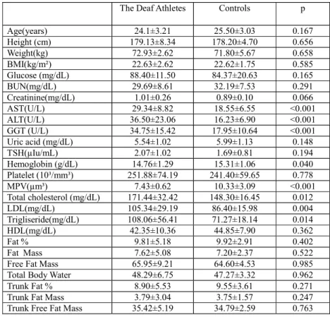
The deaf athletes and the control group did not show any difference with respect to age, anthropometric parameters and most of the laboratory data (Table 1). However liver function parameters (ALT, AST and GGT) were found to be increased in the deaf athletes though being in normal ranges in both of the groups. Hemoglobin was found to be lower in the deaf athlete group (p = 0.04) but it was still in normal ranges and mean platelet volume was significantly increased in the controls (p< 0.001). Total cholesterol, LDL and trigliseride levels were signi-ficantly increased in the deaf athletes (p< 0.05), but they were still found to be in normal ranges according to age and risk factor profile. The athletes also did not show any difference according to the parameters of the segmental analysis as shown in Table 1.
Table 1. Anthropometric, laboratory and segmental analysis parameters
BMI: body mass index; BUN: blood urea nitrogen; AST: aspartate amino transferase; ALT: alanine amino transferase; GGT: gamma glutamyl transpeptidase; TSH: thyroid stimulating hormone; MPV: mean platelet volume; LDL: low density lipoprotein; HDL: high density lipoprotein
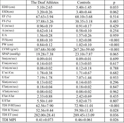
Two-dimensional (2D), Doppler and TDI Echocardiography
Among the conventional echocardiographic parameters, end-diastolic diameter and left ventricular mass index were found to be significantly increased in the control group when compared with the deaf athletes (5.40±1.45 cm, 5.10±2.65 cm and 267.26±59.60 g/m² , 187.68±30.00 g/m² respectively, p< 0.001). When TDI was taken into consideration, however, the myocardial systolic and diastolic velocities measured from both sides of the mitral annulus did not show any difference between the groups (Table 2). MPI, calculated from TDI-derived va-riables was found to be significantly decreased in the deaf athlete group (0.41±0.073 vs. 0.46±0.061 respectively, p = 0,026) as also presented in Table 2.
The Deaf Athletes Controls p Age(years) 24.1±3.21 25.50±3.03 0.167 Height (cm) 179.13±8.34 178.20±4.70 0.656 Weight(kg) 72.93±2.62 71.80±5.67 0.658 BMI(kg/m²) 22.63±2.62 22.62±1.75 0.585 Glucose (mg/dL) 88.40±11.50 84.37±20.63 0.165 BUN(mg/dL) 29.69±8.61 32.19±7.53 0.291 Creatinine(mg/dL) 1.01±0.26 0.89±0.10 0.066 AST(U/L) 29.34±8.82 18.55±6.55 <0.001 ALT(U/L) 36.50±23.06 16.23±6.90 <0.001 GGT (U/L) 34.75±15.42 17.95±10.64 <0.001 Uric acid (mg/dL) 5.54±1.02 5.99±1.13 0.148 TSH(µIu/mL) 2.07±1.02 1.69±0.81 0.194 Hemoglobin (g/dL) 14.76±1.29 15.31±1.06 0.040 Platelet (10³/mm³) 251.88±74.19 241.40±59.65 0.778 MPV(µm³) 7.43±0.62 10.33±3.09 <0.001 Total cholesterol (mg/dL) 171.44±32.42 148.30±16.45 0.012 LDL(mg/dL) 105.34±29.19 86.40±15.98 0.004 Trigliseride(mg/dL) 108.06±56.41 71.27±18.14 0.014 HDL(mg/dL) 42.35±10.36 44.85±7.90 0.362 Fat % 9.81±5.18 9.92±2.91 0.402 Fat Mass 7.62±5.08 7.20±2.37 0.522
Free Fat Mass 65.95±9.21 64.60±4.53 0.985
Total Body Water 48.29±6.75 47.27±3.32 0.962
Trunk Fat % 8.90±5.53 9.55±3.61 0.271
Trunk Fat Mass 3.79±3.04 3.75±1.57 0.247
Trunk Free Fat Mass 35.42±5.19 34.79±2.59 0.763
Table 2. Basal 2-Dimension, Doppler and TDI echocardiographic parameters
Exercise Stress Testing
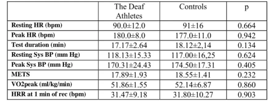
Resting heart rate and heart rate at peak exercise in both of the groups were similar. Likewise resting and peak systolic blood pressures did not differ between the deaf and the normal controls. Exercise workload in metabolic equivalents (METS) and peak oxygen consumption (VO2) values of the deaf athletes were not statistically different from the METS and VO2 values of controls. Heart rate recovery at 1 minute also did not show any difference in the deaf athlete group when compared with the control group (Table 3).
The Deaf Athletes Controls p EDD (cm) 5.10±2.65 5.40±1.45 0.033 ESD(cm) 3.20±0.26 3.40±0.44 0.062 EF (%) 67.63±3.94 68.10±3.68 0.514 FS (%) 37.88±3.26 38.35±3.18 0.483 E (m/sec) 0.96±0.19 0.91±0.17 0.429 A (m/sec) 0.62±0.14 0.58±0.10 0.254 E/A 1.56±0.28 1.57±0.26 0.959 IVS(cm) 0.88±0.10 1.02±0.08 <0.001 PW (cm) 0.84±0.12 1.02±0.10 <0.001 LVMI (g/m²) 187.68±30.00 267.26±59.60 <0.001 SPAP (mmHg) 19.28±7.38 23.10±7.87 0.065 Sm(cm/sec) 0.09±0.01 0.09±0.01 0.699 E'm(cm/sec) 0.14±0.03 0.13±0.03 0.617 A'm(cm/sec) 0.08±0.02 0.12±0.18 0.788 E’m/A’m 1.78±0.38 1.71±0.67 0.682 E/Em' 7.19±1.78 7.07±1.44 0.933 Slat(cm/sec) 0.13±0.02 0.14±0.03 0.296 E'lat(cm/sec) 0.18±0.04 0.18±0.02 0.847 A'lat(cm/sec) 0.08±0.02 0.08±0.02 0.962 E'lat/A'lat 2.33±0.88 2.42±0.69 0.435 E/E'lat 5.50±1.69 5.02±0.73 0.807 TDI IVRT(ms) 62.56±7.90 72.90±11.01 <0.001 TDI ICT(ms) 52.56±9.44 59.50±11.83 0.028 TDI ET (ms) 282.00±28.41 289.45±13.09 0.036 TDI MPI 0.41±0.073 0.46±0.061 0.026
Table 3. Exercise Stress Testing Parameters
Bpm: beat per minute; METS: metabolic equivalents.
Discussion
Deaf athletes are different from all the other athletes because of their special communication needs on the sports field. That is why the International Olympic Commitee organize the Deaflympics as a separate sports organization for the athlete with a hearing loss. The Deaflympics are ‘run by deaf people for deaf athletes’ (Stewart & Ammons, 2001). Scheetz described this distinguished indi-viduals as a ‘sociolinguistic community’ where membership is gained through the acquisition of sign language and desire to embrace the values of deaf culture (Easterbrooks & Scheetz, 2004).
On the other side both endurance and resistance-trained athletes demonstrate larger left ventricular (LV) structures than sedentary controls with greater dimen-sions in endurance athletes suggestive of an eccentric hypertrophy. Both athlete groups had a larger LV wall thickness, chamber dimensions and mass than the control group and this findings support the existence of a morphological athlete’s heart (AH) (Pluim et al., 2000). The endurance-trained athletes had marginally larger LV mass and significantly greater LV diastolic diameter and LV end-diastolic volume than resistance athletes, so the endurance athletes tend to present with the largest LV dimensions (Naylor et al., 2008). The underlying mechanism of this training induced changes in LV morphology in endurance athletes is poorly understood, however a hemodynamic volume overload is widely quoted (George, Wolfe & Burggraf, 1991). According to the echocardiographic parameters, both LV E/A and LV E’ are found to be significantly greater in endurance athletes than controls reflecting an improved diastolic filling at rest. Both IVS wall thickness and posterior wall thickness are greater in resistance-trained athletes than controls and are similar to values in endurance athletes. Cavity dimension is greater in resistance athletes than controls but smaller than in endurance athletes. As a result both athlete groups represent a similar qualitative cardiac adaptation to training with greater cardiac dimensions in endurance athletes reflecting a greater overall
The Deaf
Athletes
Controls p
Resting HR (bpm) 90.0±12.0 91±16 0.664
Peak HR (bpm) 180.0±8.0 177.0±11.0 0.942
Test duration (min) 17.17±2.64 18.12±2,14 0.134
Resting Sys BP (mm Hg) 118.13±15.33 117.00±16,25 0.624 Peak Sys BP (mm Hg) 170.31±24.43 174.50±17.31 0.405 METS 17.89±1.93 18.55±1.41 0.232 VO2peak (ml/kg/min) 51.86±1.55 52.14±6.87 0.860 HRR at 1 min of rec (bpm) 31.47±9.18 31.80±10.27 0.903
In this study we compared the deaf athletes with the normal controls, both of whom were all endurance-trained athletes. The athletes with a hearing loss con-sider themselves as a part of the deaf community, a community with a unique language and culture. To the best of our knowlodge, there has been insufficient data in the literature about their differences from the normal athletes in terms of cardiac structure and function. When the laboratory data are taken into consi-deration ALT, AST and GGT were higher in the deaf group though being in normal ranges. MPV was found to be smaller in the deaf athlete group than the controls. Likewise Banfi et al. found that platelet count and mean platelet volume were significantly increased in normal elite athletes but they were all in the physiological reference ranges (Banfi et al., 1995). The difference between the deaf and normal athletes, however, needs to be clarified by further research. Although sport activities induce a blood lipid profile superior to that of sedentary subjects, in our study the deaf athletes had higher total cholesterol, LDL and trigliseride levels when compared to the controls. However all the lipid parameters were already in the normal ranges for both of the groups. The differences between athletes and sedentary subjects are found to be mainly due to HDL cholesterol in physically active individuals (Banfi et al., 1995). In another study performed on elite athletes in Olympic sports, football, basketball, volleyball and all the disci-plines taken together showed significantly lower LDL (Tsopanakis, Kotsarellis, & Tsopanakis, 1986). The higher levels of lipid parameters in the deaf group is an interesting finding and it may be attributable to the dietary habits of the deaf population. But before making a generalization, this issue has to be investigated in a larger group of deaf athletes.
The deaf athletes also did not show any difference when the parameters of the segmental analysis were taken into consideration. They were all endurance trained athletes who were subject to the same training and diet programme. The cardiac evaluation of the AH has always been a matter of concern for the investigators. However the cardiac differences of the disabled athlete has not been investigated that much and we do not have sufficient data in the literature. In our study we found that end-diastolic diameter was smaller in the deaf group than the normal controls. Left ventricular mass index was also found to be decreased than the normal athletes. These two echocardiographic differences of the deaf athlete’s heart may indicate a transition zone between the normal heart and the athlete’s heart. The reason may be attributable to the intensity and duration of exercise of a disabled group, though not being any physical disablement.
HRR has been proposed as a marker of autonomic function and training status in athletes. According to a systematic review, most longitudinal studies observed a corresponding increase in HRR and training status of the athletes(Daanen et al., 2012). Lee et al. also suggest that as a result of the relationship between HRR and
2012). In our study, the deaf athletes did not show any difference with respect to neither the HRR at 1 minute of exercise nor the other exercise stress testing parameters. Their aerobic capacities as assessed by peak VO2 were also similar to the normal athletes. These data suggest that the deaf athletes can show the same physical performance with the same aerobic capacity and they are also not diffe-rent in terms of cardiac autonomic function.
TDI analysis has evolved as a new quantitative tool for the assessment of cardiac systolic and diastolic function. Left ventricular transmitral filling pattern can be altered by changes in preload or left atrial pressure, because of this reason TDI derived parameters are considered to be more sensitive than mitral Doppler indexes in the assessment of left ventricular relaxation. For many disease states, TDI-derived MPI has been a more sensitive parameter of global LV function when compared with Doppler-derived MPI (Farias et al., 1999). In our study we measured TDI-derived MPI in order to assess global LV function in both of the athlete groups and found out that TDI-derived MPI was significantly increased in the normal athletes when compared with the deaf group. TDI-derived MPI is more sensitive to alterations in LV stiffness or LV end-diastolic pressure. This may be explained by the fact that IVRT or ICT measured by TDI coincides with myocardial movement and may account for the altered LV function because of many disease states (Ayhan et al., 2012). Impairment of LV relaxation contribute to the increased sum of IVRT and ICT and also to decreased ET of LV, all of which results in a relative increase in TDI-derived MPI. According to the results of our study, the normal elite athletes were found to have decreased global LV performance as a reflection of MPI when compared to the deaf group and this difference can be attributable to the impaired LV relaxation dynamics as the conventional systolic parameters of all the athletes were in normal ranges. This echocardiographic finding also reinforces the data that puts the deaf athlete’s heart somewhere in-between the normal and the athlete’s heart.
Conclusions
In conclusion, beyond the loss of hearing, the deaf athletes have many cardiac differences from their normal counterparts in terms of left ventricular structure and function. The deaf athlete is physically able-bodied and can be involved in competitive sports without marked restrictions. Regular physical activity and sport is important for enhanced physical fitness and skill performance of the deaf athletes. Individuals who are deaf and hard of hearing prefer to compete against other deaf individuals rather than against those who can hear (Kurkova, Scheetz, & Stelzer, 2010). The cultural identity of having a hearing loss involves more than just competing with each other – in another definition it is a celebration of community (McKee, Schlehofer, & Thew, 2013). Since 1932, hearing students
have been described as being more fit than deaf students (Stewart, Ellis, 1999). This difference has been attributed to a lack of motivation, insufficient commu-nication during the tests or both of these factors (Hattin et al., 1986; Dunn & Potincelli, 1998). As far as we know, there has not been any research about cardiac differences of the deaf athletes in the literature. From this pont of view, our study elucidated that the deaf athletes are not only sportsmen with hearing loss sharing a unique cultural identity, but also they may exhibit some differences of cardiac structure and global left ventricular performance which are difficult to explain only with their special disablement. Our study revealed that although they resemble the athlete’s heart according to many structural and functional features in common, the deaf athlete’s heart is standing in a gray zone in-between the normal heart and the athlete’s heart. Many features may be involved in this outcome – physical exercise, dietary habits, cultural characteristics of a unique community, the idea of having a disablement, communication problems and psychological issues of having hearing loss may all contribute to this difference. In order to elucidate the physical and psychological determinants of this diffe-rence, the results of further research involving larger cohort of the deaf athletes should have to be awaited.
References
Ammons, D. K. (2009). The International Committee on Sports for the Deaf and the Deaflympics. In: D. Moores & M. Miller, eds. Deaf people around the world:
Educational and Social perspectives: Washington, DC: Gallaudet University Press,
pp. 373-386.
Ayhan, S.S., Ozdemir, K., Kayrak, M., Bacaksiz, A., Vatankulu, M.A., Eren, O. et al. (2012). The evaluation of doxorubicin-induced cardiotoxicity: comparison of Doppler and tissue Doppler-derived myocardial performance index. Cardiology
Journal, 19(4), 363-268.
Banfi, G., Marinelli, G., Roi, G.S., Giacometti, M. (1995). Platelet ind ces in athletes performing a race in altitude environment. Journal of Clinical Laboratory
Analy-sis, 9(1), 34-36.
Daanen, H.A., Lamberts, R.P., Kallen, V.L., Jin, A., Van Meeteren, N.L. (2012). A systematic review on heart rate recovery to monitor changes in traiining status in athletes. International Jounal of Sports Physiology and Performance, 7(3), 251-260.
Danermark, B., Granberg, S., Kramer, S.E., Selb, M., & Moller, C. (2013). The creation of a comprehensive and a brief core set for hearing loss using the International Classification of Functioning. Disability and Health. American Journal of
Au-diology, 22(2), 323-328.
Dunn, J.M., Potincelli, J. (1998). The effect of two different communication modes on motor performance test scores of hearing impaired children. In Abstracts of
re-Easterbrooks, S.R., & Scheetz, N.A.(2004). Applying critical thinking skills to character education and values clarification with students who are deaf or hard of hearing.
American Annals of Deaf, 149(3), 255-263.
Farias, C.A., Rodriguez, L., Garcia, M.J., Sun, J.P., Klein, A.L., & Thomas, J.D. (1999). Assessment of diastolic function by tissue Doppler echocardiography: Comparison with standard transmitral and pulmonary venous flow. Journal of the American
Society of Echocardiography, 12(8), 699-717.
George, K.P., Wolfe, L.A., & Burggraf, G.W. (1991). The ‘athletic heart syndrome’. A critical review. Sports Medicine, 11(5), 300-330.
Hattin, H., Fraser, M., Ward, G., & Sheppard, R.J. (1986). Are deaf children unusually fit ? A comparison of fitness between deaf and blind children. Adapted Physical
Activity Quarterly. 3, 268-275.
Hoffman, H.J., Dobie, R.A., Ko, C.W., Themann, C.L., & Murphy, W.J. (2010). Ame-ricans hear as well or better today compared with 40 years ago: hearing threshold levels in the unscreened adult population of the United States, 1959-1962 and 1999-2004. Ear and Hearing, 31(6), 725-734.
Kurkova, P., Scheetz, N.A., & Stelzer, J. (2010). Health and physical education as an important part of school curricula: A comparison of schools for the deaf in the Czech Republic and the United States. American Annals of the Deaf, 155(1), 78-87.
Lee, C.M, & Mendoza, A. (2012). Dissociation of heart rate variability and heart rate recovery in well-trained athletes. European Journal of Applied Physiology, 112(7), 2757-2766.
McKee, M., Schlehofer, D., & Thew, D. (2013). Ethical issues in conducting research with deaf populations. Americam Journal of Public Health, 103(12), 2174-2178. Naylor, L.H., George, K., O’Driscoll, G., & Green, D.J. (2008). The athlete’s heart: a contemporary appraisal of the ‘Morganroth hypothesis’. Sports Medicine, 38(1), 69-90.
Pluim, B.M., Zwinderman, A.H., Van Der Laarse, A., & Van Der Wall, E.E. (2000). The Athlete’s Heart: A meta-analysis of cardiac structure and function. Circulation
Journal, 101(3), 336-344.
Rakowski, H., Appleton, C., Chan, K. L., Dumesnil, J.G., Honos, G., Jue, J. et al. (1996). Canadian consensus recommendations for measurement and reporting of diastolic dysfunction by echocardiography. Journal of the American Society of
Echo-cardiography, 9(5), 736-760.
Stewart, D.A., & Ammons, D.K. (2001). Future directions of the Deaflympics. Palaestra,
17(3), 45-49.
Stewart, D.A., & Ellis, M.K. (1999). Physical education for deaf students. American
Annals of Deaf, 144(4), 315-319.
Tei, C. (1995). New non-invasive index for combined systolic and diastolic ventricular function. Journal of Cardiology, 26(2), 135-136.
Tsopanakis, C., Kotsarellis, D., & Tsopanakis, A.D. (1986). Lipoprotein and lipid profiles of elite athletes in Olympic sports. International Journal of Sports Medicine, 7(6), 316-321.