Alterations in Corneal Biomechanical and Topographic
Features After Accelerated Crosslinking: 1-Year Results
Address for correspondence: Ahmet Kirgiz, MD. Saglik Bilimleri Universitesi, Oftalmoloji Anabilim Dali, Prof. Dr. N. Resat Belger Beyoglu Goz Egitim ve Arastirma Hastanesi, Istanbul, Turkey
Phone: +90 505 397 46 83 E-mail: [email protected]
Submitted Date: November 07, 2018 Accepted Date: July 09, 2019 Available Online Date: August 08, 2019 ©Copyright 2019 by Beyoglu Eye Training and Research Hospital - Available online at www.beyoglueye.com OPEN ACCESS This work is licensed under a Creative Commons Attribution-NonCommercial 4.0 International License.
Introduction
Keratoconus is a progressive, usually bilateral corneal ecta-sia characterized by corneal thinning and protrusion, pro-gressive myopia, and irregular astigmatism (1). The only way shown to stop progression is the corneal collagen crosslink-ing (CXL) (2, 3).
CXL is a minimally invasive procedure that increases the biomechanical strength of the cornea through the induction of photochemically triggered cross-links within the colla-gen network using riboflavin and ultraviolet-A radiation (4, 5). Conventional CXL was determined as a safe and effi-cient way of treatment in keratoconus in many studies (5, Objectives: To determine the biomechanical and topographic alterations within the first year after accelerated
crosslink-ing (CXL) treatment in patients with keratoconus.
Methods: In this prospective study, 52 eyes of 52 patients with progressive keratoconus underwent accelerated CXL were included. All patients had a detailed preoperative ophthalmologic examination, including slit-lamp evaluation, Gold-mann tonometry, fundoscopy, topography by Scheimpflug imaging (Sirius), and corneal biomechanical evaluation with a biomechanical waveform analysis device (ORA). Alterations in visual acuity and topographic findings were evaluated before the treatment and at 12 months follow-up. Corneal biomechanical features were obtained before the treatment, and at 1st, 3rd, 6th and 12th months.
Results: Uncorrected-visual acuity and best-corrected visual acuity both statistically significantly improved at 12th month
(p=0.001). There were no statistically significant differences in keratometry values, whereas maximum K (AKfront) and symmetry index front (SIfront) decreased significantly (p=0.015 and p=0.009, respectively). Corneal thinnest point and volume also decreased significantly at 12th month (p=0.001 for both). Goldmann-correlated intraocular pressure (IOPg)
and corneal compensated IOP (IOPcc) values transiently increased in the first three months, while corneal hysteresis (CH) and the corneal resistance factor (CRF) transiently decreased, with the difference not statistically significant (p>0.05). However, central corneal thickness significantly decreased at the end of the 12th month (p=0.001).
Conclusion: Accelerated CXL seems to be effective in stopping the progression of keratoconus. Our findings showed transient alterations in biomechanical features, which will end with the preoperative values at the end of the 12th month.
Further studies are needed to demonstrate the changes in corneal biomechanics in vivo.
Keywords: Accelerated crosslinking, corneal biomechanics, corneal topography, corneal hysteresis, keratoconus.
Ahmet Kirgiz,1 Sevil Karaman Erdur,2 Kubra Serefoglu Cabuk,1 Kursat Atalay,3
Senay Asik Nacaroglu3
1Department of Opthalmology, University of Health Sciences, Prof. Dr. N.Resat Belger Beyoglu Eye Training and
Research Hospital, Istanbul, Turkey
2Department of Opthalmology, Istanbul Medipol University, Istanbul, Turkey
3Department of Opthalmology, University of Health Sciences, Bagcilar Training and Research Hospital, Istanbul, Turkey
6). However, the procedure lasts approximately one hour. Theoretically, the same energy dose can be delivered in a shorter time with an accelerated modality (7, 8). In accel-erated CXL, reported as the rapid, effective and safe al-ternative in keratoconus treatment, the same energy dose ensuring a proportional biological effect is delivered in a shorter exposure time by setting different UV-A powers (9, 10). The UV-A power could be set to 30, 18 or 9 mW/ cm2 to radiate cumulative dose of 5.4 J/cm2 in a shorter
time, such as 3, 5 or 10 minutes, respectively. With the increasing use of these accelerated CXL protocols, studies on the long-term effects have begun to be published in re-cent years (11, 12).
Although it is shown in ex vivo that the rigidity of the cornea is increased 4.5 times in the human cornea by CXL (13), it is important to evaluate the changes in corneal biome-chanics after the treatment in vivo to understand the effi-cacy of the procedure. It is theoretically possible to evaluate the corneal biomechanical properties that may occur in the cornea with the ocular response analyzer (ORA) designed to obtain in vivo measurements (14). While several studies report the biomechanical changes after conventional CXL with the ORA device (15–21), the data about the results of accelerated CXL regarding the biomechanical features of the cornea are limited in the literature (22). In this study, we aimed to investigate the biomechanical and topographic al-terations in 12 month period after accelerated collagen CXL treatment among patients with keratoconus.
Methods
In this prospective study, 52 eyes of 52 patients with pro-gressive keratoconus who underwent accelerated CXL between July 2014 and January 2015, in Bagcilar Education and Research Hospital, Istanbul, Turkey were included. Pa-tients who were followed up for 12 months regularly were included in this study. This study was approved by the local Ethics Committee. Progressive keratoconus was defined as an increase of 1.00 diopter (D) or more in the steepest ker-atometry (K) measurement or the loss of at least two lines of best-corrected distance visual acuity in last 1 year (23). The exclusion criteria were as follows: patients younger than 18 or older than 40 years of age, patients with a corneal thickness less than 400 µ at the thinnest point, patients with corneal scarring or severe dry eye, patients having a history of corneal surgery, or pregnant or lactating women during the course of this study.
All patients had a detailed preoperative ophthalmo-logic examination, including slit-lamp evaluation, Goldmann tonometry, fundoscopy, topography by Scheimpflug imag-ing (Sirius, Schwind Eye-Tech-Solutions GmbH, Germany), corneal biomechanical properties and intraocular pressure
evaluation by Ocular Response Analyzer (Reichert Tech-nologies, Buffalo, NY, USA). Visual acuity was obtained with Snellen charts and converted to LogMAR units for analysis. Uncorrected visual acuity (UCVA) and best-corrected visual acuity (BCVA) were both recorded.
The Sirius, a topographic device consisting of a combina-tion of two rotating Scheimpflug cameras and a Placido disk, was used for topographical and central corneal thickness (CCT) measurements. Flat keratometry value (K1), steep keratometry value (K2), average keratometry value (Kavg), topographic cylindrical value (Cyl), apical keratoscopy front
(AKfront), apical keratoscopy back (AKback), symmetry index
front (SIfront), symmetry index back (SIback), corneal thinnest point (CTP), and corneal volume (CV) were recorded be-fore the treatment and at 12 months follow-up. Apical ker-atoscopy front (AKfront) was defined as the steepest point of the anterior corneal surface, while apical keratoscopy back (AKback) was defined as the steepest point of the posterior corneal surface. Symmetry index front (SIfront), the symme-try index of the anterior curvature, was defined as the dif-ference of the mean anterior tangential curvature of two circular zones centered on the vertical axis in the inferior and superior hemispheres. Symmetry index back (SIback), the symmetry index of the posterior curvature, was defined as the difference of the mean posterior tangential curvature of two circular zones centered on the vertical axis in the inferior and superior hemispheres. All measurements were taken with the manufacturer’s guideline by the same trained examiner.
The Ocular Response Analyzer (ORA) reports two in-traocular pressure values: Goldmann-correlated IOP (IOPg) and corneal compensated IOP (IOPcc). Two different bidi-rectional (inward and outward) applanation pressure mea-surements were recorded by ORA. The difference between these two pressures is the corneal hysteresis (CH), which is an indicator of corneal viscosity. Corneal resistance factor (CRF) is considered as the indicator of the overall resistance of the cornea that is mainly associated with the elastic prop-erties of the cornea.
Alterations in visual acuity and topographic findings of study participants were evaluated before the treatment and at 12th month of follow-up. Corneal biomechanical proper-ties were obtained before the treatment and at 1, 3, 6, and 12 months following accelerated CXL.
Surgical Technique
Accelerated crosslinking has been performed to all patients. Initially, a topical anesthetic agent (Alcaine; Alcon Labora-tories, Inc.) was administered and the central 8.0 mm ep-ithelium was removed with a blunt spatula. Riboflavin with dextran (0.1% in 20% dextran solution, Medicross,
Ger-many) was administered topically every three minutes for 30 minutes. The cornea was aligned and exposed to UV-A 365 nm light for five minutes at an irradiance of 18 mW/ cm2 (Peschke Meditrade, GmbH) (11). During UV-A expo-sure, riboflavin administration was continued every minute. After the treatment, the eye surface was washed with 20.0 mL of a balanced salt solution. Postoperatively, antibiotic (Vigamox; Alcon Co., Inc.), corticosteroid drops (Lotemax; Bausch & Lomb) and artificial tears (Refresh; Allergan) were administered; a soft contact lens bandage was placed. The contact lens was removed after the closure of the ep-ithelial defect. Antibiotics and corticosteroid drops were continued four times daily for one week and two weeks, respectively.
Statistical Analysis
All analyses were performed with the Statistical Package for Social Sciences software (SPSS, Windows version 21.0; SPSS Inc.; Chicago, IL, USA). A P value of less than 0.05 was considered statistically significant. Topographic findings (K1,
K2, Kavg, AKfront, AKback, SIfront, SIback, Cyl, CTP, and CV), BCVA
and UCVA were compared using the paired Student-t test. Corneal biomechanical properties were compared using re-peated-measures ANOVA with post hoc Tukey test. The correlations between CCT at 12 months, and each periop-erative variable were evaluated using the Pearson correlation coefficient.
Results
Totally 52 eyes (28 right and 24 left) of 52 patients (18 fe-male, 34 male) with a mean age of 24.15±6.75 (range: 18-39 years) years were included. Visual acuities and topographic findings of patients before the treatment and at 12 months follow-up are summarized in Table 1.
There was a statistically significant improvement in the UCVA and BCVA after the 12-month visit (both p=0.001). The AKfront and SIfront values showed a significant decrease after 12 months compared with the preoperative values (p=0.015, and p=0.031, respectively). The CTP and CV also revealed a significant decrease at the end of 12-month visit (both p=0.001).
Alterations in corneal biomechanical properties during the 12-month follow-up are given in Table 2. Regarding these alterations, IOPcc and IOPg values transiently increased in the first three months; whereas CH and CRF values tran-siently decreased in first three months, although the changes were statistically insignificant. However, CCT significantly decreased at the end of 12-month visit (p=0.001) (Fig. 1).
As regards the correlations, there was significant corre-lation between the mean CCT at 12th month and baseline
CCT, CTP, and CV (r=0.888, r=0.769, r=0.523, respectively, p=<0.001 for all); whereas no significant correlation was found between the mean CCT at 12th month and other
pa-rameters (p>0.05 for all).
Baseline 12th month P* Mean±SD Mean±SD UCVA (LogMAR) 0.71±0.32 0.55±0.32 <0.001 BCVA (LogMAR) 0.42±0.22 0.26±0.14 <0.001 K1 (D) 45.4±1.8 45.4±2 0.805 K2 (D) 48.6±2.2 48.6±2.4 0.837 Kavg (D) 46.9±1.9 46.9±2.1 0.944 Cyl (D) 3.2±1.4 3.1±1.4 0.198 AKfront (D) 55.1±4.1 54.6±4.2 0.015 AKback (D) 79.6±8.8 83.9±11.2 0.001 SIfront (D) 6.1±2.8 5.8±2.8 0.031 SIback (D) 1.5±0.7 1.6±0.7 0.013 CTP (µm) 456±32 424±48 0.001 CV (mm³) 55.8±3.3 54.4±3.5 0.001
SD: standard deviation; UCVA: uncorrected visual acuity; BCVA: best corrected visual acuity; K1: flat keratometry
value; K2: steep keratometry value; Kavg: average keratometry value; Cyl: topographic cylindrical value; AKfront:
apical keratoscopy front; AKback: apical keratoscopy back; SIfront: symmetry index front; SIback: symmetry index back;
CTP: corneal thinnest point; CV: corneal volume. *Paired samples t test.
Table 1. Comparison of visual acuity and topographic findings between the preoperative and the 12 months of follow-up
Discussion
This study is one of the few studies in the literature evalu-ating biomechanical features along with topography changes following accelerated CXL reporting at least 1-year results (22). Both UCVA and BCVA statistically significantly im-proved in 12th months of follow up with significant
alter-ations in AKfrontand SIfront values of the cornea, demonstrating the efficacy of this method in controlling keratoconus. On the other hand, CH and CRF remained unchanged after 12
months of the treatment suggesting these parameters may not represent the exact biomechanical effects of accelerated CXL.
In CXL treatment, reactive oxygen species are thought to be produced, leading to the formation of covalent cross-links between collagen molecules, with a resulting stiffening of the stromal tissue. This strengthening of the corneal stroma de-celerates the progression of keratoconus and ectasia and, in many cases, improves patients’ visual, refractive, and topo-graphic outcomes (5, 24). In that aspect, some alterations especially in corneal biomechanical properties are expected. In the relevant literature, there are several studies about the alterations in topographic and corneal biomechanical properties after conventional CXL. Goldich et al. (15) an-alyzed 10 eyes in the first six months after conventional CXL treatment and consistent with our results, they also reported transient nonsignificant elevations in CH and CRF values after CXL. Sedaghat et al. (16) compared CH and CRF values in the first six months after conventional CXL for ker-atoconus in 56 eyes of 51 patients and did not determine any significant alterations in CH, CRF, CCT and IOPg, but IOPcc was reported to be decreased significantly. In another study of Goldich et al. (17), 14 patients were assessed after con-ventional CXL for 24 months and a significant improvement in BCVA with a decrease in the mean cylinder without any Table 2. Alterations in corneal biomechanical properties during follow-up after accelerated
crosslinking
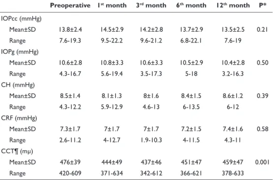
Preoperative 1st month 3rd month 6th month 12th month P*
IOPcc (mmHg) Mean±SD 13.8±2.4 14.5±2.9 14.2±2.8 13.7±2.9 13.5±2.5 0.21 Range 7.6-19.3 9.5-22.2 9.6-21.2 6.8-22.1 7.6-19 IOPg (mmHg) Mean±SD 10.6±2.8 10.8±3.3 10.6±3.3 10.5±2.9 10.4±2.8 0.50 Range 4.3-16.7 5.6-19.4 3.5-17.3 5-18 3.2-16.3 CH (mmHg) Mean±SD 8.5±1.4 8.1±1.3 8±1.6 8.4±1.5 8.6±1.2 0.39 Range 4.3-12.2 5.9-12.9 4.6-13 6-13.5 6-12 CRF (mmHg) Mean±SD 7.3±1.7 7±1.7 7±1.7 7.2±1.5 7.4±1.6 0.58 Range 2.6-11.2 4-12.7 1.9-10.3 4-11.5 4.3-11 CCT¶ (mμ) Mean±SD 476±39 444±49 437±46 451±47 459±47 0.001 Range 420-609 371-634 342-612 366-621 378-633
SD: standard deviation; IOPcc: corneal compensated intraocular pressure; IOPg: Goldmann-correlated intraocular pressure; CH: corneal hysteresis; CRF: corneal resistance factor; CCT: central corneal thickness ¶ measured with Sirius device. *Repeated measures ANOVA, post-hoc Tukey test.
Figure 1. Alterations in central corneal thickness (CCT) at postoper-ative 12 months after accelerated crosslinking.
410 420 430 440 450 460 470 480
Preop 1st month 3rd month 6th month 12th month
mµ
significant alterations in minimal corneal thickness, endothe-lial cell density, corneal hysteresis, or corneal resistance factor was reported. Greenstein et al. (18) investigated the corneal biomechanical changes in 69 eyes after conventional CXL for one year, and consistent with our results, they re-ported that there were not any alterations in CH or CRF values. In this study, we determined transient alterations in the first three months in IOPcc, IOPg, CRF and CH after accelerated CXL which were not statistically significant but a significant decrease in CCT in the 12th month of follow-up.
In another recent study investigating corneal topograph-ical and biomechantopograph-ical properties in the 1st year after con-ventional CXL in 97 eyes and compatible with our results they also reported a significant improvement in UCVA and BCVA values with a significant decrease in CCT and corneal volume without any significant alterations in CH and CRF. However, diversely from our results, they reported a sig-nificant decrease in average keratometry value (19). Inter-estingly, Beckman et al. (20) investigated the biomechanical effects of conventional CXL in 29 eyes of 28 patients and reported increased CH values with applanation resonance tonometer but not with ORA. They also determined a sig-nificant decrease in CCT in 1st and 6th months of follow-up
after treatment. Terai et al. (24) defined ORA as providing invaluable information for defining biomechanical conditions of the cornea. We used ORA to determine the corneal biomechanical properties in this study and did not deter-mine any significant alterations in the corneal biomechanical properties during the first year after accelerated CXL. Con-sistent with our results, Çağıl et al. (25) did not find any sig-nificant alterations in CH and CRF but observed a sigsig-nificant decrease in CCT at postoperative six months after conven-tional CXL. In a recent and the only one study investigating the corneal biomechanical alterations after accelerated CXL, Uzel et al. (22) found out that second-generation parame-ters (p1 area, p2 area, h2, and dive 2) were more sensitive than CH and CRF with a new ORA software (version 2.04). CH is an indicator of corneal viscosity, and CRF is consid-ered as the indicator of the overall resistance of the cornea that is mainly associated with the elasticity of the cornea. After CXL, accelerated or conventional, decrease in CH and/ or CRF values is expected since CXL increases the biome-chanical strength of the cornea. However, in this study, we determined that, though transient alterations in CH or CRF might be seen in first months after accelerated CXL, there was not a statistically significant increase at the end of 12th
month compared with the preoperative values. There may be some reasons for these insignificant results. Firstly, the decrease in CCT may have prevented the accurate measure-ment of corneal biomechanics. Secondly, the ORA device may be inadequate in evaluating the corneal biomechanical
properties due to the changing in corneal collagen configu-ration after CXL. The transient alteconfigu-rations seen in the first three months in CH and CRF may be due to the insufficient ORA signals which were probably affected by optical irreg-ularity.
The data about the effects of accelerated CXL are in-creasing in the literature. Elbaz et al. (26) assessed the topo-graphical, visual, and refractive effects of accelerated CXL in keratoconus at 6th and12th months of follow-up and reported
that there was not a statistically significant alteration in cor-rected distance visual acuity and mean refractive cylinder, but uncorrected distance visual acuity was improved. More-over, all corneal parameters, including Ksteep, Kflat, average K, corneal astigmatism, were stable at 6 and 12 months in all patients. Mita et al. (27) evaluated the effectiveness of accel-erated CXL for keratoconus in first six months after treat-ment in 39 eyes of 22 patients and reported a statistically significant improvement in UCVA and a significant decrease in the mean maximum keratometry readings without any changes in the endothelial cell density. Although alterations in 1st year after accelerated CXL were investigated in a few studies before, there are also some recent studies reporting the differences between conventional CXL and accelerated CXL. Chow et al. (28) compared one-year outcomes of 2 CXL methods in patients with progressive keratoconus and reported insignificantly flattened maximum and minimum keratometry values and significantly reduced corneal thick-ness in both groups. Moreover, similar to our results sig-nificant improvements in UCVA and BCVA were reported. Hashemi et al. (29) compared the six months results of 2 CXL methods in 62 eyes and reported similar clinical effi-cacies. Tomita et al. (30) reported that there were no sta-tistically significant differences in the postoperative changes in the keratometric readings or the corneal biomechanical responses between the accelerated and conventional CXL.
In correlation analysis, we found no statistically signifi-cant relation between CCT at 12 months versus preopera-tive corneal biomechanical properties. However, there was a significant correlation between CCT at 12 months versus preoperative CCT, CTP and CV. In that aspect, in all pa-tients, preoperative CCT, CTP and CV should be evaluated carefully. Unlike with our results, Viswanathan et al. (31) de-termined a positive correlation of both CH and CRF with CCT in the normal, keratoconic and crosslinked eyes.
Maximum keratometry value (AKfront) was defined as one of the topographic indicators of CXL success and reported to be stable or decreased after CXL (32). Likewise, signif-icantly decreased AKfront values were also reported in this study in 12th-month control after the treatment showing
that accelerated CXL is effective in stopping the progression of keratoconus.
The main limitations of our study were the small sample size and lack of a control group consisting of patients who underwent conventional CXL. However, the investigation of both topographic and corneal biomechanical properties in the 12 month period is the strength of this study.
In conclusion, in the 12th month after accelerated CXL,
determination of improved UCVA and BCVA, together with the stable K values, decreased AKfront and SIfront suggests the long-term efficacy of this procedure. Our findings showed transient alterations in biomechanical features, which will end with the preoperative values at the end of 12th month.
Further studies with larger groups and longer follow-up pe-riods are required to determine the exact effects of acceler-ated CXL on corneal topography and advanced technologies are needed to assess the corneal biomechanical alterations more accurately.
Disclosures
Ethics Committee Approval: Bagcilar Training and Research
Hospital Clinical Research Ethics Committee, May 17, 2016, no: 459.
Peer-review: Externally peer-reviewed. Conflict of Interest: None declared.
Authorship Contributions: Involved in design and conduct of
the study (AK, SKE, KSC); preparation and review of the study (AK, SKE, SAN); data collection (AK, KSC, KA); and statistical analysis (AK, KA, SAN).
References
1. Romero-Jiménez M, Santodomingo-Rubido J, Wolffsohn JS. Ker-atoconus: a review. Cont Lens Anterior Eye 2010;33:157–66. 2. Farjadnia M, Naderan M. Corneal cross-linking treatment of
keratoconus. Oman J Ophthalmol 2015;8:86–91.
3. Wollensak G, Spoerl E, Seiler T. Riboflavin/ultraviolet-a-induced collagen crosslinking for the treatment of keratoconus. Am J Ophthalmol 2003;135:620–7.
4. Raiskup-Wolf F, Hoyer A, Spoerl E, Pillunat LE. Collagen crosslinking with riboflavin and ultraviolet-A light in kerato-conus: long-term results. J Cataract Refract Surg 2008;34:796– 801.
5. Wittig-Silva C, Chan E, Islam FM, Wu T, Whiting M, Snibson GR. A randomized, controlled trial of corneal collagen cross-linking in progressive keratoconus: three-year results. Ophthal-mology 2014;121:812–21.
6. Caporossi A, Mazzotta C, Baiocchi S, Caporossi T. Long-term results of riboflavin ultraviolet a corneal collagen cross-linking for keratoconus in Italy: the Siena eye cross study. Am J Oph-thalmol 2010;149:585–93.
7. Brindley GS. The Bunsen-Roscoe law for the human eye at very short durations. J Physiol 1952;118:135–9.
8. Wernli J, Schumacher S, Spoerl E, Mrochen M. The efficacy of corneal cross-linking shows a sudden decrease with very high intensity UV light and short treatment time. Invest Ophthalmol
Vis Sci 2013;54:1176–80.
9. Schumacher S, Oeftiger L, Mrochen M. Equivalence of biome-chanical changes induced by rapid and standard cornealcross-linking, using riboflavin and ultraviolet radiation. Invest Oph-thalmol Vis Sci 2011;52:9048–52.
10. Bouheraoua N, Jouve L, Borderie V, Laroche L. Three Differ-ent Protocols of Corneal Collagen Crosslinking in Kerato-conus: Conventional, Accelerated and Iontophoresis. J Vis Exp 2015;105.
11. Kurt T, Ozgurhan EB, Yildirim Y, Akcay BI, Cosar MG, Bozkurt E, et al. Accelerated (18 mW/cm(2)) Corneal Cross-Linking for Progressive Keratoconus: 18-Month Results. J Ocul Pharmacol Ther 2016;32:186–91.
12. Ting DSJ, Rana-Rahman R, Chen Y, Bell D, Danjoux JP, Morgan SJ, et al. Effectiveness and safety of accelerated (9 mW/cm2) corneal collagen cross-linking for progressive keratoconus: a 24-month follow-up. Eye (Lond) 2019;33:812–8.
13. Wollensak G, Spoerl E, Seiler T. Stress-strain measurements of human and porcine corneas after riboflavin-ultraviolet-A-in-duced cross-linking. J Cataract Refract Surg 2003;29:1780–5. 14. Luce DA. Determining in vivo biomechanical properties of the
cornea with an ocular response analyzer. J Cataract Refract Surg 2005;31:156–62.
15. Goldich Y, Barkana Y, Morad Y, Hartstein M, Avni I, Zadok D. Can we measure corneal biomechanical changes after collagen cross-linking in eyes with keratoconus?--a pilot study. Cornea 2009;28:498–502.
16. Sedaghat M, Naderi M, Zarei-Ghanavati M. Biomechanical pa-rameters of the cornea after collagen crosslinking measured by waveform analysis. J Cataract Refract Surg 2010;36:1728–31. 17. Goldich Y, Marcovich AL, Barkana Y, Mandel Y, Hirsh A, Morad
Y, et al. Clinical and corneal biomechanical changes after colla-gen cross-linking with riboflavin and UV irradiation in patients with progressive keratoconus: results after 2 years of follow-up. Cornea 2012;31:609–14.
18. Greenstein SA, Fry KL, Hersh PS. In vivo biomechanical changes after corneal collagen cross-linking for keratoconus and corneal ectasia: 1-year analysis of a randomized, controlled, clinical trial. Cornea 2012;31:21–5.
19. Sedaghat M, Bagheri M, Ghavami S, Bamdad S. Changes in corneal topography and biomechanical properties after colla-gen cross linking for keratoconus: 1-year results. Middle East Afr J Ophthalmol 2015;22:212–9.
20. Beckman Rehnman J, Behndig A, Hallberg P, Lindén C. Increased corneal hysteresis after corneal collagen crosslinking: a study based on applanation resonance technology. JAMA Ophthalmol 2014;132:1426–32.
21. Küçümen RB, Şahan B, Yıldırım CA, Çiftçi F. Evaluation of Corneal Biomechanical Changes After Collagen Crosslinking in Patients with Progressive Keratoconus by Ocular Response Analyzer. Turk J Ophthalmol 2018;48:160–5.
22. Uzel MM, Koc M, Can C, Polat S, Yılmazbaş P, Ileri D. Effect of accelerated corneal crosslinking on ocular response analyzer waveform-derived parameters in progressive keratoconus. Arq Bras Oftalmol 2019;82:18–24.
23. Sorkin N, Varssano D. Corneal collagen crosslinking: a system-atic review. Ophthalmologica 2014;232:10–27.
24. Terai N, Raiskup F, Haustein M, Pillunat LE, Spoerl E. Identifi-cation of biomechanical properties of the cornea: the ocular response analyzer. Curr Eye Res 2012;37:553–62.
25. Çağıl N, Saraç Ö, Akçay E, Aksoy B, Uğurlu N, Ayan M. Kera-tokonus hastalarında korneal kollajen çapraz bağlama tedavisinin kısa dönemde kornea biyomekaniği üzerine etkileri. MN Oftal-moloji 2014;21:152–6.
26. Elbaz U, Shen C, Lichtinger A, Zauberman NA, Goldich Y, Chan CC, et al. Accelerated (9-mW/cm2) corneal collagen crosslink-ing for keratoconus-A 1-year follow-up. Cornea 2014;33:769– 73.
27. Mita M, Waring GO 4th, Tomita M. High-irradiance acceler-ated collagen crosslinking for the treatment of keratoconus:
six-month results. J Cataract Refract Surg 2014;40:1032–40. 28. Chow VW, Chan TC, Yu M, Wong VW, Jhanji V. One-year
out-comes of conventional and accelerated collagen crosslinking in progressive keratoconus. Sci Rep 2015;5:14425.
29. Hashemi H, Fotouhi A, Miraftab M, Bahrmandy H, Seyedian MA, Amanzadeh K, et al. Short-term comparison of acceler-ated and standard methods of corneal collagen crosslinking. J Cataract Refract Surg 2015;41:533–40.
30. Tomita M, Mita M, Huseynova T. Accelerated versus conven-tional corneal collagen crosslinking. J Cataract Refract Surg 2014;40:1013–20.
31. Viswanathan D, Kumar NL, Males JJ, Graham SL. Relationship of Structural Characteristics to Biomechanical Profile in Normal, Keratoconic, and Crosslinked Eyes. Cornea 2015;34:791–6. 32. Raiskup F, Theuring A, Pillunat LE, Spoerl E. Corneal collagen
crosslinking with riboflavin and ultraviolet-A light in progres-sive keratoconus: ten-year results. J Cataract Refract Surg 2015;41:41–6.