1
ORIGINAL INVESTIGATION
J Crit Intensive Care 2021;12:1−7https://doi.org/10.37678/dcybd.2021.2583
Predictors of Intraventicular
Hemorhage in the Neonatal
Intensive Care Unit
Hakan ONGUN1 , Sariye Elif OZYAZICI OZKAN2
ABSTRACT
Aim: To analyze intraventricular hemorrhage (IVH) incidence, clinical features and risk factors in extremely
low birth weight infants.
Materials and Method(s): It is a single-center, retrospective cohort in infants delivering neonatal intensive
care between 2010 and 2019. Infants of birth weight <1000 grams who had transfontanel-ultrasounds in the first week of life were included. Postnatal transfers, central nervous system anomalies, antenatal-diagnosed hemorrhage, death before obtaining transfontanel-ultrasound and incomplete dataset were excluded from the study. Infants were categorized as mild IVHs (grades 1-2), severe IVHs (grade-3, periventricular-hemorrhagic infarct) and control group. Ante-perinatal characteristics, clinical and laboratory features were recorded. SPSS-23 program utilized analysis for three groups.
Results: Overall IVH incidence was 22.9% in 455 neonates (52.9%, 49%, 31.5%, 13.4%, 6.7% at ≤25, 26,
27, 28 and 29 gestational weeks. Seizure was the first sign in 32.7% of the infants. Neonatal resuscitation, umbilical-cord blood pH, chorioamnionitis, patent ductus arteriosus were associated with developing mild IVH, but did not influence the progression to severe hemorrhage (p=0.782, p=0.109, p=0.566, p=0.111). Gestational age, invasive mechanical ventilation, inotrope-required hypotension was related to high-grade IVHs (p<0.001, p=0.025, p=0.013). The predictive strength of platelets to define IVH was poor at intensive care admission (sensitivity: 74%, specificity: 68.3%). Inotrope-required hypotension and low-umbilical-cord blood pH were determined as independent risk factors of high-grade IVHs (OR: 6.678, 95% CI: 2.557-17.443 and OR: 3.554, 95%CI: 1.089–11.602). Four infants necessitated ventriculo-peritoneal shunting. Mortality was 23.1% in infants developing IVH.
Conclusion: Inotrope-required hypotension is the strongest predisposing factor for high-grade ventricular
hemorrhage in extremely low birth weight infants.
Keywords: Intraventricular hemorrhage, prematurity, extremely low birth weight, risk factors
1İstinye University Faculty of Medicine Antalya Medical Park Hospital, Department of Pediatrics, Division of Neonatology, Antalya, Turkey 2University of Health Sciences, Antalya Research and Training Hospital, Department of Pediatrics, Division of Neonatology, Antalya, Turkey
Cite this article as: Ongun H, Ozyazici
Ozkan SE. Predictors of Intraventicular Hemorhage in the Neonatal Intensive Care Unit. J Crit Intensive Care 2021;12:1−7
Corresponding Author: Hakan Ongun E mail: [email protected]
©Copyright 2020 by Turkish Society of Medical and Surgical Intensive Care Medicine - Available online at www.dcyogunbakim.org
Received: Dec 21, 2020 Accepted: Dec 28, 2020 Available online: Jan 8, 2021
Introduction
Intraventricular hemorrhage (IVH) is a serious complication of the preterm newborn with significant morbidity and mortality (1). The incidence ranges between 20% and 25% in birth weights less than 1500 grams (2,3). High-grade IVHs (grade-3 and periventricular hemorrhagic infarction - PVHI) remain to be a problem in neonatal morbidity (increased risk of cerebral palsy and neurodevelopmental disorders) and develop at a rate of 22% in infants born less than 1000 grams (4-6).
Most IVHs originate from the germinal matrix that consists of a fragile vasculature leading to structural immaturity. Any fluctuations in the cerebral blood flow (CBF) to this site appear
to be the major cause of bleeding. Following the initial hemorrhage, production of reactive oxygen species and proinflammatory cytokines increase the permeability of the vasculature. The consequent result is the expansion of hemorrhage in the neighboring white matter and parenchyma. However, the physiopathology is believed to be multifactorial: immature cerebral vessels, fluctuations in the blood flow, platelet abnormalities and coagulopathy contribute to IVH development (2,6-8).
Several predisposing factors have been identified in the development of hemorrhage (5,8). Low Apgar score, instrumental birth delivery, severe respiratory distress syndrome (RDS), air leakage syndromes, hypoxia, hypercapnia, patent ductus arteriosus (PDA), sepsis, thrombocytopenia and
coagulopathy are some of the clinical features associated with IVH (2,9). Thrombocytopenia is common in extremely low birth weight (ELBW: birth weight <1000 grams) infants and platelet infusions are prophylactically used to avoid the risk of bleeding. However, the lack of evidence for platelet transfusion thresholds leads to 15% of transfusion rates and increased risk of transfusion reactions in this vulnerable population (6,10).
Data suggests that wide variations occur in predisposing factors and controversies exit in clinical management and daily transfusion practice (6,10). Each neonatal care center should assess regional risk factors and establish their local protocols to avoid IVH in babies born less than 1000 grams. Regarding this fact, we conducted an observational cohort to evaluate IVH incidence, maternal and neonatal characteristics and risk analysis in ELBW preterm infants at a highly-referral, tertiary neonatal intensive care unit (NICU).
Materials and Methods
The study was designed as a single-center, retrospective cohort in ELBW (<1000 grams) neonatal admissions to one of the largest, level III, university-affiliated NICU with 34 incubators around the Mediterranean region between 2010 and 2019.
The patient records were extracted from the hospital's electronic database. The inclusion criteria were neonatal intensive care unit admissions i) born in the same perinatal center where the neonatal care will be delivered, ii) birth weight <1000 grams, iii) who had a transfontanel ultrasound (TFU) in the first seven days of life. Exclusion criteria were, i) birth delivery at another facility and transferred postnatally, ii) major central nervous system (CNS) anomalies, iii) antenatal diagnosed IVH, iv) newborns who died before obtaining TFU and v) incomplete patient dataset.
Based on 2018 Volpe’s ventricular hemorrhage grading classification, IVH severity was defined as, Grade-1 (bleeding in the germinal matrix), Grade-2 (bleeding <50% in the ventricle, without any expansion into the ventricular system, Grade-3 (bleeding ≥ 50% of the ventricle with expansion) and PVHI (5,11). The infants developing IVHs were categorized as mild IVH (grades-1 and 2 IVHs), severe IVH (grade-3 IVH and PVHI) and the control group (infants who did not present any hemorrhage on cranial imaging). The statistical analysis assessed the maternal factors and neonatal characteristics including maternal anticoagulant use (aspirin, low molecular weight heparin and heparin), preeclampsia (hypertension and proteinuria) (12), chorioamnionitis (clinical and/or histological), antenatal steroid administration, intrauterine growth restriction (IUGR: birth weight <10th percentile) (13). The clinical variables were examined for resuscitation at birth delivery; low-umbilical -cord blood pH, echocardiographically-confirmed and medically-intervened PDA, inotrope-required hypotension. Of note, the study center follows the recommendations of the Turkish Neonatal Society on neonatal hemodynamics and management of hypotension (the use of intravenous fluids, inotropes, vasopressors, and steroids when necessary) (14). According to the national guideline, the definition of hypotension is, i) mean blood pressure (BP) below the gestational week or at the <3rd (or 10th) percentile according to the gestational
age; ii) systolic, diastolic, and mean BP <95% CI according to the gestational week; iii) mean BP of <30 mm Hg in the first 72 hours in preterm babies with gestational age of <32 weeks; iv) BP value that disrupts tissue perfusion. Other clinical variables such as RDS, invasive mechanical ventilation (MV), bronchopulmonary dysplasia (BPD: oxygen requirement in the postmenstrual 36th week) (15), early-onset sepsis (suspected or confirmed sepsis in the first 48 hours of life), necrotizing enterocolitis (NEC), duration of NICU stay and survival were extracted from the medical files.
The routine TFUs were performed by the staff radiologists using General Electric LOGIQ S6 2007 device (C3/5L/10 L MHz probs) within the first 24 hours, third day, 7th day and at least once a week in subsequent weeks until the end of the first month. Sequential TFUs were performed for follow-up once a week for four weeks and at the time of discharge in grade I-II IVH, and two times a week and until the time of discharge in grade III-IV IVHs. If the initial IVH grade progresses, the final stage of the hemorrhage was recorded and grouped accordingly. The decision of surgical intervention depended on the risk stratification of the patient defined by El-Dib et al. (red zone – high-risk patient: ventricular index >97th percentile +4 mm and anterior horn width >10 mm and/or thalamo-occipital distance >25 mm in TFU measurements in addition to clinical findings such as the growth of head circumference >2cm per week, separated sutures and bulging fontanelles) (16).
Laboratory data consisted of platelet counts obtained in the first and third days of life. Coagulation tests (prothrombin time, activated partial thromboplastin time – aPTT, the international normalized ratio - INR and levels of fibrinogen) at NICU admission were also collected from the database. We defined coagulopathy if fibrinogen was < 5th % (<71 mg/dL), prothrombin time >95th% (>21 sec), aPTT > 95th% (>4 seconds) (17), and thrombocytopenia as platelet counts <100x109/L (18). In respect to transfusion guidelines of the Turkish Neonatal Society, babies born less than 1000 grams deliver platelet transfusions in presence of severe thrombocytopenia (platelets counts <50x109/L) to avoid bleeding complications (18). The authors also recorded whether if the newborn received platelet transfusion or not.
SPSS–23 program was utilized for descriptive analysis of variables, central tendency and dispersion of quantitative variables. Kolmogorov-Smirnov test was applied for normality assumption, then we used Kruskal Wallis or One-way analysis of variance (followed by posthoc comparisons by Tamhane test) were performed for analysis within three groups. Receiver operating characteristic (ROC) assessed the area under the curve (AUC) and the cutoff values of the platelets using Medcalc statistical program (free trial version). Univariate logistic regression was performed to predict potential risk factors of developing IVH. The multivariate logistic regression determined independent risk factors of severe IVHs. All data were expressed as percentage (%), median (interquartile range - IQR) and odds ratios (OR) and 95% confidence intervals (CI). A p-value of less than 0.05 was considered significant.
The study was approved by the Institutional Review Board of Istinye University Antalya Medical Park Hospital (no: 2019/10, date: 23.10.2019). We did not perform a power analysis due to the retrospective nature of the study.
Results
Total of 455 ELBW infants out of 592 NICU admissions with birth weight <1000 grams were enrolled in the study (exclusions due to birth in another facility in 109 infants, lack of patient data in twelve infants, CNS anomaly in eight infants, death before obtaining TFU in five infants and antenatal-diagnosed IVH with CNS anomaly in three infants). One hundred and four (22.9%) ELBW infants developed IVH (Grade-1 IVH: 11.7%, Grade-2 IVH: 5.7%, Grade-3 IVH: 3.7%, PVHI: 1.8%). The majority of IVHs occurred within 72 hours of life (day-1 22.1%, day-3 61.5%, day-7 16.4%). In respect to gestational weeks, the incidence of IVHs at ≤25, 26, 27,28 and 29 weeks were 52.9% (18/34), 49% (24/49), 31.5% (23/73), 13.4% (38/284) and 6.7% (1/15) (Table 1). The neonatal seizure was the first sign of IVH in 32.7% (34/104) of the infants.
Maternal and neonatal characteristics can be observed in Table 2. The presence of chorioamnionitis, neonatal resuscitation at birth delivery, low-umbilical-cord blood pH, PDA were all associated with developing IVH (p<0.001, p=0.001, p<0.001,
p<0.001); but once the bleeding started, they did not have any impact on proceeding to severe IVH (mild IVH → severe IVH; p=0.782, p=0.109, p=0.566, p=0.111) (Table 3). On the contrary, gestational age, invasive MV and inotrope-required hypotension have been identified as three variables associated with expanding severe hemorrhage (p<0.001, p=0.025, p=0.013; Table 3)
Infants developing IVH have exhibited significant thrombocytopenia at day-1 and day-3 (p=0.005, p=0.023; Table 3); but the platelet counts in the first 72-hours of life have shown low sensitivity and specificity for developing IVH (sensitivity: 74%, 45.9%; specificity: 68.3%, 52.1%; Figure 1). The predictive strength of platelets in developing severe IVH was also weak: the highest area under the curve for the severe hemorrhage was observed with platelet counts day-1 with a negative predictive value (NPV) of 97%.
Several risk factors for developing IVH can be observed in Table 4. After adjusted for birth weight, the multivariate logistic regression model has identified inotrope-required hypotension
Table 1. IVH and radiological diagnosis based on gestational weeks
Gestational weeks ≤25 weeks (n=18) 26 weeks (n=24) 27 weeks (n=23) 28 weeks (n=38) 29 weeks (n=1) Overall IVHs (n=104) Grade 1 IVH* 5 (27.8%) 10 (41.7%) 8 (34.8%) 29 (76.3%) 1 (100%) 53 (51%) Grade 2 IVH* 4 (22.2%) 7 (29.2%) 9 (39.1%) 6 (15.8%) - 26 (25%) Grade 3 IVH* 7 (38.9%) 4 (16.7%) 4 (17.4%) 2 (5.3%) - 17 (16.3%) PVHI 2 (11.1%) 3 (12.5%) 2 (8.7%) 1 (2.6%) - 8 (7.7%)
*IVH refers to intraventricular hemorrhage, PVHI: periventricular hemorrhagic infarction
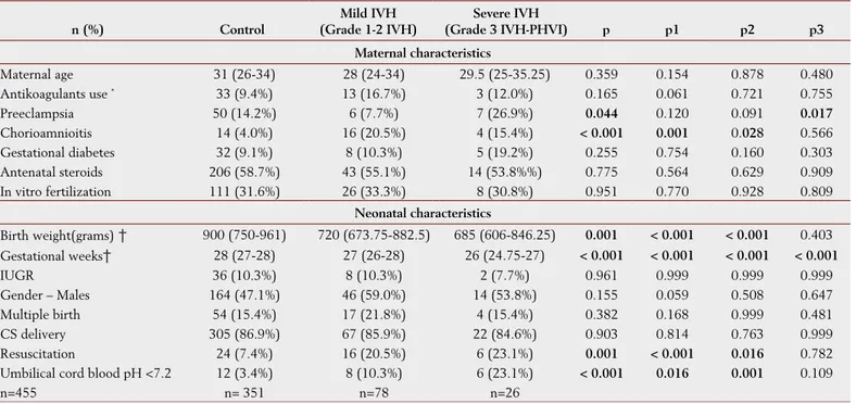
Table 2. Perinatal and demographic data of the study population
n (%) Control Mild IVH (Grade 1-2 IVH) Severe IVH (Grade 3 IVH-PHVI) p p1 p2 p3 Maternal characteristics Maternal age 31 (26-34) 28 (24-34) 29.5 (25-35.25) 0.359 0.154 0.878 0.480 Antikoagulants use * 33 (9.4%) 13 (16.7%) 3 (12.0%) 0.165 0.061 0.721 0.755 Preeclampsia 50 (14.2%) 6 (7.7%) 7 (26.9%) 0.044 0.120 0.091 0.017 Chorioamnioitis 14 (4.0%) 16 (20.5%) 4 (15.4%) < 0.001 0.001 0.028 0.566 Gestational diabetes 32 (9.1%) 8 (10.3%) 5 (19.2%) 0.255 0.754 0.160 0.303 Antenatal steroids 206 (58.7%) 43 (55.1%) 14 (53.8%%) 0.775 0.564 0.629 0.909 In vitro fertilization 111 (31.6%) 26 (33.3%) 8 (30.8%) 0.951 0.770 0.928 0.809 Neonatal characteristics Birth weight(grams) † 900 (750-961) 720 (673.75-882.5) 685 (606-846.25) 0.001 < 0.001 < 0.001 0.403 Gestational weeks† 28 (27-28) 27 (26-28) 26 (24.75-27) < 0.001 < 0.001 < 0.001 < 0.001 IUGR 36 (10.3%) 8 (10.3%) 2 (7.7%) 0.961 0.999 0.999 0.999 Gender – Males 164 (47.1%) 46 (59.0%) 14 (53.8%) 0.155 0.059 0.508 0.647 Multiple birth 54 (15.4%) 17 (21.8%) 4 (15.4%) 0.382 0.168 0.999 0.481 CS delivery 305 (86.9%) 67 (85.9%) 22 (84.6%) 0.903 0.814 0.763 0.999 Resuscitation 24 (7.4%) 16 (20.5%) 6 (23.1%) 0.001 < 0.001 0.016 0.782 Umbilical cord blood pH <7.2 12 (3.4%) 8 (10.3%) 6 (23.1%) < 0.001 0.016 0.001 0.109 n=455 n= 351 n=78 n=26
IUGR: intrauterin growth restriction, CS: Cesarean section*Anticoagulants refer to aspiririn, low molcular weight heparin and heparin delivery at pregnancy; † median (interquartile range: 25%-75%). Statistics for numerical variables were obtained by Kruskall Wallis and pairwise comparisons by Tamhane test; P1: control – mild IVH, P2: control – severe IVH, p3: mild-severe IVH.
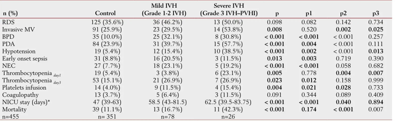
Table 3. The clinical features of the study population n (%) Control Mild IVH (Grade 1-2 IVH) Severe IVH (Grade 3 IVH–PVHI) p p1 p2 p3 RDS 125 (35.6%) 36 (46.2%) 13 (50.0%) 0.098 0.082 0.142 0.734 Invasive MV 91 (25.9%) 23 (29.5%) 14 (53.8%) 0.008 0.520 0.002 0.025 BPD 35 (10.0%) 25 (32.1%) 8 (30.8%) < 0.001 < 0.001 < 0.001 0.257 PDA 84 (23.9%) 31 (39.7%) 15 (57.7%) < 0.001 0.004 < 0.001 0.111 Hypotension 19 (5.4%) 12 (15.4%) 10 (38.5%) < 0.001 0.002 < 0.001 0.013
Early onset sepsis 31 (8.8%) 16 (20.5%) 3 (11.5%) 0.013 0.003 0.719 0.390 NEC 27 (7.7%) 18 (23.1%) 5 (19.2%) < 0.001 < 0.001 0.058 0.682 Thrombocytopenia day1 19 (5.4%) 3 (3.8%) 6 (23.1%) 0.005 0.778 0.004 0.007
Thrombocytopenia day3 53 (15.1%) 21 (26.9%) 7 (26.9%) 0.023 0.012 0.158 0.999
Platelets infusion 14 (4.0%) 9 (11.5%) 4 (15.4%) 0.004 0.021 0.028 0.733 Coagulopathy 13 (3.7%) 5 (6.4%) 3 (11.5%) 0.091 0.344 0.089 0.409 NICU stay (days)* 47 (39-63) 58.5 (43-81.5) 62.5 (39.5-83.75) < 0.001 < 0.001 0.040 0.894
Mortality 39 (11.1%) 13 (16.7%) 11 (42.3%) < 0.001 0.174 < 0.001 0.007 n=455 n= 351 n=78 n=26
*median (IQR); statistics for numerical variables were obtained by Kruskall Wallis and pairwise comparisons by Tamhane test; P1: control – mild IVH, P2: control – severe IVH, p3: mild-severe IVH. Abbreviations: RDS: respiratory distress syndrome, MV: mechanical ventilation, BPD: broncho pulmonary dysplasia, PDA:patent ductus arteriozus NEC: necrotizing enterocolitis, Thrombocytopenia: platelet counts <100 x 109 /L, Coagulopathy: fibrinogen < %5th %: < 71 mg/dL, prothrombin time > 95th % >21 seconds and aPTT >95th % >4 seconds.
Figure 1A-B. The predictive strength of platelets in ventricular
hemorrhage
Platelet counts and IVH Development (Platelets day1: AUC: 0.605[0.546- 0.664], sensitivity: 74.0% [64.5% - 82.1%)], specificity: 45.9% [40.6% - 51.2%], LH+: 1.37 [1.2 - 1.6], PPV: 28.8% [23.5% - 34.7%], NPV: 85.6% [79.8% - 90.3%]; Platelets day3: AUC: 0.619 [0.559 - 0,679], sensitivity: 68.3% [58.4% - 77.1%], specificity: 52.1% [46.8% - 57.5%], LH+: 1.43 [1.2 - 1.7], PPV: 29.7% [24% - 35.9%], NPV: 84.7% [79.2% -89.2%])
Statistics were obtained by ROC analysis. PPV: positive predictive value, NPV: negative predictive value, LH: likelihood ratio.
A
B
A
B
Figure 2A-B. The predictive strength of platelets in severe ventricular
hemorrhage
Platelet counts and Severe IVH Development (Platelets day1: AUC: 0.635[0.503 - 0.766], sensitivity: 65.4 [44.3% - 82.8%], specificity: 68.8% [64.1%) - 73.1%], LH+: 2.1 [1.5 - 2.9], PPV: 11.3% [6.7% -17.4%] f NPV: 97.0% [94.5% - 98.6%]; Platelets day3: AUC: 0.558 [0.434 - 0,683], sensitivity: 50.0% [29.9% - 70.1%], specificity: 65.5% [60.8%) - 70.0%)], LH+: 1.45 [1.0 - 2.2], PPV: 8.1% [4.4% -13.4%], NPV: 95.6% [92.6% - 97.6%]).
Statistics were obtained by ROC analysis. PPV: positive predictive value, NPV: negative predictive value, LH: likelihood ratio.
Table 4. Risk factors associated with developing IVH*
OR (95% CI) p
Birth weight 0.993 (0.991 - 0.995) < 0.001
Gestational weeks 0.505 (0.414 - 0.616) < 0.001
Resuscitation 3.354 (1.809 - 6.217) < 0.001
Umbilical cord pH <7.2 4.394 (1.964 - 9.832) < 0.001
Invasive mechanical ventilation 1.578 (0.989 - 2.517) 0.056
Medical PDA 2.521 (1.594 – 3.986) < 0.001
Hypotension 4.688 (2.424 – 9.068) < 0.001
Chorioamnionitis 5.731 (2.780 – 11.817) < 0.001
Early onset sepsis 2.307 (1.242 - 4.285) 0.0086
Thrombocytopeniaday1 1.655 (0.725 – 3.778) 0.231
Thrombocytopeniaday3 2.071 (1.228 - 3.493) 0.006
Platelets infusion 3.439 (1.561 – 7.574) 0.002
Coagulopathy 2.167 (0.873 – 5.380) 0.096
*IVH includes all grades of intraventricular hemorrhage. Statistics were obtained by univariate logistic regression. Medical PDA: Patent ductus arteriozus that requires medical intervention using ibuprofen or indomethacin
and low-umbilical-cord blood pH as independent risk factors of severe hemorrhage (OR: 6.678, 95% CI: 2.557-17.443 and OR: 3.554, 95% CI: 1.089–11.602; Table-5).
Four infants developed progressive post-hemorrhagic ventricular dilatation (PHVD) necessitating a cerebrospinal fluid (CSF) diverting procedure (ventriculo-peritoneal shunt – VP shunt). Twenty-two convulsive infants (22/104, 21.2%) received anti-seizure medications at hospital discharge. All patients developing any IVH grade were taken into neurological follow-up after hospital discharge, but the long-term morbidity was not included because the study period was limited to neonatal intensive care unit. Overall mortality in babies born less than 1000 grams was 13.9% due to miscellaneous prematurity-related causes (nosocomial sepsis and NEC). The mortality rate has increased to 23.1% in the infants developing any hemorrhage IVH (16.7% in patients with mild IVH, 42.3% in severe IVH)
Discussion
This observational cohort in ELBW preterm infants to define neonatal characteristics and risk factors for developing IVHs has elucidated several outcomes such as, i) the rate of developing IVH was 22.9%, ii) the hemorrhage has expanded to the surrounding tissues (severe IVHs: grade-3 and PVHI) in 24% of 104 bleeding patients, iii) chorioamnionitis, neonatal resuscitation, low-umbilical-cord blood pH and PDA were associated hemorrhage with developing IVH, but once the bleeding started, they did not influence the proceeding hemorrhage from mild to severe IVH, iv) the predictive strength of platelets in the first 72-hours of life in developing hemorrhage was weak, v) hypotension necessitating the use of inotropes and low-umbilical-cord blood pH were identified as independent risk factors for severe IVH.
IVH remains to be an important issue in the vulnerable population of ELBW neonates (5,19). The incidence ranges from 15% to 40%, depending on the NICU standard care (20). Literature data suggest that approximately 22% of the ELBW neonates develop high degrees of IVH (6). A recent prospective cohort including 69 Turkish NICUs reported 8.6% overall incidence for grade >2 and higher IVHs in infants of birth weight ≤1000 grams (14.4% and 6.6% in birth weight of 501-750 grams and 751-1000 grams, respectively) (21). The present study demonstrated a 22.9% overall IVH rate including grade-1 IVHs and the hemorrhage has progressed to more severe grades in 24% of the patients. The outcome reflected the incidence of low-birth-weight infants who
were born in the same birth center where intensive care would be delivered. However, the authors have concerns about the true incidence of ELBW infants delivering intensive care unit. To eliminate the risk of subjectivity in birth and/or postnatal transport data, the outborn infants (born in another birth facility and transferred to intensive care) were not included in the study data. There is evidence that birth outside the perinatal center and postnatal transports are associated with severe brain injury and adverse neonatal outcomes in infants of gestational age less than 29 weeks (22). Excluding the ones who died before obtaining cranial ultrasound might also affect the outcome by missing massive IVHs that resulted in sudden death in the present study. Both concerns raise the suspicion of increased incidence of IVH development in this vulnerable population.
The majority of IVHs occur in the first week of life and in 10% of patients, the onset of hemorrhage is within 12 hours following birth (5,8). Compared to previous ultrasound findings, the onset of diagnosis within the first day was significantly higher (22.1%) in this study, possibly due to our institutional policy. The practice is to obtain early diagnostic imaging within the first day of life once an infant with extremely lower gestation is admitted to intensive care unit. Ten out of 23 (43.5%) IVHs occurred in infants with gestational age ≤26 weeks. These infants were more likely to develop easy-to-diagnose high-grade hemorrhage.
Immature vascularity, hemodynamic instability and the consequent fluctuations in CBF, platelet abnormalities and coagulopathy contribute to IVH development (2,6-8). Several predisposing factors have been identified in the etiology and severity of IVHs. Lower gestational age and birth weight, IUGR, mode of birth (vaginal delivery), instrumental delivery, cardiopulmonary resuscitation, Apgar score, acidosis, feto-maternal infections, sepsis, RDS, PDA, air leak syndromes, hypoxemia, hypercapnia, seizures, thrombocytopenia have been shown to play a role in the pathogenesis (2,5,9,19,20,22,23). Our findings indicated inotrope-required hypotension as the most significant risk factor for developing severe IVH. CBF relies upon two cardiovascular components: cardiac output and regional vascular resistance regulated by autoregulatory mechanisms (24,25). Previous investigations demonstrated lower CBF in infants developing IVH (24,26). The limited autoregulatory capacity of the preterm and fluctuations in arterial blood pressure influence the perfusion of the brain, which lead to capillary vessel ruptures and the consequent hemorrhage (5,24-27). German Neonatal Network and Neocirculation project postulated increased risk of IVH in
Table 5. Multivariate Logistic Regression for Severe IVH
95% C.I. for EXP(B)
Exp (B) Lower limit Upper limit p
Hypotension 6.678 2.557 17.443 <0.001 Umbilical cord pH <7.2 3.554 1.089 11.602 0.036 Chorioamnionitis 1.156 0.294 4.545 0.835 Platelet infusion 1.351 0.345 5.286 0.666 Invasive MV 2.352 0.971 5.695 0.058 MV: mechanical ventilation
infants with gestational age less than 28 weeks in the presence of inotrope necessitating hypotension (28). However, there are controversial data on the topic of BP. Rather than measuring BP itself, monitoring the signs of perfusion should be the goal of neonatal intensive care (5,29). The selection of inotrope also influences the risk of bleeding. Vesoulis et al. reported an increase in mean blood pressure over 10 mmHg with Dopamine reduced the risk of IVH(30). On the contrary, the PROGRESS study consortium and Nationales Genomforschungsnetz Deutschland demonstrated that the use of catecholamines was more likely to develop severe IVH (31). Due to the retrospective observational design of the present study, the authors focused on risk analysis of IVH in infants born less than 1000 grams. The influence of inotrope selection in hemodynamically unstable infants was beyond the scope of this investigation. Yet, there is no doubt, future clinical research is needed to clarify whether hypotension itself or a sudden elevation in blood flood due to inotropic support is the cause of a high-grade hemorrhage. Inotrope selection and CBF monitorization should be the mainstay of clinical investigations to minimize short and long-term IVH morbidity.
Coagulopathy and thrombocytopenia are two predisposing factors related to IVH (1,6,10,23). Platelet counts less than 150x109/L are very common in very low birth weight neonates that leave them vulnerable to hemorrhage (10). Lower platelet counts were frequently observed in the study population: 8.7% and 26.9% at NICU admission and 3rd day of intensive care unit. Of note, the infants were defined thrombocytopenic if the platelet counts were less than 100x109/L and received platelet transfusions when the values were below 50x109/L to minimize the risk of IVH. The study center’s transfusion policy is compliant with the national recommendations of the Turkish Society of Neonatology to administer platelet transfusions if the platelet counts less than 50x109/Lin infants of birth weight less than 1000 grams (18). Approximately in 12.5% (13 out of 104) of the IVHs, the platelet counts were below 50x109/L and these infants had to deliver platelet transfusions. Despite the calculated risks for lower platelet counts, ROC analysis has shown poor predictive strength in determining IVH development. The subject of platelet infusion also remains to be a matter of debate: did the increased IVH risk occur due to very low platelet counts that were already below 50x109/L and leave them vulnerable to bleeding; or an immune response was catalyzed because of the transfusion itself since all transfusion product are known for their inflammatory-triggering effects (32,33). Still, controversies exist in the literature and the absence of definitive thresholds for platelet transfusion creates significant variability in clinical practice among NICUs (6,10,20,32). Coagulopathy, as another hemostatic abnormality, was not described as a potential risk factor in this study, possibly
due to frequent of fresh frozen plasma as soon as an abnormality was detected. Yet, the potential benefits of all blood product transfusions need to be balanced against the risks of adverse transfusion reactions.
All IVHs delivered neurological follow-up after hospital discharge. Twenty-two convulsive infants (21.2%) received anti-seizure medication at discharge. Progressive post-hemorrhagic ventricular dilatation (PHVD) that necessitated VP-shunting was developed in four of twenty-five (16%) severe IVHs in the present study. PVHD is reported between 30% and 50% of high-grade IVHs and 25% of the infants with progressive PVHDs necessitate CSF diverting procedures (34). However, the timing of intervention is disputable; the decision of surgery is left to the attending neurosurgeon and therapeutic approaches vary greatly among NICUs (16,35). Unfortunately, the absence of post-ICU follow-ups has restrained the estimation of long-term VP-shunting rates and neurological outcome. Overall mortality in babies born less than 1000 grams was 13.9%. In the case of a developing IVH, the mortality has increased to 23.1%.
The limitations of the present study were the retrospective design leading to a potential risk of bias. Keeping the onset of ultrasound-diagnosis restricted to one week was another limitation. Even if many IVHs occur during the first week of life, there was the risk of unnoticing hemorrhage that might occur in the following weeks. The study era also consisted of intensive care unit period that limited the estimation of the long-term neurodevelopmental outcome. Despite the limitations, planning this retrospective cohort in a very specific group of newborns (birth weight < 1000 grams) and excluding the ones born in another perinatal center have created homogeneity in the study population. Hence the results have enabled us to analyze all maternal and neonatal characteristics of ELBW infants and the true incidence of IVH outcomes of this highly-referral, level III NICU.
Conclusion
Hemodynamic instability requiring inotropic support is the main predisposing factor in developing severe IVH. Monitoring cerebral blood flow and perfusion should be the focus of future investigations. Despite poor predictive strength of platelet counts, timely intervention for thrombocytopenia is essential to avoid intraventricular hemorrhage of ELBW infants.
Acknowledgment
Authors would like to thank Dr. Meltem DEMİR for her assistance in obtaining laboratory data.
Ethics Committee Approval: Institutional Review Board of İstinye University Antalya Medical Park Hospital - approval code: 2019/10, date: 23.10.2019 Peer-review: Externally peer-reviewed.
Conflict of Interest: Authors have no conflicts of interest to declare. Financial Disclosure: The authors declared that this study has received no financial support.
AUTHOR CONTRIBUTIONS:
Concept: HO, SEOO; Design: HO, SEOO; Supervision: HO, SEOO; Fundings: HO, SEOO; Materials: HO, SEOO; Data Collection and/or Processing: HO, SEOO; Analysis and/or Interpretation: HO, SEOO; Literature Search: HO, SEOO; Writing Manuscript: HO, SEOO; Critical Review: HO, SEOO.
References
1. Neary E, Okafor I, Al-Awaysheh F, et al. Laboratory Coagulation Parameters in Extremely Premature Infants Born Earlier than 27 Gestational Weeks upon Admission to a Neonatal Intensive Care Unit. Neonatology 2013; 104: 222-7.
2. Howes A, Hilditch C, Keir A. What Clinical Practice Strategies Have Been Shown to Decrease Incidence Rates of Intraventricular Haemorrhage in Preterm Infants? J Paediatr Child Health 2019; 55: 1269-78.
3. Szpecht D, Szymankiewicz M, Nowak I, et al. Intraventricular hemorrhage in neonates born before 32 weeks of gestation retrospective analysis of risk factors Childs Nerv Syst 2016; 32: 1399-404.
4. Ancel PY, Goffinet F, Kuhn P, et al; EPIPAGE-2 Writing Group. Survival and morbidity of preterm children born at 22 through 34 weeks’ gestation in France in 2011: results of the EPIPAGE-2 cohort study. JAMA Pediatr 2015; 169: 230–8.
5. Leijser LM, de Vries LS. Preterm brain injury: Germinal matrix– intraventricular hemorrhage and post-hemorrhagic ventricular dilatation. Editor(s): Linda S. de Vries, Hannah C. Glass. Handbook of Clinical Neurology. Elsevier. 1st ed. 2019: 173-99.
6. Grevsen AK, Hviid CVB, Hansen AK, et al. The Role of Platelets in Premature Neonates with Intraventricular Hemorrhage: A Systematic Review and Meta-Analysis. Semin Thromb Hemost. 2020; 46: 366-78.
7. Neary E, McCallion N, Kevane B, et al. Coagulation indices in very preterm infants from cord blood and postnatal samples. J Thromb Haemost 2015; 13: 2021–30.
8. Korkmaz L, Bastug O, Ozdemir A, et al. Can Platelet Mass Index Be a Parameter to Predict Intraventricular Hemorrhage in Very-Low-Birth-Weight Newborns? Am J Perinatol 2019; 36: 1188-97. 9. Elfarargy MS, Eltomey MA, Soliman NA. Early predictors of neonatal
intraventricular hemorrhage. Electron Physician 2017; 9: 4946–51. 10. Moore CM, Curley A. Platelet Transfusion Thresholds in Neonatal
Medicine. Early Hum Dev 2019; 138: 104845.
11. Inder TE, Perlman JM, Volpe JJ. Preterm intraventricular hemorrhage / posthemorrhagic hydrocephalus. Editor(s): Volpe JJ, Inder TE, Darras BT, de Vries LS, du Plessis AJ, Neil JJ. Volpe's neurology of the newborn. Elsevier. 6th ed. 2018: 637e98.e21.
12. ACOG Committee on Obstetric Practice. ACOG practice bulleting. Diagnosis and management of preeclampsia and eclampsia. Number 33, January 2002. American College of Obstetricians and Gynecologists. Int J Gynaecol Obstet 2002; 77: 67–75.
13. Bernstein IM, Horbar JD, Badger GJ, et al; The Vermont Oxford Network. Morbidity and mortality among very low- birth-weight neonates with intrauterine growth restriction. Am J Obstet Gynecol 2000; 182: 198–206.
14. Dilli D, Soylu H, Tekin N. Neonatal hemodynamics and management of hypotension in newborns. Turk Pediatri Ars 2018; 53: 65-75. 15. Narang I. Review series: what goes around, comes around: childhood
influences on later lung health? Long-term follow-up of infants with lung disease of prematurity. Chron Respir Dis 2010; 7: 259–69. 16. El-Dib M, Limbrick DD Jr, Inder T, et al. Management of
Post-hemorrhagic Ventricular Dilatation in the Infant Born Preterm. J Pediatr 2020; 30: S0022-3476(20)30978-1.
17. Christensen RD, Baer VL, Lambert DK, et al. Reference intervals for common coagulation tests of preterm infants. Transfusion 2014; 54: 627-32.
18. Çetinkaya M, Atasay B, Perk Y. Turkish Neonatal Society guideline on the transfusion principles in newborns. Turk Pediatri Ars 2018; 53: 101-8.
19. Huang J, Meng J, Choonara I, et al. Antenatal infection and intraventricular hemorrhage in preterm infants. Medicine (Baltimore) 2019; 98: e16665.
20. Mitsiakos G, Papathanasiou AE, Kyriakidis I, et al. Intraventricular Hemorrhage and Platelet Indices in Extremely Premature Neonates. J Pediatr Hematol Oncol 2016; 38: 533-38.
21. Koc E, Demirel N, Bas AY, et al. Early neonatal outcomes of very-low-birth-weight infants in Turkey: A prospective multicenter study of the Turkish Neonatal Society. PLoS ONE 2019; 14: e0226679. 22. Amer R, Moddemann D, Seshia M, et al; Canadian Neonatal
Network and Canadian Neonatal Follow-up Network Investigators. Neurodevelopmental Outcomes of Infants Born at <29 Weeks of Gestation Admitted to Canadian Neonatal Intensive Care Units Based on Location of Birth. J Pediatr 2018; 196: 31-37.e1.
23. Williams AG, Odd D, Bates S, et al. Elevated International Normalized Ratio (INR) Is Associated With an Increased Risk of Intraventricular Hemorrhage in Extremely Preterm Infants. J Pediatr Hematol Oncol 2019; 41: 355-60.
24. Lim J, Hagen E. Reducing Germinal Matrix-Intraventricular Hemorrhage: Perinatal and Delivery Room Factors. Neoreviews 2019; 20: 452-63.
25. Brew N, Walker D, Wong FY. Cerebral vascular regulation and brain injury in preterm infants. Am J Physiol Regul Integr Comp Physiol 2014; 306: 773-86.
26. Barzilay E, Gadot Y, Koren G. Safety of vaginal delivery in very low birthweight vertex singletons: a meta-analysis. J Matern Fetal Neonatal Med 2016; 29: 3724–9.
27. Vesoulis ZA, Mathur AM. Cerebral autoregulation, brain injury, and the transitioning premature infant. Front Pediatr 2017; 5: 64. 28. Faust K, Härtel C, Preuß M, et al; Neocirculation project and the
German Neonatal Network (GNN). Short-term Outcome of Very-Low-Birthweight Infants with Arterial Hypotension in the First 24 H of Life. Arch Dis Child Fetal Neonatal Ed 2015; 100: 388-92. 29. Dempsey EM, Al Hazzani F, Barrington KJ. Permissive hypotension
in the extremely low birthweight infant with signs of good perfusion. Arch Dis Child Fetal Neonatal Ed 2009; 94: 241–4.
30. Vesoulis ZA, Ters NE, Foster A, et al. Response to dopamine in prematurity: a biomarker for brain injury? J Perinatol 2016; 36: 453-8.
31. Poryo M, Boeckh JC, Gortner L, et al; PROGRESS study consortium and NGFN-Nationales Genomforschungsnetz Deutschland. Ante-, peri- and postnatal factors associated with intraventricular hemorrhage in very premature infants. Early Hum Dev 2018; 116: 1–8.
32. Stolla M, Refaai MA, Heal JM, et al. Platelet transfusion - the new immunology of an old therapy. Front Immunol 2015; 6: 28. 33. McFadyen JD, Kaplan ZS. Platelets are not just for clots. Transfus
Med Rev 2015; 29: 110-9.
34. Brouwer AJ, Groenendaal F, Benders MJ, et al. Early and late complications of germinal matrix-intraventricular haemorrhage in the preterm infant: what is new? Neonatology 2014; 106: 296-303. 35. Brouwer AJ, Brouwer MJ, Groenendaal F, et al. European perspective
on the diagnosis and treatment of posthaemorrhagic ventricular dilatation. Arch Dis Child Fetal Neonatal Ed 2012; 97: F50-5.