The effects of microwave frequency electromagnetic fields on the
development of
Drosophila melanogaster
EMEL ATLI & HACER U
¨ NLU
¨
Hacettepe University, Faculty of Science, Department of Biology, Ankara, Turkey (Received 25 July 2005; accepted 9 May 2006)
Abstract
Purpose: To investigate the effects of microwave frequency electromagnetic fields (EMF) on the development of Drosophila melanogaster.
Materials and methods: Larvae of D. melanogaster were exposed to 10 GHz EMF continuously (3 h, 4 h and 5 h) and discontinuously (3 h exposureþ 30 min interval þ 3 h exposure). The percentages and times of transition from larvae to pupae and from pupae to adults were determined, and the mean offspring number was examined using the offspring of the females which had been exposed as larvae.
Results: No differences were found in the transition percentages from larvae to pupae and from pupae to adults (p4 0.05). However, it was found that the mean pupation time was delayed linearly with an increasing electromagnetic field (EMF) exposure period (p5 0.05). In the 3þ 3-h exposed group (E3 þ 3), the mean offspring number was significantly less than that of the control (p5 0.05).
Conclusions: 10 GHz EMF can cause developmental delay and decrease the number of offspring in D. melanogaster.
Keywords:Drosophila melanogaster, development, electromagnetic fields, mean offspring number
Introduction
Over the last decade, the exponential growth of mobile communications has been accompanied by a parallel increase in the density of electromagnetic fields (EMF). As such, the continued expansion of mobile communications raises important questions because EMF have long been suspected of having biological effects. It has been claimed that EMF, especially microwave frequency EMF, cause biolo-gical effects by increasing the temperature, changing the chemical reactions, or inducing an electrical current (World Health Organization [WHO] Inter-national EMF Project 1997, Brent 1999, Somosy 2000, Banik et al. 2003). The effects of EMF have been handled by using different features in various organisms by many researchers and different results have been obtained. In these studies, determining the effects of EMF upon development was also considered. In the publications dealing with the effects of EMF, the effects are not always in the same direction. For example, Drosophila melanogaster
exposed to a 80mT field differed from the control flies in their time of eclosion. Six days after oviposition, 58.8% of the flies exposed to the 80mT had eclosed, while only 26.7% of the flies in the control had done so (Graham et al. 2000). In contrast, Delgado et al. (1982) reported delayed and arrested development of chick embryos exposed to a 1.2mT field at 100 Hz. Developmental delay has also be reported in fish (Cameron et al. 1985).
In this study, the developmental effects of 10 GHz EMF were examined by taking into account the changes in developmental stages and the differences in mean offspring number in D. melanogaster.
Materials and methods
The organism and environmental conditions
In this study, the wild type Oregon strain of D. melanogaster was used. The flies were kept in a Drosophila culture room (Hacettepe University, Ankara/Turkey) at 25+ 18C and relative humidity
Correspondence: Emel Atli, Hacettepe University, Faculty of Science, Department of Biology, Division of Genetics, 06800, Beytepe, Ankara, Turkey. E-mail: [email protected]
ISSN 0955-3002 print/ISSN 1362-3095 online Ó 2006 Taylor & Francis DOI: 10.1080/09553000600798849
of 50 – 60% and in 8 h light, 16 h dark periods on a standard Drosophila medium described by Bozcuk (1978).
Virgin Oregon females and males of the same age were crossed in culture bottles. Individuals were then removed from the culture bottles after 8 h. Some 72+ 4 h later, the third instar larvae were collected using a 20% sodium chloride (Merck; Durmstadt, Germany) solution (Idaomar et al. 2002).
EMF exposures
EMF exposures were done by using an electro-magnetic radiation source (antenna) at 25+ 18C temperature in Hacettepe University, Microwave and Aerial Laboratory of Electricity and Electronic Engineering Department (Ankara, Turkey). The dosage used was determined taking into account previous publications and our pilot experiments. Oregon strain (w.t.) larvae of D. melanogaster were exposed to a 10 GHz EMF for 3, 4 and 5 h con-tinuously and 3 hþ 3 h discontinuously with a 30-min interval in between. The groups were organized according to the dose levels to which they were exposed.
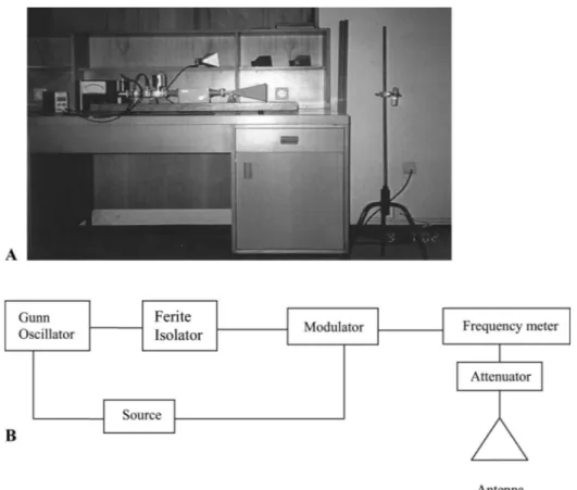
The EMF source was a horn type antenna (Figure 1) which produced a pulsed (modulated) square wave (1 kHz), approximately 5 mW (lowered power). This had an X-band frequency range
8.2 – 12.4 GHz with a gain of 16 dB. The frequency was fixed at 10 GHz. The power density of the antenna was 0.0156 Watt/m2, electric field intensity was 3.42 V/m and SAR (Specific Absorption Rate) was approximately 9.8 mW/kg (Dalgic 2003, Dalgic & Bozcuk 2004).
During the EMF exposure, larvae were placed in glass tubes (2.567.5 cm) containing drying papers that had absorbed a 5% sucrose (Merck; Durmstadt, Germany) solution. The experimental tubes were kept just opposite the antenna at a distance of 1 m, and stabilized by a rectangular holder. The larvae in the control group were kept away from the EMF source in the same laboratory conditions during the exposure period.
Observation of developmental stages
EMF exposed and non-exposed (control group) larvae were placed in 250 ml glass bottles that con-tained a standard Drosophila medium. The develop-ment of all experidevelop-mental groups was observed at 4-h intervals by recording the number of individuals passing from larvae to pupae and from pupae to adults, and the transition periods. After the adults began to hatch, the 4-hourly observations ended, but the counting of adults continued until all adults had hatched from the pupae. From the adults that emerged, virgin females were collected in order to
use them in the ‘‘determination of the mean offspring number’’ experiment.
Determination of the mean offspring number
In order to determine the effects of the 10 GHz EMF on the daily mean offspring number, virgin females, hatched from the exposed larvae were used. An exposed female and 3 non-exposed males of same age were crossed. Parents were removed when the first pupa was seen. After the first adults began to hatch, the number of offspring emerging in 10 days were counted at 24-h intervals.
Statistical methods
The statistical analysis of the results was carried out using the SPSS 10.0 programme. In the experiment of the observation of developmental stages, the determination of the significance of the transition percentages from larvae to pupae and from pupae to adults were obtained by an analysis of variance, and the comparison of the transition period from larvae to pupae was done by a two-variable t-test. The daily mean offspring number was calculated with the analysis of variance (ANOVA) test.
Results
Observation of developmental stages
Effect of the EMF on the transition percentages from larvae to pupae. The pupated larvae from those exposed and non-exposed to the EMF were counted seperately and their transition percentages were determined (Table I). Pupation percentages were found to be 95% in the control group, 98.67% in the 3-h exposed group (E3), 93.75% in the 4-h exposed group (E4), 92% in the 5-h exposed group (E5) and 100% in the 3þ 3 h exposed group (E3 þ 3). As seen, all of the larvae were pupated in the E3þ 3. Due to the fact that this situation does not allow variance analysis (as the group variance is 0) the data of this group were not included in the chart. The result of a one variable variance analysis showed that
EMF exposures did not have any effect in terms of the number of pupated larvae (p4 0.05).
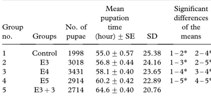
Effects of the EMF on the mean pupation time. Table II shows the effect of different EMF doses on mean pupation time. The mean pupation time was 55 h for the non-exposed control group. However, the mean pupation time increased to 56.8, 58.1, 60.2 and 64.6 in the E3, E4, E5 and E3þ 3 exposure groups, respectively. This clearly shows that the mean pupation time was extended according to the EMF exposure period in comparison to the control group and that this increase was significant (p5 0.05).
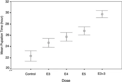
The changes in mean pupation time are shown on the same graph in Figure 2. As can be seen from this Figure, the longer the exposure period (dose) becomes, the greater the extent of the larval period is, so the mean pupation time is delayed. In other words, EMF exposures caused a developmental delay by extending the transition period from larvae to pupae.
Further analysis has shown that the delay occuring in the first 36 h caused an increase in the mean pupation time. This situation is shown in Table III using a t-test, realized in terms of 36-h development for the control and the exposed groups. As seen in this table, all the differences between the groups were found to be statistically significant (p5 0.05). As seen in Figure 3, the mean pupation time of the exposed groups were delayed compared to the control in the first 36 h, and this increase depended on the EMF-exposure period.
The same studies were repeated after the first 36-h developmental period (60th h, 80th h, 96th h) and no reasonable difference was found statistically between mean pupation times (p4 0.05). The fact that the differences in the further phases disappeared indicates that this delay is an early developmental delay. Effects of the EMF on adult formation. The transition rates of EMF exposed and non-exposed flies from pupae to adult were determined and then compared statistically. As seen from Table IV, the adult
Table I. The changes of transition percentages from larvae to pupae depending on EMF exposure period.
Groups No. of larvae No. of pupae
Pupation percentages+ SE SD Control 100 95 95.00+ 1.92 3.83 E3 148 146 98.67+ 0.84 2.07 E4 189 177 93.75+ 2.25 6.36 E5 174 160 92.00+ 3.49 9.24
E, Exposed group (3, 4, 5 h); SE, Standard error; SD, Standard deviation.
Table II. The changes of mean pupation time depending on EMF exposure period. Group no. Groups No. of pupae Mean pupation time (hour)+ SE SD Significant differences of the means 1 Control 1998 55.0+ 0.57 25.38 1 – 2* 2 – 4* 2 E3 3018 56.8+ 0.44 24.16 1 – 3* 2 – 5* 3 E4 3431 58.1+ 0.40 23.65 1 – 4* 3 – 4* 4 E5 2914 60.2+ 0.42 22.89 1 – 5* 4 – 5* 5 E3þ 3 2714 64.6+ 0.40 20.76
E, Exposed group (3, 4, 5 h, 3þ 3 h); SE, Standard error; SD, Standard deviation;*p 5 0.05.
Figure 2. 95% confidence intervals of mean pupation time in the control (non-exposed) and the exposed groups (E3, E4, E5 and E3þ 3). Mean pupation time of the exposed groups was extended compared to the control group depending on EMF exposure period.
Table III. The statistical comparisons of mean pupation time in the control and the exposed groups in the first 36 h.
Groups df T Significance C – E3 271 2.65 0.008** C – E4 271 2.49 0.014* C – E5 271 4.41 0.000*** C – E3þ 3 271 10.33 0.000*** E3 – E4 719 2.38 0.017* E3 – E5 554 5.21 0.000*** E3 – E3þ 3 342 11.68 0.000*** E4 – E5 554 2.91 0.004** E4 – E3þ 3 342 8.18 0.000*** E5 – E3þ 3 342 6.25 0.000***
C, Control (non-exposed) group; E, Exposed group (3, 4, 5 h, 3þ 3 h); df, degrees of freedom; t, t-distribution; *p 5 0.05; **p 5 0.01; ***p 5 0.001.
Figure 3. 95% confidence intervals of mean pupation time in the control (non-exposed) and the exposed groups (E3, E4, E5 and E3þ 3) in the first 36 h. Mean pupation time of the exposed groups increased compared to the control group depending on EMF exposure period (dose).
Table IV. The changes of transition percentages from pupae to adult depending on EMF exposure period.
Groups No. of pupae No. of adult
Adult percentages+ SE SD Control 95 82 86.50+ 2.02 4.04 E3 146 127 87.00+ 2.52 6.16 E4 177 162 91.63+ 1.75 4.96 E5 135 105 77.83+ 6.72 0.17 E3þ 3 174 151 86.86+ 2.26 5.98
E, Exposed group (3, 4, 5 h, 3þ 3 h); SE, Standard error; SD, Standard deviation.
formation rate was found to be 86.5% in the control group, 87% in the 3-h exposed group (E3), 91.63% in the 4-h exposed group (E4), 77.83% in the 5-h exposed group (E5) and 86.86% in the 3þ 3 h exposed group (E3þ 3). A variance analysis showed that the differences between the groups were insig-nificant at a level of p4 0.05.
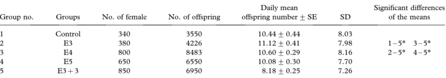
The effects of the EMF on daily mean offspring numbers Table V shows the effect of different EMF-exposure periods on the daily mean offspring number. The mean daily offspring number per female was 10.44 for the non-exposed control group. However, the mean daily offspring number was 11.12, 10.60, 10.08 and 8.18 in the E3, E4, E5 and E3þ 3 exposure group, respectively (Table V).
As seen in Table V, there was a statistically significant reduction in the daily mean offspring number in the E3þ 3 compared not only to the control but also to the other exposed groups (p5 0.05). The other minor deviations, observed in the other groups, were not significant statistically (p4 0.05).
Discussion
In this study, the effects of 10 GHz EMF on the development of D. melanogaster were investigated. Under the experimental conditions, the factors that might have affected the development were kept stable. The heat increase in the system during exposure was calculated and found to be too small to cause a significant heating effect. Thus, the differences in the results were interpreted as being caused by the EMF used.
In our experiments, it was seen that EMF expo-sures did not have any effect on the pupation rate, but that mean pupation time was delayed according to the increase of exposure period (Figure 2). This situation implies that exposure to a 10 GHz EMF causes developmental delay.
In publications dealing with the effects of EMF, specific effects of exposure have not been clearly shown, and EMF effects have not always occurred in the same direction. While in some publications it
was reported that EMF accelerated development (Graham et al. 2000), in others it was reported that EMF caused developmental delay (Delgado et al. 1982, Michel & Gutzeit 1999).
Michel and Gutzeit (1999) found that development was considerably delayed in transgenic Drosophila embryos exposed to 50 Hz EMF under mild thermal stress. In another study, fertilized fish eggs were exposed to 60 Hz EMF to examine the effects of EMF on embryonic development of vertebrates and a significant developmental delay was reported (Camer-on et al. 1985). In another work, it was reported that 100 Hz EMF slowed down the development of chicken embryos (Delgado et al. 1982). Similarly, Cecconi et al. (2000) observed that the development of 33 Hz EMF exposed mouse follicles were sig-nificantly reduced compared with controls. These studies show that EMF have a retarding effect upon developmental time, and support our findings which suggest that EMF might have caused an extension of the larval process and a pupation delay.
It is known that developmental time is affected by many external and internal factors. For example, several studies have found that mildly stressful deve-lopmental conditions, e.g., larval crowding, short-term high temperature exposure and restricted diet, may lead to increased developmental time (Sorensen & Loeschke 2004). Lints and Lints (1971b) reported that changes in environmental factors cause a delay in developmental phases, from larval growth to death, by affecting the vital programmes and functions of molecules.
Many chemical and physical stimuli are known to drive such responses, including the induction of oxidative stress and heat shock. One source of a stress stimulus which has received less attention, but which is rapidly coming into focus from a biological and human health perspective, is that of non-ionizing electromagnetic fields. Increasing use of mobile telephony in our society has brought a focus on the potential for microwave electromagnetic radiation to elicit biological stress responses, in association with potentially detrimental effects to human health (Cotgreave 2005).
In the studies dealing with the effects of EMF, it is emphasized that exposure to non-ionizing
Table V. The effect of EMF exposure on daily mean offspring number of Drosophila melanogaster.
Group no. Groups No. of female No. of offspring
Daily mean offspring number+ SE SD Significant differences of the means 1 Control 340 3550 10.44+ 0.44 8.03 2 E3 380 4226 11.12+ 0.41 7.98 1 – 5* 3 – 5* 3 E4 800 8483 10.60+ 0.29 8.16 2 – 5* 4 – 5* 4 E5 650 6550 10.08+ 0.30 7.70 5 E3þ 3 850 6950 8.18+ 0.25 7.26
EMFs could induce stress and damage to the cells (Laurence et al. 2000, Kwee et al. 2001, Weisbrot et al. 2003). In order to protect itself, the cell repairs the proteins damaged due to stress or increases the production of special proteins. Some of these proteins are known as ‘‘heat shock proteins (hsp)’’ (Han et al. 1998, Roberts & Feder 1999, Neven 2000, Kwee et al. 2001, Morrow & Tanguay 2003). In a study carried out to determine the effects of stress factors such as heat, application of a stress factor (heat) caused hsp70 to increase and extended the developmental time. It was stated that the exten-sion in the developmental time was a response to stress (Sorensen & Loeschke 2004). And in another study, it was shown that when the hsp70 transcrip-tion increased the delay in developmental time was reduced (Krebs & Feder 1998).
It one accepts that EMF cause cellular stress, it is reasonable to expect a similar increase in the quantity of hsp in EMF-exposed cells. It has been shown that hsp can be induced by EMF at a very low fre-quency and at microwave frefre-quency (Kwee et al. 2001, Weisbrot et al. 2003, Cotgreave 2005). For example, during the last 10 years, a number of studies in in vitro systems have indicated that continuous wave (CW) radio frequency (RF) emis-sions, in the range between 750 MHz and 2.4 GHz, are able to induce the expression of hsp in a large variety of eukaryotic cell systems (Cotgreave 2005). In our study, EMF exposure caused developmental delay that was observed mainly in the first 36 h of pupation. This change in pupation occuring mainly in the first 36 h may be due to changes in the transcription of hsp caused by stress conditions.
Krebs and Feder (1998) reported that the function of hsp is dependent on the cellular concentration of hsp. At low to moderate concentrations, hsp behave as molecular chaperones to protect the cell. On the other hand, they may harm organismal growth and development in high concentrations. Hsp transcrip-tion increases under stress conditranscrip-tions, but decreases as soon as repair is completed. The EMF that we applied as a stress factor might have spoilt the usual development by causing an increase in hsp expres-sion. Then the disapperance in the pupation delay after first 36 h could be explained by the return of the hsp concentration to its previous level after the stress factor was eliminated.
During the larval-pupal metamorphosis, the ex-pression of hsp is regulated by the steroid molting hormone ecdysone and there is a mutual interaction between the gene areas where they are coded (Morrow & Tanguay 2003). Thus, hsp synthesis, induced under stress conditions, might have affected development by affecting the synthesis of ecdysone, which is an important hormone in pupation.
In the experiment to determine the daily mean offspring number of a female, it was observed that this number for the 3þ 3 h exposed group (E3 þ 3) was less than that of the control group (p5 0.05). While this value was 10.44 female/day for the control group, it was 8.18 female/day for the E3þ 3 (Table V). This effect may be related to the length of the exposure period. It is known that there are trade-offs between a fast developmental time and other life history traits. For example, it is possible that slower developing flies suffer from reduced fitness in some traits as a consequence of exposure to food source deterioration (Nunney 1996, Sorensen & Loeschke 2004). In the experiment to observe developmental stages, we found that the larvae in the E3þ 3 pupated more slowly compared to other groups (p5 0.05). In this group, the significant reduction in the offspring number may be related to the developmental delay. Apart from this, there are some studies reporting that EMF applications cause damage in imaginal discs (Mirabolghasemi & Azarnia 2002). EMF exposure to 72+ 4 h larvae might have affected genital imaginal discs, thus as a result of this effect a problem in mating might have occurred. If there is such an effect it cannot be strong enough to reduce mating to very low levels.
The effects of EMF on the number of offspring were investigated in previous works. Marec et al. (1985) investigated the effect of 2375 MHz micro-wave irradiation on D. melanogaster and found a significant decrease in the number of offspring. On the other hand, it was found that a disconti-nuous radio frequency signal produced by a GSM multiband mobile phone (900/1900 MHz; SAR*1.4 W/kg) increased the number of offspring in D. melanogaster (Weisbrot et al. 2003). In these publications, the effects of EMF on the number of offspring were in opposite directions, but it was clear that EMF exposure affected the number of offspring in Drosophila.
In general, it is clear that 10 GHz EMF exposure has effects upon development. Based on the present report we conclude that microwave frequency EMF can cause developmental delay and decrease the number of offspring. But, the developmental effects of EMF should be examined in detail at the bio-chemical and molecular level to clarify the effect mechanisms.
Acknowledgements
The authors would like to thank TUBITAK (The Scientific & Technological Research Council of Turkey) (Project No: TBAG-1976) for their finan-cial support.
References
Banik S, Bandyopadhyay S, Ganguly S. 2003. Bioeffects of microwave – a brief review. Biosource Technology 87:155 – 159.
Bozcuk AN. 1978. The effects of some genotypes on the longevity of adult Drosophila. Experimental Gerontology 13:279 – 286. Brent RL. 1999. Reproductive and teratologic effects of
low-frequency electromagnetic fields, A review of in vivo and in vitro studies using animal models. Teratology 59:261 – 286. Cameron IL, Hunter KE, Winters WD. 1985. Retardation of embriyogenesis by extremely low frequency 60 Hz electro-magnetic fields. Physiological Chemistry and Physics and Medical NMR 17:135 – 138.
Cecconi S, Gualtieri G, Bartolomeo AD, Troiani G, Cifone MG, Canipari R. 2000. Evaluation of the effects of extremely low frequency electromagnetic fields on mammalian follicle devel-opment. Human Reproduction 15(11):2319 – 2325.
Cotgreave IA. 2005. Biological stress responses to radio frequency electromagnetic radiation: are mobile phones really so (heat) shocking? Archives of Biochemistry and Biophysics 435:227 – 240.
Dalgic BS. 2003. The effects of electromagnetic radiation in microwave frequency on longevity of some Drosophila melano-gaster mutants. MSc Thesis. Hacettepe University The Institute for Graduate Studies in Pure and Applied Sciences, Ankara.
Dalgic BS, Bozcuk AN. 2004. The effects of a short-term microwave exposure on the life span Drosophila melanogaster mutants. Hacettepe Journal of Biology and Chemistry 33:111 – 117.
Delgado JMR, Leal J, Monteagudo JL, Gracia MG. 1982. Embryological changes induced by weak, extremely low frequency electromagnetic fields. Journal of Anatomy 134(3): 533 – 551.
Graham JH, Fletcher D, Tigue J, McDonald M. 2000. Growth and developmental stability of Drosophila melanogaster in low frequency magnetic fields. Bioelectromagnetics 21:465 – 472.
Han L, Lin H, Head M, Jin M, Blank M, Goodman R. 1998. Application of magnetic field-induced heat shock protein 70 for presurgical cytoprotection. Journal of Cellular Biochemistry 71:577 – 583.
Idaomar M, El Hamss R, Bakkali F, Mezzoug N, Zhiri A, Baudoux D, Mun˜ oz-Serrano A, Liemans V, Alonso-Moraga A. 2002. Genotoxicity and antigenotoxicity of some essential oils evaluated by wing spot test of Drosophila melanogaster. Mutation Research 513:61 – 68.
Krebs RA, Feder ME. 1998. Hsp 70 and larval thermotolerance in Drosophila melanogaster : how much is enough and when is more too much? Journal of Insect Physiology 44:1091 – 1101. Kwee S, Raskmark P, Velizarov S. 2001. Changes in cellular proteins due to enviromental non-ionizing radiation. I. Heat-shock proteins. Electro- and Magnetobiology 20(2):141 – 152. Laurence JA, French PW, Lindner RA, McKenzie DR. 2000. Biological effects of electromagnetic fields – mechanisms for the effects of pulsed microwave radiation on protein con-formation. Journal of Theoretical Biology 206:291 – 298. Lints FA, Lints CV. 1971b. Relationship between growth and
aging in Drosophila. Nature 229:86 – 88.
Marec F, Ondracek J, Brunnhofer V. 1985. The effect of repeated microwave irradiation on the frequency of sex-linked recessive lethal mutations in Drosophila melanogaster. Mutation Research 157:163 – 167.
Michel A, Gutzeit HO. 1999. Electromagnetic fields in combina-tion with elevated temperatures affect embryogenesis of Drosophila. Biochemical Biophysical Research Communica-tions 265:73 – 78.
Mirabolghasemi G, Azarnia M. 2002. Developmental changes in Drosophila melanogaster following exposure to alternating electromagnetic fields. Bioelectromagnetics 23:416 – 420. Morrow G, Tanguay RM. 2003. Heat shock proteins and aging in
Drosophila melanogaster. Seminars in Cell & Developmental Biology 14:291 – 299.
Neven LG. 2000. Physiological responses of insects to heat. Postharvest Biology and Technology 21:103 – 111.
Nunney L. 1996. The response to for fast larval development in Drosophila melanogaster and its effect on adult weight: an example on a fitness trade-off. Evolution 50:1193 – 1204. Roberts SP, Feder ME. 1999 Natural hyperthermia and
expres-sion of the heat shock protein Hsp70 affect developmental abnormalities in Drosophila melanogaster. Oecologia 121:323 – 329.
Somosy Z. 2000. Radiation response of cell organels. Micron 31:165 – 181.
Sorensen JG, Loeschke V. 2004. Effects of relative emergence time on heat stress resistance traits, longevitiy and hsp 70 expression level in Drosophila melanogaster. Journal of Thermal Biology 29(4 – 5):195 – 203.
Weisbrot D, Lin H, Ye L, Blank M, Goodman R. 2003. Effects of mobile phone radiation on reproduction and development in Drosophila melanogaster. Journal of Cellular Biochemistry 89:48 – 55.
WHO International EMF Project. 1997. www.who.int/entity/ peh-emf/en.