ORIGINAL PAPER
Distal partial gluteus maximus musculocutaneous V-Y flap:
a simplified technique for reconstruction of ischial pressure sores
Mustafa Nışancı1&Mehmet Altıparmak1Received: 28 May 2019 / Accepted: 23 August 2019
# Springer-Verlag GmbH Germany, part of Springer Nature 2019
Abstract
Background Ischial pressure sores are mostly seen in wheelchair-bound patients and they have a high risk of recurrence due to a multitude of reasons compared with sacral and trochanteric ulcers. Various treatment modalities have been presented but high recurrence rates maintain its importance.
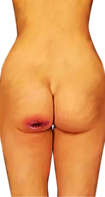
Methods Between 2009 and 2016, a total of 14 patients with spinal cord injuries were operated on for a total of 17 Shea stage IV ischial ulcers with the described technique. Demographic variables were recorded. Postoperative outcomes were categorized as early and late complications. A partial distal portion of gluteus maximus muscle (not the whole) was dissected and freed from the femoral insertion. The muscle was used as a carrier for the triangular flap lying above. The muscle was transposed medially and the triangular flap was advanced in a V-Y fashion to cover the defect.
Results The follow-up periods ranged between 15 and 61 months. Among all the 14 patients, only one (7.1%) had a recurrence after 1 year postoperatively which needed an additional debridement and advancement of the previous flap. This patient had a non-recurrent period of 16 months after the second operative procedure. Wound dehiscence, as an early complication, was seen in one patient which healed secondarily.
Conclusions The distal partial gluteus maximus musculocutaneous V-Y flap is a simple and new approach with low complication rates for reconstruction of ischial pressure sores. Although there is still not an ideal flap for this challenge, the present technique could be a reliable option for the reconstructive armamentarium.
Level of evidence: Level IV, therapeutic study.
Keywords Ischial pressure sore . Myocutaneous flap . Reconstruction . Gluteus maximus
Introduction
Ischial pressure sores are mostly seen in wheelchair-bound patients and they have a high risk of recurrence due to a mul-titude of reasons. These risk factors are shear force, spasticity, contractures, infections, and prolonged pressure on bony prominences [1]. Compared with sacral and trochanteric ul-cers, ischial sores are considered the most difficult to treat with low success rates [2]. Spinal cord injuries represent the leading
cause of pressure sores. It is shown that approximately one-third of patients with spinal cord injuries develop pressure sores and most of them have more than one ulcer. Not only do they create an economical burden but they also constitute a medical burden for healthcare providers [1]. Thus, minimizing recurrences is a primary objective in ischial pressure ulcer treatment.
Flap selection is one of the major influences in reducing the re-operation rates [3]. Although there are various techniques described for reconstruction of ischial pressure sores, there is still a long-lasting debate on an optimal method of treatment. In pressure sore treatments, surgical techniques should be se-lected carefully by considering the following features: repro-ducible, simple, efficient, and use of flaps with low morbid-ities and low recurrence rates, fill dead space, and provide enough padding for the ischial tuberosity [4]. An ideal flap should ensure these features without a steep learning curve. Reconstructive surgeons should aim to approach this optimum
* Mehmet Altıparmak
[email protected]; [email protected] Mustafa Nışancı
1 Muğla Sıtkı Koçman University, Faculty of Medicine, Department of
Plastic, Rekonstructive and Aesthetic Surgery, Marmaris yolu üzeri, 48000 Muğla, Turkey
/ Published online: 5 September 2019
Check for updates
method. Because recurrence rates of ischial pressure sores are high, simplicity and effectiveness should be the main determi-nant in flap selection so as to recover easily. Regarding the biomechanics of the ischial region, this study presents a new and simple modification in covering and reinforcing the mo-bile area.
Patients and methods
Between 2009 and 2016, a total of 14 patients with spinal cord injuries were operated on for a total of 17 Shea [5] stage IV ischial ulcers with the described technique. Demographic var-iables including age, sex, co-morbidities (diabetes, immune deficiencies, etc.), preoperative hemoglobin and albumin levels, previous reconstructions, concurrent ulcers, and pres-ence of osteomyelitis were recorded and presented on Table1. All patients were either consulted from other clinics or applied from home-care facilities. Malnutritional status (albumin < 3.0 g/dL, Hgb < 10 g/dL, etc.), spasticity, and poor intellect of patient caretakers were exclusion criterions for surgery. Urethral catheterization was applied to all patients during sur-gery and maintained for 3 weeks post-operation. Postoperative outcomes were categorized as early and late complications. Early complications were defined as wound breakdown, dehiscence, hematoma, seroma, and partial or to-tal flap necrosis. Late complications were characterized as ulcer recurrence, which was defined as separation of tissues occurring 1 year or more after the operation [6].
Operative technique
Pressure sores with eschars or with poor granulation were debrided and tissue biopsies taken during outpatient visits. Patients with exposed ischial tuberosity and positive bone cultures were assumed to have osteomyelitis and were treated with intravenous antibiotics for 6 weeks. All
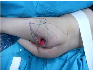
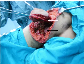
patients received a rectal enema the day before surgery. The operation was performed under general anesthesia with the patient placed in the 30° flexed prone position. Ischial tissue defects were debrided aggressively in a man-ner of malignant tumor extirpation so as to remove all fibrotic tissue. Care was taken to complete the bursectomy and blunt the ischial tuberosity conservatively. Bone wax was used for bone hemostasis. The overall dimension of the defect was measured and an appropriate triangular flap was drawn above the gluteal crease next to the defect (Figs. 1 and2). Proportionate to the defect size, a partial distal portion of gluteus maximus muscle (not the whole) was dissected and freed from the femoral insertion with an electrocautery. The muscle was used as a carrier for the triangular flap lying above (Fig.3). The muscle was trans-posed medially and the triangular flap was advanced in a V-Y fashion to cover the defect (Fig. 4). A drain was placed underneath the flap extending along the donor area. Care was taken to avoid any dead spaces and to maintain 3-layer closures over the ischial tuberosity. Simple sutures with 2/0 obtuse monofilament polyglyconate synthetic ab-sorbable sutures (Maxon, Covidien, Mansfield, MA) were used to approximate the muscle fibers to the periosteum and/or gluteal plate. Skin closure was completed with sta-ples. At the end, the tail and one side of the Y should fit on the gluteal crease. Surgical demonstration is shown in in-traoperative views (Figs.5,6,7, and8).
Postoperative care
In the immediate postoperative period, patients were allowed to lie in a supine position until reanimation
Table 1 Characteristics of patients with ischial pressure ulcers Number of patients 14 (12 male, 2 female) Number of flaps 17
Number of previous surgery 8 patients (57.1%) Age (years) 56.8 (38–74) Number of osteomyelitis treated 5
Range of defect size 8 × 6–12x11cm Range of flap size 10 × 7–14 × 10 cm Mean hospital stay 12.6 days Duration of surgery 100–135 min Range of follow-up time 15–61 months
Early complication 7.1% (wound dehiscence) Late complication/recurrence 7.1% (healed with re-advancement)
was completed. All patients were placed in the prone position on air-fluidized beds for 3 weeks. They re-ceived a pulpless high-protein, high-calorie diet during this period. Dressings were cleaned with povidone io-dine and changed daily. Drains were removed once the output was below 30 ml per day. Staples were removed after 3 weeks and patients were monitored by physical therapy and rehabilitation. Patients were discharged in this period unless they received intravenous antibiotics for the treatment of osteomyelitis. Meanwhile, the posi-tion was changed every 2 h for the bedridden patient. A strict sitting protocol was described to both the patients
and their caretakers starting a month after surgery. One hour of sitting was permitted every day and was in-creased by 30 min every week until a total of 2 h of daily sitting was reached. Patients were allowed 10 min of sitting with 10 s of resting during this period. Monthly follow-ups were recommended for 1 year post-operatively and yearly afterwards.
Results
Two female and 12 male patients were included in this study. The mean age of the study population was 56.8 (38–74) years. All wounds were Shea stage IV with three having the ischial tuberosity exposed. Two patients had a positive biopsy culture and, in addition to them, a total of
Fig. 2 Triangular flap was drawn above the gluteal crease
Fig. 3 Muscle used as a carrier
Fig. 4 Triangular flap was advanced in a V-Y fashion to cover the defect
five patients received osteomyelitis treatment. After wound debridement, the average size of the defects ranged from 8 × 6 to 12 × 11 cm. The mean hospital stay was 12.6 days and the duration of surgery was between 100 and 135 min. Three patients had bilateral ischial ul-cers, while one had both ischial and trocanteric pressure ulcers. Some of the patients (8 patients; 57.1%) had pre-vious surgery for reconstruction of various pressure ul-cers. The follow-up periods ranged between 15 months to 5 years and 1 month. Among all 14 patients, only one (7.1%) had a recurrence after 1 year post-operation, which needed an additional debridement and advancement of the previous flap. This patient had a non-recurrent pe-riod of 16 months after the second operative procedure. Early complications such as hematomas and seromas were not seen, except in one patient who had a 6 cm2wound dehiscence which healed with secondary intention. Patient demographics are shown in Table1.
Discussion
All procedures in the literature have their own advan-tages and disadvanadvan-tages. Despite the fact that there are numerous treatment alternatives, ischial pressure sores are still the most recurrent pressure sores and most pa-tients have a history of more than one previous surgery for this reason [7]. Thus, alternative methods should be simple and re-applicable which could cover large ischial defects with enough padding and low morbidities. The authors believe that the present method could be a reli-able treatment modality for ischial pressure sores.
Ischial pressure sore recurrence rates differ from 18.1 to 41.4% in the literature [6,8,9]. According to a review, ischial pressure ulcers treated with myocutaneous flaps have a recur-rence rate of 8.9% while fasciocutaneous flaps have 11.2% [10]. Combination of muscle and fasciocutaneous flaps in ischial ulcer reconstruction is shown to have lower recurrence rates [11,12]. This may be due to distribution of pressures over the ischial region by opposing tissues. However, the main disadvantage of the combined method is the long duration of operations. Our modification reduces the operational time and maintains low recurrence rates. Besides, being able to re-advance a flap is an advantage in cases of recurrence.
The importance of filling dead spaces with muscles in chronic cavities to prevent further infections and treat osteomyelitis has been emphasized in various studies [13]–[14]. Using gluteus muscle as a coverage of ischial regions provides low complication rates [12]. However, dissecting the whole muscle not only elongates the op-erational time but also disrupts probable ambulation. Thus, our modification reduces both disadvantages. In our experience, gluteus muscle was noted to be already atrophic in paraplegic patients. Partial detachment of the muscle would not only simplify the procedure but also
Fig. 7 Flap elevated with muscle carrier. White arrow indicates gluteus maximus muscle underneath. Black arrow indicates bony prominence going to be covered with myocutaneous flap
Fig. 6 The distal portion of gluteus maximus muscle is freed.Note the mobility of the flap compared with classic fasciocutaneous V-Y flaps
Fig. 8 Early postoperative view. Defect coverage with minimal donor site morbidity
prevent from further atrophic degeneration. Nişancı et al. [15] have demonstrated promising results with a similar modification of this flap for coverage of trochan-teric ulcers. In the present technique, only an adequate division of the muscle is transposed which does not need extensive dissection and does not deprive the ma-jor feeding vessels. Triangular skin island corresponding with the wound area also facilitates tension-free cover-age of the donor area.
Combined techniques can be useful in preventing recur-rences. It enables effective and durable coverage but is also time consuming. The combined techniques have a longer du-ration of surgery (3 to 4 h) with greater dissection areas com-pared with our method [16]. Di Pompeo et al. [17] described the anterolateral thigh flap for ischiatic sore reconstruction having a mean operational time of 212 min and covers medium-sized defects. Gracilis muscle flaps were also used in combination with fasciocutaneous flaps in order to obliter-ate dead space [18]. Although it necessitates additional inci-sion sites which elongates the overall surgery time, it could be useful as an alternative. Our modification has reasonably low-er oplow-eration times and is easily learned.
Retrospective design and small patient population are some of the limitations of this study. Pressure ulcer patients are not always good candidates for surgery. Thus, planning a study with a large population of ischial sore reconstruction cases is relatively hard. Nevertheless, low complication rates and op-erational time make this modification a promising option.
Conclusion
Having a large repertoire of treatment methods is useful in various situations like recurrent pressure ulcers. Ischial pres-sure sores are privileged cases based on high recurrence rates. There are numerous treatment methods which could be de-signed with regard to the patient characteristics. The distal partial gluteus maximus musculocutaneous V-Y flap is a sim-ple and new approach with low complication rates for recon-struction of ischial pressure sores. Although there is still not an ideal flap for this challenge, the present technique could be a reliable option for the reconstructive armamentarium.
Compliance with ethical standards
Funding information Not applicable.
Conflict of interest Mustafa Nışancı and Mehmet Altıparmak declare that they have no conflict of interest.
Ethical approval This study was approved by“The Ethical Committee of Muğla Sıtkı Koçman University.”
Informed consent Not applicable.
Participation consent statement Mustafa Nışancı: creation of the idea, executing the operations, collecting data.
Mehmet Altıparmak: executing the operations, collecting data, writing the manuscript.
References
1. Eslami V, Saadat S, Habibi Arejan R, Vaccaro AR, Ghodsi SM, Rahimi-Movaghar V (2012) Factors associated with the develop-ment of pressure ulcers after spinal cord injury. Spinal Cord 50(12): 899–903
2. Kua EHJ, Wong CH, Ng SW, Tan KC (2011) The island pedicled anterolateral thigh (pALT) flap via the lateral subcutaneous tunnel for recurrent ischial ulcers. J Plast Reconstr Aesthet Surg 64(1): e21–e23
3. Otto A, Wechselberger G, Schoeller T (1998) Ischial pressure sore coverage: a rationale for flap selection. Br J Plast Surg 51(3):260– 261
4. Ichioka S, Okabe K, Tsuji S, Ohura N, Nakatsuka T (2004) Triple coverage of ischial ulcers with adipofascial turnover and fasciocutaneous flaps. Plast Reconstr Surg 114(4):901–905 5. Shea JD (1975) Pressure sores: classification and management. Clin
Orthop Relat Res (112):89–100
6. Jordan SW, De la Garza M, Lewis VL (2017) Two-stage treatment of ischial pressure ulcers in spinal cord injury patients: technique and outcomes over 8 years. J Plast Reconstr Aesthet Surg 70(7): 959–966
7. Keys KA, Daniali LN, Warner KJ, Mathes DW (2010) Multivariate predictors of failure after flap coverage of pressure ulcers. Plast Reconstr Surg 125(6):1725–1734
8. Unal C, Ozdemir J, Yirmibesoglu O, Yucel E, Agir H (2012) Use of inferior gluteal artery and posterior thigh perforators in manage-ment of ischial pressure sores with limited donor sites for flap cov-erage. Ann Plast Surg 69(1):67–72
9. Tavakoli K, Rutkowski S, Cope C, Hassall M, Barnett R, Richards M, Vandervord J (1999) Recurrence rates of ischial sores in para-and tetraplegics treated with hamstring flaps: an 8-year study. Br J Plast Surg 52(6):476–479
10. Sameem M, Au M, Wood T, Farrokhyar F, Mahoney J (2012) A systematic review of complication and recurrence rates of musculocutaneous, fasciocutaneous, and perforator-based flaps for treatment of pressure sores. Plast Reconstr Surg 130(1):67–77 11. Demirseren ME, Ceran C, Aksam B, Demiralp CO (2016) Clinical
experience with the combination of a biceps femoris muscle turn-over flap and a posterior thigh fasciocutaneous hatchet flap for the reconstruction of ischial pressure ulcers. Ann Plast Surg 77(1):93– 96
12. Ahluwalia R, Martin D, Mahoney JL (2010) The operative treat-ment of pressure wounds: a 10-year experience in flap selection. Int Wound J 7(2):103–106
13. Acartürk TO (2009) Treatment of large ischial ulcers communicat-ing with the hip joint with proximal femoral resection and recon-struction with a combined vastus lateralis, vastus intermedius and rectus femoris musculocutaneous flap. J Plast Reconstr Aesthet Surg 62(11):1497–1502
14. J. Hong, T. Goh, D. Choi,… J. K.-P. and, and undefined 2017, “The efficacy of perforator flaps in the treatment of chronic osteomyelitis, ”ingentaconnect.com
15. Hong JP, Goh TLH, Choi DH, Kim JJ, Suh HS (2017) The efficacy of perforator flaps in the treatment of chronic osteomyelitis. Plast Reconstr Surg 140(1):179–188
16. Nisanci M, Sahin I, Eski M, Alhan D (2015) A new flap alternative for trochanteric pressure sore coverage: Distal gluteus maximus
musculocutaneous advancement flap. Ann Plast Surg 74(2):214– 219
17. Santanelli Di Pompeo F, Longo B, Pagnoni M, Laporta R (2015) Sensate anterolateral thigh perforator flap for ischiatic sores recon-struction in meningomyelocele patients. Microsurgery 35(4):279– 283
18. Lee S-S, Huang S-H, Chen M-C, Chang K-P, Lai C-S, Lin S-D (2009) Management of recurrent ischial pressure sore with gracilis
muscle flap and V-Y profunda femoris artery perforator-based flap. J Plast Reconstr Aesthet Surg 62(10):1339–1346
Publisher’s note Springer Nature remains neutral with regard to jurisdictional claims in published maps and institutional affiliations.