doi:10.21010/ajtcam.v14i2.7
60
THE BIOLOGICAL ACTIVITIES OF MOLTKIA AUREA BOISS., AN ENDEMIC SPECIES TO TURKEY
Neslihan Balpinar
1*, Gulten Okmen
21
Department of
Biology, Faculty of Arts and Science, Mehmet Akif Ersoy University, Burdur 15030,
Turkey
2Department of
Biology, Faculty of Science, Mugla Sıtkı Kocman University, Mugla 48000,
Turkey.
*
Corresponding author E-mail: [email protected]
Abstract
Background: Staphylococcus aureus is the direct reason of mastitis. Mastitis is a disease characterized by pathological
changes in mammary glands as well as physical, chemical, bacteriological changes in milk. This disease causes loses in milk yield and quality. In recent years, it is reported that mastitis pathogens have developed a resistance to antibiotics as a natural consequence of widespread use of it. Today’s researches are focused on discovering and using new antibiotics against bacteria. The aim of this paper is to examine the antibacterial properties of Moltkia aurea Boiss.][o] (an endemic species to Turkey), and its other biological activities.
Materials and Methods: All of the extracts were tested by disc diffusion assay in order to screen antibacterial activity.
MIC values were evaluated as antibacterial activities of plant extracts. The non-enzymatic antioxidative activities including DPPH radical scavenging effects were studied in vitro.
Results and Conclusions: Results shown that the extracts had strong antibacterial effects on three bacteria (S.
aureus-17, S. aureus-18 and CNS-37) and the range of inhibition zone was 4-6mm. This three bacteria screened the lowest sensitivity to 65000 µg /mL concentration. Besides, the extracts were tested for non-enzymatic antioxidant activities. As a result, the methanol extract of the flower displayed a strong antioxidant activity. The various extracts of Moltkia
aurea have different antibacterial and antioxidant properties.
Key words: Antibacterial activity, Antioxidant activity, Mastitis, Moltkia aurea
Introduction
Staphylococcus aureus is naturally found in mucosal epithelia, human and animal skin. However, the bacteria
are the direct reason of mastitis. It is contagious and located at teat canals and mammary glands of infected cows. The toxins produced by S. aureus cause demolition of cell membranes and milk-producing tissue (Petersson-Wolfe, et al., 2010). Therefore, mastitis can degrade milk production and fluctuate its quality, and even cause systematic diseases (Le Marechal, et al., 2011). There are two forms of it: subclinical form characterized by change in milk abnormalities, and clinical form characterized by the presence of inflammation signs (Gruet, et al., 2001). Subclinical pathogens are
Pseudomonas, Coagulase-Negative Staphylococci (CNS), S. aureus, S. agalactiae, S. dysgalactiae and E. coli, and
clinical ones are S. aureus, S. dysgalactiae, S. agalactiae and E. coli (Amir, 2013). Among of them, CNS are considered as the most common pathogen in many countries (Pitkälä, et al., 2007). Besides, 10-12 percent of all clinical mastitis infections are due to S. aureus (Tenhagen, et al., 2009).
It is reported that the cows infected by S. aureus must be segragated from non-infected ones because the infected ones do not give good response to antibiotic therapy (Roberson, et al., 1994). That makes treatment more difficult and sometimes unsuccessful (Sommerhäuser et al., 2003). Most of antibiotics in use are broad-spectrum antibiotics which are effective againts Gram-positive/negative bacteria (NCCLS, 2002).
The systematic studies of medicinal plants arose in the 1970s. This action was encouraged by the World Health Organisation (WHO) via the medicinal plants evaluation programme conducted in 1978. Many African plants (more than 20000 species) were identified in that study (Farnsworth, 1994). In recent years, there seems to be an inclination that studies on medicinal plants, which are basis for the development of new biologically active molecules for pharmaceuticals, have rapidly increased. Recent studies associated with medicinal plants make possible to better understand their properties, safety and efficiency.
In this study, Moltkia aurea Boiss. classified within Boraginaceae family will be examined. Boraginaceae is a family containing about 150 genera and 2700 species concentrated in the Mediterranean region (Hickey and King, 1997), and spreaded in tropical and temperate zones (Heywood, et al., 2007). Boraginaceae comprises high number of medicinal plants. Different plant parts of this family contain secondary metabolites, flavonoids, terpenoids, viz.-alkaloids, fatty acids, glycoisides, phytosterols and various proteins (Sharma, et al., 2009). Some compounds of this plant like shikonin demonstrate to possess antibacterial, antifungal, antiinflammatory effect. In addition wound healing property is known today (Terada, et al., 1990). The family comprises 342 specific and subspecific taxa belonging to 34 genera in Turkey, among these the Mediterranean genus Moltkia is represented with 4 species, one of them is M. aurea which is endemic to Anatolia. M. aurea is a perennial herb which grows at calcareous steppe, skirts of hills, 300-1300m
doi:10.21010/ajtcam.v14i2.7
61
altitude (Davis, 1978). There are only few studies on medicinal properties of Moltkia species (Končić et al., 2010), and there is barely one previous study which concentrated on antioxidant and cytotoxic effects of M. aurea (Harput, et al., 2012).
In this study, we provide an analysis on biological activities of M. aurea from Burdur Province (SW Turkey). We examine the antibacterial activity of the various extracts of M. aurea against mastitis pathogens which has not been studied before, and analyse non-enzymatic antioxidant activities of the extracts, which is less studied comparing to its other biological activities.
Materials
and Methods Plant MaterialPlant samples belonging to M. aurea were collected from 10kilometers South of Burdur in May 2015, 1201meters, 37 41.375' N 030 20.312' E. The authenticated materials which were identified according to Davis (1978) were stored at the Botanical Research Laboratory of Mehmet Akif Ersoy University, Turkey. Taxonomical identification of plant was performed by Dr. Neslihan Balpınar from the same university.
Plant Extraction
The plant parts, which consist of stem, leaf and flower, were washed under fresh flowing water and sterile distilled water. These parts were air-dried, and then powdered by using a laboratory mill. All powdered material had been maintained at room temperature until initial sample preparation was done and then all of the prepared samples were stored at 4°C until analysis. The powdered plant parts (50g) were extracted with various solvents by Soxhlet device. The extracts were evaporated and all of extracts were stored in themselves solvents. The obtained extracts were stored under refrigerated conditions in sterile opaque bottles until the request for analysis (200 mg/mL).
Microorganisms and Cultivation
Mastitis pathogens were provided from the project performed by Dr. Zafer Cantekin, Mustafa Kemal University, Turkey (Project number: 1101 M 0103; Ethics council number: 2010/02-30:12). Seven bacteria were utilized in the present study. These are including two Staphylococcus aureus and five CNS. These bacteria were cultivated on Mueller-Hinton Broth (Merck) at 37°C (Bauer, et al., 1966), and identified by traditional biochemical tests (Quinn, et al., 1994). The extracts of the plant parts were tested one by one against mastitis pathogens.
Determination of Antibacterial Activity
The various extracts were tested by disc diffusion assay. Bauer-Kirby method was used for determining
antibacterial activity. The bacteria were grown on Mueller-Hinton Agar plates at 37°C for 24 hours. Bacterial cultures
were adjusted to 0.5 Mc Farland. The extracts were saturated to sterile discs as 25 µL. The assessment of antibacterial activity was based on measurement of the diameters of the inhibition zones around the discs after 24 hours. In this study, solvents and ampicillin (10 μg) were used, respectively as negative and positive control. The tests were realized in triplicate and the mean values were given.
Determination of Minimum Inhibitory Concentration (MIC)
MICs were evaluated as antibacterial activity. After incubation, MICs were taken as the lowest concentration which it has inhibited growth. The broth dilution analysis was carried out in the standards of CLSI (2003, 2006). The analysis was performed at final concentrations of the extracts (65000; 32500; 16250; 8125; and 4062.5 µg/mL).
Determination of Non-enzymatic Antioxidant Activity
Non-enzymatic antioxidant properties of extracts were determined by free radical DPPH. In order to find out free radical activities of the stem, flower and leaf extracts, the stable radical 2,2-diphenyl-1-picrylhydrazyl-hydrate was used. Each of the extracts (0.1 mL) were added to 3.9 mL of a methanolic DPPH solution (0.1 mM). After incubation for 30 minutes, the absorbances of the extracts were measured at 515 nm by spectrophotometer. Methanolic DPPH solution and Methanol were used in this study, respectively as control and methanol as a blank. Trolox was utilized as reference to assess antioxidant activity. The DPPH scavenging capacity indicated with percentage (%) was calculated from the formula (Brand-Williams, et al., 1995).
Results
The antibacterial activities of the various extracts were evaluated in vitro against mastitis-causing pathogens. Table 1 shows the antibacterial activities of stem, flower and leaf extracts of M. aurea on the test bacteria. These
doi:10.21010/ajtcam.v14i2.7
62
activities were recorded as zone of inhibition in mm. Our results show that the extracts inhibited the growth of three bacteria (S. aureus-17, S. aureus-18 and CNS-37), and the inhibition zones were 4-6 mm. Most of the extracts supressed the growth of S. aureus-17. The highest inhibition zone was shown in S. aureus-17, and its zone was 6 mm. None of the extracts did not affect against four test bacteria. These bacteria are CNS-22, 32, 33 and 36. Ampicillin was used as positive control and it was seen that the growths of the test bacteria were strongly inhibited by this antibiotic.
Table 1: Antibacterial activities of the various extracts of M. aurea (200mg/mL) Inhibition zone diameters (mm)
Stem Extracts Flower Extracts Leaf Extracts Ampicillin Bacteria Ethanol Methanol Aqueous Ethanol Methanol Aqueous Ethanol Methanol Aqueous
S. aureus-17 4 4 - 4 - 4 4 - 6 18 S. aureus-18 - 4 5 - 4 4 4 4 - 12 CNS-22 - - - - - - - - - - CNS-32 - - - - - - - - - 10 CNS-33 - - - - - - - - - 8 CNS-36 - - - - - - - - - - CNS37 4 4 4 5
-CNS: Coagulase Negative Staphylococci; (-): No inhibition
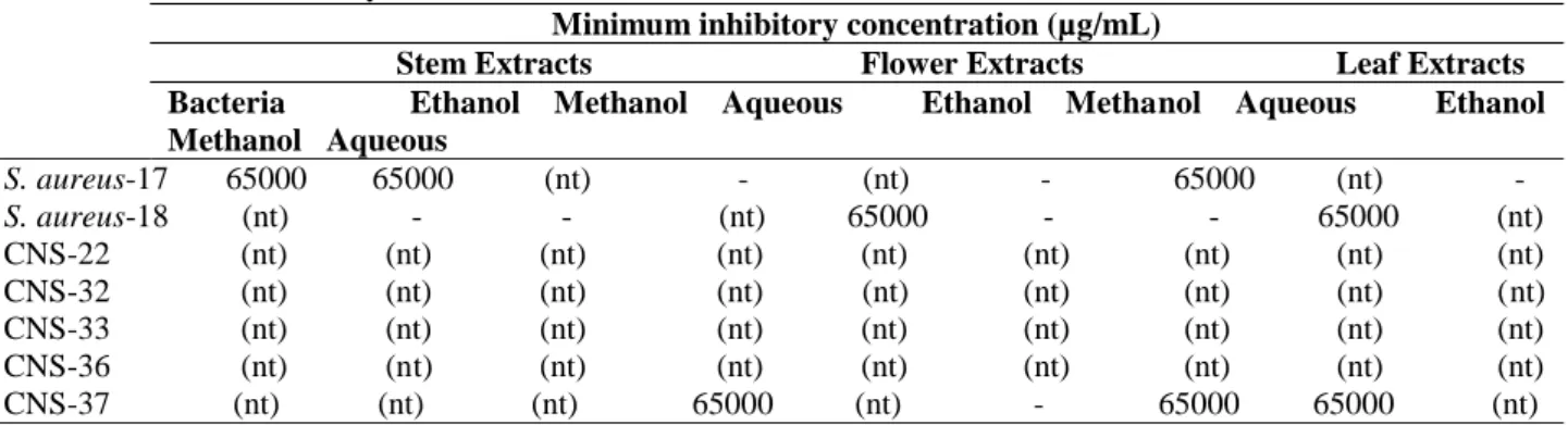
Table 2 shows the MICs of the plant extracts. Three test bacteria showed the lowest sensitivity to various extracts of M.
aurea (65000 µg/mL). These bacteria are S. aureus-17, S. aureus-18 and CNS-37. The inhibition was not found on the
other bacteria.
Table 2: Minimum inhibitory concentrations of the various extracts of M. aurea
Minimum inhibitory concentration (µg/mL)
Stem Extracts Flower Extracts Leaf Extracts Bacteria Ethanol Methanol Aqueous Ethanol Methanol Aqueous Ethanol Methanol Aqueous S. aureus-17 65000 65000 (nt) - (nt) - 65000 (nt) - S. aureus-18 (nt) - - (nt) 65000 - - 65000 (nt) CNS-22 (nt) (nt) (nt) (nt) (nt) (nt) (nt) (nt) (nt) CNS-32 (nt) (nt) (nt) (nt) (nt) (nt) (nt) (nt) (nt) CNS-33 (nt) (nt) (nt) (nt) (nt) (nt) (nt) (nt) (nt) CNS-36 (nt) (nt) (nt) (nt) (nt) (nt) (nt) (nt) (nt) CNS-37 (nt) (nt) (nt) 65000 (nt) - 65000 65000 (nt) CNS: Coagulase Negative Staphylococci; (nt): Not tested; (-): No inhibition
The non-enzymatic antioxidant activities of the extracts were evaluated by the DPPH radical scavenging capacity. The percentage of DPPH scavenging capacity with trolox is shown in Table 3. We determined that methanol extract of the flower has the highest DPPH scavenging capacity. This inhibition was 64 % at 200 mg/mL concentration. Trolox equivalent value was 2.0 mM/g DW. Furthermore, the highest radical scavenging capacity of other plant parts was determined in methanol extracts (Table 3).
Table 3: DPPH radical scavenging capacity of the various extracts of M. aurea (200 mg/mL) Plant parts Stem Flower Leaf
Extracts Ethanol Methanol Aqueous Ethanol Methanol Aqueous Ethanol Methanol Aqueous
DPPH (%) 17 57 55 30 64 29 33 57 35 TE (mM/g DW) 0.8 1.9 1.8 1.4 2 1.4 1.4 1.8 1.3
TE: Trolox equivalent; DW: dry weight
Discussion
This study confirms that various extracts of M. aurea possess antibacterial and antioxidant activities. The extracts were tested on mastitis pathogens, and its antibacterial activity was compared with ampicillin which is a penicillin antibiotic. All of the extracts were low effective on growth of the three bacteria (Table 1). Matias et al. (2013) showed that the extract from Cordia verbenacea’s leaves has no antibacterial properties. Our results are better than data of Matias. According to our results, the highest inhibition zone against S. aureus was provided by aqueous extract of the leaf (Table 1). Ahmad et al. (2009) claimed that the aqueous fraction of Onosma griffithii demonstrated moderate
doi:10.21010/ajtcam.v14i2.7
63
antibacterial activity against S. aureus (42.3%). Rancic et al. (2005) indicated that extract of Hypericum perforatum showed 5 mm inhibition zone against S. aureus. The reports support the results obtained from our study.
In this study, three test bacteria showed the lowest sensitivity to various extracts of M. aurea (65000 µg/mL) (Table 2). Matias et al. (2013) reported that S. aureus showed a MIC value greater than or equal to 1024 µg/mL, whereby the results were considered clinically irrelevant. In the study of Couladis et al. (2003), essential oil of
Hypericum rumeliacum indicated moderate activity against the tested bacteria (the range of MICs was 3.80–17.20
mg/mL). In our study, MIC value was measured as 65000 µg /mL and this result is higher than those of Couladis. No wonder that various plant groups have different antibacterial effects because of phytochemical differences among species. The reason for this may be the plant composition. It is known that some factors such as geographic distribution, phenological cycle and seasonal variation affect chemical content and composition of essential oils (Guedes, et al., 2004; Schwob, et al., 2004). Aligiannis et al. (2001) claimed that plant materials must be classified with regard to MIC results, in our study the various extracts from M. aurea can be seen as a weak inhibitor of mastitis pathogens.
It is known that antioxidant and free radical scavenging activity play the most important role in human life, especially on biological functions such as anti-carcinogenicity and anti-aging. Our results indicated that the extract obtained from M. aurea had powerful DPPH scavenging capacity. The methanol extract of the flower showed 64% inhibition at 200 mg/mL concentration. Trolox equivalent value was determined as 2.0 mM/g DW (Table 3). These results indicates that the extract contains high phenolic compounds, and this makes the plant interesting in terms of antioxidative and cytotoxic activities (Harput, et al., 2012).
Conclusions
In conclusion, various extracts of M. aurea tested in the study were determined to have potential antibacterial activities against CNS and S. aureus pathogens obtained from subclinical cow mastitis. In this study, DPPH assay was applied to determine the antioxidant potentials of the various extracts of M. aurea. Our results indicate that the leaf, flower and the stem extracts of M. aurea exhibit different antioxidant activities. Many of the extracts were proven to be notable antioxidants. Our findings suggest that M. aurea can be utilized as a natural antibacterial and antioxidant source by pharmaceutical industry especially focused on developing anti-carcinogenicity, anti-aging and antibacterial drugs.
References
1. Ahmad, B., Ali, N., Bashir, S., Choudhary, M.I., Azam, S., and Khan, I. (2009). Parasiticidal, antifungal and antibacterial activities of Onosmagriffithii Vatke. Afr. J. Biotechnol. 8(19), 5084-5087.
2. Aligiannis, N., Kalpotzakis, E., Mitaku, S., and Chinou, I.B. (2001). Composition and antimicrobial activity of the essential oils of two Origanum species. J. Agric. Food Chem. 40,4168-4170.
3. Amir, H.A.E. (2013). Mastitis in housed dairy buffaloes: incidence, etiology, clinical finding, antimicrobial sensitivity and different medical treatment against E. coli mastitis. Life Sci. J. 10(1),532-538.
4. Bauer, A.W., Kirby, W.M., Sherris, J.C., and Turck, M. (1966). Antibiotic susceptibility testing by a standardized single disk method. Am. J. Clin. Path. 45(4),493-496.
5. Brand-Williams, W., Cuvelier, M.E., and Berset, C. (1995). Use of a free radical method to evaluate antioxidant activity. Lebensm.-Wiss Technol. 28,25–30.
6. CLSI (Clinical and Laboratory Standards Institute) (2003). Methods for dilution antimicrobial susceptibility test for bacteria that grow aerobically; approved standard M7-A 6th ed. Wayne, Pennsylvania: National Committee for Clinical Laboratory Standards.
7. CLSI (Clinical and Laboratory Standards Institute) (2006). Performance standards for antimicrobial susceptibility testing. 16th Informational Supplement M100-S16. Wayne, Pennsylvania: National Committee for Clinical Laboratory Standards.
8. Couladis, M., Chinou, I.B., Tzakou, O., and Petrakis, P.V. (2003). Composition and antimicrobial activity of the essential oil of Hypericumrumeliacum subsp. apollinis (Boiss. & Heldr.). Phytother.Res. 17(2),152-154.
9. Davis, P.H. (1978). Flora of Turkey and East Aegean Islands, Vol. 6. Edinburgh: Edinburgh University Press, pp. 826.
10. Farnsworth, N. R. (1994). Ethnopharmacology and drug development. In: Prance, G. T., D. J. Chadwick and J. Marsh (Ed.), Ethnobotany and the Search for New Drugs. Chichester (London): John Wiley & Sons, pp. 42-59. 11. Gruet, P., Maincent, P., Berthelot, X., and Kaltsatos, V. (2001). Bovine mastitis and intramammary drug delivery:
review and perspectives. Adv. Drug Delivery Reviews 50,245–259.
12. Guedes, A.P., Amorim, L.R., Vicente, A., and Fernandes-Ferreira, M. (2004). Variation of the essential oil content and composition in leaves from cultivated plants of Hypericumandrosaemum L. Phytochem. Analysis 15,146-51. 13. Harput, U.S., Nagatsu, A., and Saraçoglu, I. (2012). Antioxidant and cytotoxic effects of Moltkiaaurea
Boiss. Records of Natural Products 6,62-6.
14. Heywood, V.H., Brummitt, R., Culham, A., and Seberg, O. (2007). Flowering Plant Families of the World. London: Kew Publishing, p 424.
15. Hickey, M., and King, C. (1997). Common Families of Flowering Plants. Cambridge: Cambridge University Press, pp. 226.
doi:10.21010/ajtcam.v14i2.7
64
16. Končıć, M.Z., Kremer, D., Gruz, J., Strnad, M., Biševac, G., Kosalec, I., Šamec, D., Piljac-Ţegarac, J., and Karlović, K. (2010). Antioxidant and antimicrobial properties of Moltkiapetraea (Tratt.) Griseb. flower, leaf and stem infusions. Food, Chem. Toxicol. 48(6),1537-1542.
17. Le Marechal, C., Thiery, R., Vautor, E., and Le Loir, Y. (2011). Mastitis impact on technological properties of milk and quality of milk products – a review. Dairy Sci. Technol. 91,247–282.
18. Matias, E.F.F., Alves, E.F., Santos, B.S., de Souza, C.E.S., Ferreira, J.V.A., de Lavor, A.K.L.S., Figueredo, F.G., de Lima, L.F., dos Santos, F.A.V., Peixoto, F.S.N., Colares, A.V., Boligon, A.A., Saraiva, R.A., Athayde, M.L., Rocha, J.B.T., Menezes, I.R.A., Coutinho, H.D.M., and da Costa, J.G.M. (2013). Biological activities and chemical characterization of Cordiaverbenacea DC. as tool to validate the ethnobiological usage. Evid.-Based Complem. Alternat. Medic. Article ID 164215, http://dx.doi.org/10.1155/2013/164215.
19. NCCLS (National Committee For Clinical Laboratory Standards). (2002). Performance standards for antimicrobial disk and dilution susceptibility tests for bacteria ısolated from animals. 2nd ed. Pennsylvania: CLSI.
20. Petersson-Wolfe, C., Mullarky, I. and Jones, G. (2010). Staphylococcusaureus mastitis: cause, detection, and control. College Agricult. Life Sci. 404-229. http://pubs.ext.vt.edu/404/404-229/404-229_pdf.pdf.
21. Pitkälä, A., Salmikivi, L., Bredbacka, P., Myllyniemi, A. L., and Koskinen, M. T. (2007). Comparison of tests for detection of beta-lactamase-producing Staphylococci. J. Clin. Microbiol. 45,2031-2033.
22. Quinn, P.J., Carter, M.E., Markey, B.K., and Carter, G.R. (1994). Clinical Veterinary Microbiology. London: Lynton House, pp. 648.
23. Rancic, A., Sokovic, M., Vukojevic, J., Simic, A., Marin, P., Duletic-Lausevic, S., and Djokovic, D. (2005).
Chemical composition and antimicrobial activities of essential oils of Myrrhisodorata (L.)
Scop, Hypericumperforatum L and Helichrysumarenarium (L.) Moench. J. Essent. Oil Res. 17,341–345.
24. Roberson, J.R., Fox, L.K., Hancock, D. D., Gay, J.M., and Besser, T.E. (1994). Ecology of Staphylococcusaureus isolated from various sites on dairy farms. J. Dairy Sci. 77(11),3354-3364.
25. Schwob, I., Bessiere, J.M., Masotti, V., and Viano, J. (2004). Changes in essential oil composition in Saint John's wort (Hypericumperforatum L.) aerial parts during its phenological cycle. Biochem.System. Ecol. 32(8),735-745. 26. Sharma, R.A., Singh, B., Singh, D., and Chandrawat, P. (2009). Ethnomedicinal, pharmacological properties and
chemistry of some medicinal plants of Boraginaceae in India. J. Medic. Plants Res. 3(13),1153-1175.
27. Sommerhäuser, J., Kloppert, B., Wolter, W., Zschock, M., Sobiraj, A., and Failing, K. (2003). The epidemiology of Staphylococcusaureus infections from subclinical mastitis in dairy cows during a control programme. Vet. Microbiol. 96,91-102.
28. Tenhagen, B.A., Hansen, I., Reinecke, A., and Heuwieser, W. (2009). Prevalence of pathogens in milk samples of dairy cows with clinical mastitis and in heifers at first parturition. J. Dairy Res. 76(2),179-87.
29. Terada, A., Tanone, Y., and Taniguchi, H. (1990). Chemistry of shikonin, ancient purple pigment and its derivatives. J. Synthetic Organ. Chem. 48,866-875.