Assays that measure markers of inflammation in cardio-vascular disease have become an important part of the clinical laboratory repertoire.1 Acute myocardial infarction
(AMI), is currently the leading cause of death in developed countries.2 AMI is caused by coronary artery occlusion
due to local thrombosis, precipitated by rupture of an unstable atherosclerotic plaque and by the interactions of numerous inflammatory, prothrombotic, and vasoactive factors. Acute coronary syndrome (ACS) has come to be
understood as a combination of factors involving inflam-mation and thrombosis that precede AMI.2,3
Abbreviations
ACS, acute coronary syndrome; AMI, acute myocardial infarction; Gal-3, galectin-3; IL-6, interleukin-6; TNF-α, tumor necrosis factor– alpha; CRP, C-reactive protein; CAD, coronary artery disease; ECG, electrocardiogram; ELISA, enzymatic immunoassay; LDH-2, lactate dehydrogenase 2; CK-MB, creatine kinase–myocardial band; cTnI, cardiac troponin I; SPSS, Statistical Package for Social Sciences; BMI, body mass index
1 Department of Biochemistry, Istanbul University, Cerrahpasa, Istanbul,
Turkey; 2Department of Cardiology, Cerrahpasa Medical Faculty,
Istanbul, Turkey, 3Department of Biochemistry, Faculty of Dentistry,
Marmara University, Nisantasi, Istanbul, Turkey, 4Department of
Biochemistry, Faculty of Medicine, Medipol University, Istanbul, Turkey *To whom correspondence should be addressed.
Galectin-3 and Plasma Cytokines in Patients
With Acute Myocardial Infarction
A. Ata Alturfan, PhD,
1Isık Basar, MD,
2Ebru Emekli-Alturfan, PhD,
3*Faruk Ayan, MD,
2Lale Koldas, MD,
2Nesrin Emekli, PhD
4Lab Med Fall 2014;45:332-337
DOI: 10.1309/LM3JZKBDA7D4QFOC
Galectins are a family of β-galactoside–binding lectins.4
Galectins bind to cell-surface and extracellular matrix glycans and thereby affect a variety of cellular processes. Galectins are also detectable in the cytosol and nucleus and may influence intracellular signaling pathways through interactions with cytoplasmic and nuclear proteins.5,6 Recent
studies indicate that galectins are important components of many physiological and pathological processes. Immune and inflammatory responses, tumor development and progression, neural degeneration, atherosclerosis, diabetes mellitus, and wound repair are among these processes.5,6
The wide distribution of the galectins in both species and tissues suggests that these proteins are functionally diverse.4,7,8 The presence of galectin-3 (Gal-3) in the
epithelium, and activated tissue macrophages prompted us and others4,7-9 to investigate the involvement of galectins
in inflammatory processes, including AMI8 and ACS.9
Interleukin-6 (IL-6) and tumor necrosis factor–alpha (TNF-α) have been identified as proinflammatory cytokines. TNF-α is a multifunctional circulating cytokine derived from macrophages and endothelial and smooth muscle cells. TNF-α plays a major role in the cytokine cascade because it stimulates the synthesis of the other cytokines. IL-6, is a pleiotropic cytokine with a broad
ABSTRACT
Objective: To investigate the concentrations of plasma cytokines and
Galectin-3 (Gal-3) as inflammatory markers in patients with acute myocardial infarction (AMI).
Methods: The study population consisted of 29 patients with AMI
and 29 healthy control subjects. We measured Gal-3, tumor necrosis factor–alpha (TNF-α), and interleukin-6 (IL-6) levels in plasma using enzyme-linked immunosorbent assays (ELISAs). We measured levels of C-reactive protein (CRP) via the nephelometric method.
Results: Patients with AMI showed significantly higher plasma
Gal-3, TNF-α, and IL-6 levels compared with controls. Gal-3 levels were positively and significantly correlated with plasma IL-6, TNF-α, and CRP levels in the control and patient groups.
Conclusion: Our findings suggest that Gal-3 can be a new circulating
biomarker of inflammation associated with AMI.
Keywords: Galectin-3, AMI, inflammation, cytokines, CRP, TNF-α
range of humoral and cellular immune effects relating to inflammation, host defense, and tissue injury.10,11 The
release of proinflammatory cytokines such as IL-6 and TNF-α is known to play a pivotal role in the development of systemic inflammatory response.11 We measured plasma
levels of IL-6 and TNF-α in patients with AMI to assess the inflammatory response in these patients.
C-reactive protein (CRP) is an acute and fast increasing major acute-phase protein that is released following infection and tissue injuries in humans. CRP has been used as a marker of inflammation in the setting of AMI.12-14
ACS encompasses a spectrum of coronary disease, including myocardial infarction. Early diagnosis and risk stratification are needed to correctly determine the needs for hospitalization and treatment. We measured pro-inflammatory cytokines and Gal-3 in patients with AMI.
Materials and Methods
Patients
Twenty-nine patients admitted to Cerrahpasa Medical Faculty with AMI less than 12 hours from the onset of symptoms were enrolled in the study; blood samples were drawn 24 hours from the onset of symptoms. The subjects were categorized into 2 groups, namely, a control group (n = 29), which consisted of 10 female and 19 male healthy volunteers without coronary artery disease (CAD), and the patient group (n = 29), which consisted of 8 female and 21 male patients. The study was approved by the Hospital Ethics Committee; all patients gave written informed consent to participate in the study. We excluded patients who had a history of hematological, neoplastic, renal, liver, or thyroid disease, and patients receiving treatment with anti-inflammatory drugs. Patients with infectious or autoimmune diseases or familial hyperlipidemia also were excluded from the study. Clinical and biochemical data were collected from subjects after enrollment.
Characteristics of the patient and control groups are given in Table 1. Histories and the results of physical
examination, electrocardiogram (ECG) testing, chest radiography testing, and routine laboratory tests showed no evidence of CAD in the controls.
AMI is caused by myocardial ischemia leading to myocardial injury and necrosis. The ACC criteria for AMI
are met when a rise and/or fall of cardiac biomarkers is observed, along with supportive evidence in the form of typical symptoms, suggestive changes via ECG, or imaging evidence of new loss of viable myocardium or new regional wall motion abnormality.
Blood Samples
We collected blood from patients and healthy controls into 0.109-M trisodium citrate tubes (Vacutainer; Becton, Dickinson, and Company; Franklin Lakes, NJ) by antecubital vein venipuncture. For enzymatic immunoassay assays (ELISA), blood samples were immediately centrifuged at 3000 rpm for 10 minutes at room temperature, and the aliquots of plasma were stored at −80°C for 1 month.
Determination of Concentration of Gal-3
We measured plasma Gal-3 concentration using a commercial ELISA kit for human Gal-3 (Lot no.31642003, Bender MedSystems, Vienna, Austria) according to the manufacturer’s instructions. Accordingly the limit of detection of human Gal-3, defined as the analyte concentration resulting in an absorbance significantly higher than that of the dilution medium (mean [SD] plus 2), was determined to be 0.12 ng/mL (ie, the mean [SD] of 6 independent assays). The detected human Gal-3 levels in plasma samples ranged between 4.67 and 10.30 ng/mL, with a mean level of 7.07 ng/mL (as determined by the manufacturer). The interassay coefficient of variation of the Galectin-3 ELISA is 11.4%, and the intra-assay coefficient of variation is 6.4% as determined by the manufacturer.
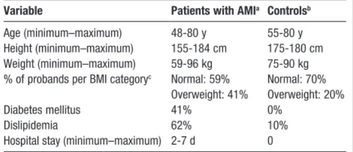
Table 1. Demographic Data of Control Individuals
and Patients With AMI
Variable Patients with AMIa Controlsb
Age (minimum–maximum) 48-80 y 55-80 y
Height (minimum–maximum) 155-184 cm 175-180 cm Weight (minimum–maximum) 59-96 kg 75-90 kg % of probands per BMI categoryc Normal: 59% Normal: 70%
Overweight: 41% Overweight: 20%
Diabetes mellitus 41% 0%
Dislipidemia 62% 10%
Hospital stay (minimum–maximum) 2-7 d 0
AIM, acute myocardial infarction; BMI, body mass index an = 29 (8 women, 21 men).
bn = 29 (10 women, 19 men).
cBody mass index is calculated as weight in kilograms divided by height in meters squared. As defined by the Centers for Disease Control and Prevention, for adults, normal BMI range is defined as 18.5-24.9, overweight as 25.0-29.9, and obese as ≥30.0. None of the participants qualified as underweight (ie, having a BMI score of <18.5).
Determination of IL-6 and TNF-
α Levels
IL-6 and TNF-α were measured using a commercially available ELISA (lot no. 7106577; Assaypro, St. Charles, MO and Life Technologies Corporation, Carlsbad, CA; lot no.062805, respectively) in plasma samples from our cohort. The minimum detectable concentration of IL-6 is <10 pg per/mL. The detection limit of TNF-α is0.7 pg/mL. The interassay coefficients of variations of TNF-α and IL-6 ELISA assays are 3.9% and 7.5%, respectively; the intra-assay coefficients of variations are 6.4% and 4.9%, respectively.
Determination of CK-MB, Troponin I,
and CRP Levels
CRP was measured by a nephelometric method (Siemens AG, Munich, Germany). We measured CK-MB with an autoanalyzer (Abbott C8000, Abbott Laboratories, Abbott Park, IL) and serum cardiac troponin I (cTnI) levels using the chemiluminescence immunoassay method via the DPC IMMULITE Analyzer (Siemens AG). We evaluated CRP, CK-MB, and cTnI concentrations after 24 hours from the onset of symptoms. All blood samples for cardiac markers were centrifuged at 3000 rpm for 5 minutes to separate the serum.
Statistical Analysis
For the descriptive statistics, after having checked the normality of the variables using the Kolmogorov-Smirnov test, we used an independent sample t-test to compare the serum levels of all variables. We used Pearson correlation testing for the correlations. Statistical analyses were performed using SPSS (Statistical Package for Social Sciences, Chicago, IL) software for Windows (version 11.5). Values are presented as mean (SD); a P value of less than .05 was considered significant.
Results
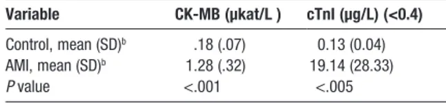
CK-MB activities were significantly higher (P <.001) in the patient group compared with the control group (mean [SD], 1.28 [.32] μkat/L and .18 [.07] μkat/L, respectively) (Table 2).
cTnI concentrations were significantly higher (P <.005) in the patient group compared with the controls (19.14 [28.33] μg/L and 0.13 [0.04] μg/L, respectively) (Table 2).
A significant increase was observed in Gal-3 levels of patients with AMI compared with healthy subjects (mean [SD], 10.14 [2.08] ng/mL and 8.24 [1.64] ng/mL, respectively;
P <.005) (Table 3). TNF-α levels increased significantly in
the plasma of patients with AMI compared with healthy
subjects (18.62 [1.68] and 12.35 [1.52] pg/mL, respectively;
P <.001) (Table 3). IL-6 levels also increased significantly
in the plasma of patients with AMI (170.21 [14.13] pg/mL) compared with healthy subjects (140.79 [11.19] pg/mL;
P <.001; (Table 3). CRP concentration increased
significantly in patients with AMI, compared with controls (87.81 [29.52] nmol/L vs 9.05 [3.24] nmol/L, respectively;P
<.001) in patients with AMI (Table 3).
In the patient group, plasma Gal-3 levels were significantly and positively correlated with plasma IL-6 and TNF-α levels (r = 0.427 and r = 0.382 respectively; P <.05). Moreover, in the patient and control groups (n = 58), plasma Gal-3 levels were significantly and positively correlated with plasma IL-6, TNF-α, and CRP levels (r = 0.488, r = 0.514, and r = 0.517, respectively; P <.01). Positive correlations were also observed between IL-6 and CRP levels and between IL-6 and TNF-α levels (r = 0.742, r = 0.682 respectively; P <.01).
Discussion
Recently, researchers have focused on the potential role of circulating markers of inflammation in ACS as risk predictors among those who have suffered an acute cardiovascular event. Our findings suggest that Gal-3 is a biomarker of inflammation in AMI. However, our knowledge of the actual role of Gal-3 in ACS has not been elucidated. Inflammation is a key component of atherosclerosis, from fatty streak formation to plaque rupture, subsequent thrombosis, and progressive mechanical and dynamic obstruction. When the fibrous cap of arterial plaque is ruptured, tissue factors (thromboplastin) in the necrotic core are released which leads to increased inflammation, cell adhesion, and coagulation cascade that at the end causes thrombus formation.15,16 Recent research has
focused on the potential role of circulating markers of inflammation in ACS as risk predictors among those who
Table 2. Mean Serum Concentrations of
Cardiac Markers
aVariable CK-MB (μkat/L ) cTnI (μg/L) (<0.4)
Control, mean (SD)b .18 (.07) 0.13 (0.04)
AMI, mean (SD)b 1.28 (.32) 19.14 (28.33)
P value <.001 <.005
CK-MB, creatine kinase–myocardial band; LDH-2, lactate dehydrogenase 2; cTnI, cardiac troponin I
aP <.05 is considered significant. bn = 29.
have experienced an acute cardiovascular event.17 We
assessed the inflammatory responses of patients with AMI by measuring IL-6, TNF-α, CRP, and Gal-3 levels.
Gal-3 is one of several tissue factors suspected to be involved in atherosclerosis. Gal-3, regulates adhesive interactions between the cell and extracellular matrix and mediates signal transduction events accordingly it participates in organogenesis, angiogenesis, inflammation, and autoimmune disorders.17 Falcone et al18 measured
plasma Gal-3 in patients with CAD, and suggested that Gal-3 could be a useful biomarker of atherosclerotic plaques and, particularly, their destabilization. Tsai et al8
found that circulating Gal-3 levels were significantly higher in patients with AMI compared with controls; Grandin et al9
found that Gal-3 is associated with the risk of developing heart failure after ACS.
Nachtigal et al17 investigated the potential contribution
of Gal-3 to the development and progression of atherosclerotic lesions in mice. They found intense immunostaining for Gal-3 in foam cells that were present in the atheromatous plaques in the artery walls. Galectins produced by circulating blood cells or endothelial cells have been reported18-21 to induce leukocyte attachment
to the endothelium by cross-linking carbohydrates on the respective cell surfaces. The expression of Gal-3 is highly upregulated when monocytes differentiate into macrophages, a step in atherogenesis that follows monocyte recruitment from the peripheral blood to the artery wall and also occurs when in vivo or in vitro macrophages or aortic smooth muscle cells are loaded with lipids and transformed into foam cells. Like the activated macrophages, plaque foam cells may secrete Gal-3, a potent chemoattractant for monocytes and macrophages, and thus enhance the recruitment of these cells to the artery wall.17,22,23
The localization of Gal-3 within the tissue microenvironment may be extracellular, cytoplasmic, or nuclear; Gal-3 has
a concentration-dependent ability to be monomeric or to form oligomers. These properties impart great flexibility, making Gal-3 a specific regulator of many biological systems, including inflammation. Therefore, Gal-3 can be viewed as a regulatory molecule that acts at various stages along the continuum, from acute inflammation to chronic inflammation and tissue fibrogenesis.24 In our
study, Gal-3 levels were significantly increased in the plasma of patients with AMI compared with controls. The increase might be due to upregulation of Gal-3, especially at inflammatory sites, as a result of disruption of arteries and macrophage production and secretion. Consistent with this finding, certain studies in the literature5,11,17,20
have reported the presence of an increased level of Gal-3 in physiological and pathological processes, including immune and inflammatory responses, wound repair, and atherosclerosis. However, knowledge about the contribution of Gal-3 in patients with AMI is still scarce; further studies are necessary to elucidate this mechanism.
In the human body, several cytokines are involved in the inflammatory processes; these cytokines have overlapping, antagonistic, and synergic effects on many cell types and upregulate and downregulate the production of other cytokines and inflammatory mediators.24-26 The release of
proinflammatory cytokines, such as IL-6 and TNF-α, has been reported25 to play a pivotal role in the development
of systemic inflammation. The production of IL-6 from macrophages is induced by IL-1 and TNF-α. Endothelial cells are capable of producing IL-6 when stimulated with a variety of inflammatory mediators.26 IL-6 levels have been
shown to be elevated with infections and inflammation. High IL-6 levels are associated with unfavorable outcomes in different disease processes, including unstable angina and septic shock.26 IL-6 acts on hepatic cells to produce
acute-phase proteins, such as fibrinogen and CRP. A strong correlation has been shown27 between IL-6 and CRP levels.
In our study IL-6 levels were positively correlated with CRP levels (r = 0.742; P <.01). Also, IL-6 has procoagulant properties that may influence the course of ACS.28-30
Table 3. Mean Plasma Concentrations of Gal-3, TNF-
α, IL-6, and Serum CRP in Patients With AMI
aVariable Gal-3 (ng/mL) TNF-α (pg/mL) IL-6 (pg/mL) CRP (nmol/L)
Control, mean (SD)b 8.24 (1.64) 12.35 (1.52) 140.79 (11.19) 9.05 (3.24)
AMI, mean (SD)b 10.14 (2.08) 18.62 (1.68) 170.21 (14.13) 87.81 (29.52)
P value <.005 <.001 <.001 <.001
Gal-3, Galectin-3; TNF-α, tumor necrosis factor––alpha; IL-6, interleukin-6; CRP, C-reactive protein.
aP <.05 is considered significant. bn = 29.
Biasucci31 demonstrated the elevation of IL-6 in unstable
angina and the prognostic value of cytokine levels in the course of hospitalization. Neumann32 found elevated
concentrations of IL-6 in the coronary sinus blood of patients with AMI before and after recanalization, compared with arterial blood. He suggested that the vascular endothelium was the predominant source of this cardiac IL-6. Further, one should assume the possibility of important systemic effects if cardiac liberation of IL-6 is ongoing.31 Therefore, our finding of elevated IL-6 levels
in patients with AMI is consistent with those of other investigators.
TNF-α, a polypeptide with hormone-like properties, is derived from leukocytes/macrophages but not from endothelial cells. Elevated TNF-α levels have been found in patients with advanced congestive heart failure.32
Moreover, it has been shown26 that TNF-α is cardiotoxic
and can produce cardiomyopathy, left ventricular remodeling, and pulmonary edema. In AMI, significant changes in TNF-α have mainly been associated with extensive myocardial damage, signs of heart failure, and the presence of heart-rhythm disturbances.26,35,36
Wang et al37 reported significantly higher levels of TNF-α
in patients with ACS compared with healthy controls. Also, Vaddi35 reported that secretion of TNF-α by
mononuclear leukocytes increased progressively during a 48-hour period and was consistently higher in patients with AMI compared with controls. Similarly, in our study, TNF-α values were significantly higher in patients with AMI compared with controls. According to our results and those of other researchers, we speculate that TNF-α and IL-6 production may be involved in the development of AMI.
CRP is a useful inflammatory marker for ACS as it has been reported to be present in the atherosclerotic plaques, in monocytes and tissue macrophages of the necrotic core of the lesions. CRP in the necrotic core of lesions that are prone to plaque rupture may promote atherogenesis. CRP also stimulates monocyte release of proinflammatory cytokines such as IL-1, IL-6, and TNF-α;35,38 also, it has
been reported26 that the production of CRP in the liver is
mainly induced by IL-6. Several reports12-14 have suggested
that serum CRP can predict increased risk of coronary heart disease. Serum concentration of CRP has been interpreted as an indication of overall inflammatory activity within the body. Also, CRP amplifies the underlying cytokine signal in the disease process.39 Accordingly, in the
present study, CRP levels were significantly increased in the patient group.
In this study, Gal-3 levels were slightly, but significantly increased in a small cohort study of patients with AMI compared with controls. Moreover, our report of positive correlations determined between Gal-3 and IL-6, TNF-α, and CRP levels may contribute to the growing body of literature in this field suggests that this marker is associated with inflammation. Limitations of our study included a small sample size, and the absence of Gal-3 measurements before AMI. Accordingly, more studies in larger populations are necessary to strengthen the notion that Gal-3 can be a new biomarker indicating the occurreance of AMI. LM
References
1. Pearson TA, Mensah GA, Alexander, RW, et al. Markers of inflammation and cardiovascular disease: application to clinical and public health practice: a statement for healthcare professionals from the Centers for Disease Control and Prevention and the American Heart Association. Circulation. 2003;107:499-411.
2. Dabek J, Kulach A, Smolka G, et al. Expression of genes encoding kinin receptors in peripheral blood mononuclear cells from patients with acute coronary syndromes. Intern Med J. 2008;38:892-896. 3. Utuk O, Şekuri C, Bayturan O, et al. The effects of clarithromycin
treatment on cardiac events in acute coronary syndrome patients [in Turkish]. Anadolu Kardiyol Derg. 2004;4:213-216.
4. Almkvist J, Karlsson A. Galectins as inflammatory mediators.
Glycoconj J. 2004;19:575-581.
5. Yang R-Y, Rabinovich GA, Liu F-T. Galectins: structure, function and therapeutic potential. Expert Rev Mol Med. 2008;13:e17.
6. Arar C, Gaudin J-C, Capron L, Legrand A. Galectin-3 gene (LGALS3) expression in experimental atherosclerosis and cultured smooth muscle cells. FEBS Lett. 1998;430:307-311.
7. Li Y-J, Kukita A, Teramachi J, et al. A possible suppressive role of galectin-3 in upregulated osteoclastogenesis accompanying adjuvant-induced arthritis in rats. Lab Invest. 2009;89:26-37. 8. Tsai TH, Sung PH, Chang LT, et al. Value and level of Galectin-3
in acute myocardial infarction patients undergoing primary percutaneous coronary intervention. J Atheroscler Thromb. 2012;19:1073-1082.
9. Grandin EW, Jarolim P, Murphy SA, et al. Galectin-3 and the development of heart failure after acute coronary syndrome: pilot experience from PROVE IT-TIMI 22. Clin Chem. 2012;58(1):267-273. 10. Van Snick J. Interleukin-6: an overview. Annu Rev Immunol.
1990;8:253-278.
11. Lind L. Circulating markers of inflammation and atherosclerosis.
Atherosclerosis 2003; 169:203-214.
12. Howard BV, Comuzzie A, Devereux RB, et al. Cardiovascular disease prevalence and its relation to risk factors in Alaska Eskimos. Nutr
Metab Cardiovasc Dis. 2010;20:350-358.
13. Labonté M-E, Dewailly E, Chateau-Degat M-L, Couture P, Lamarche B. Population-based study of high plasma C-reactive protein concentrations among the Inuit of Nunavik. Int J Circumpolar Health. 2012;71. Published online October 17, 2012. doi: 10.3402/ijch. v71i0.19066.
14. Zorkun C, Akkaya E, Zorlu A, Tandogan I. Determinants of coronary collateral circulation in patients with coronary artery disease [in Turkish]. Anadolu Kardiyol Derg. 2013;13:146-151.
15. Tziakas DN, Chalikias GK, Kaski JC, et al. Inflammatory and anti-inflammatory variable clusters and risk prediction in acute coronary syndrome patients: a factor analysis approach. Atherosclerosis 2007;193:196-203.
16. Napoleão P, Santos MC, Selas M, Viegas-Crespo AM, Pinheiro T, Ferreira RC. Variations in inflammatory markers in acute myocardial infarction: a longitudinal study. Rev Port Cardiol. 2007;26:1357-1363. 17. Nachtigal M, Ghaffar A, Mayer EP. Galectin-3 gene inactivation
reduces atherosclerotic lesions and adventitial inflammation in ApoE-deficient mice. Am J Pathol. 2008;172:247-255.
18. Falcone C, Lucibello S, Mazzucchelli I, et al. Galectin-3 plasma levels and coronary artery disease: a new possible biomarker of acute coronary syndrome. Int J Immunopathol Pharmacol. 2011;24(4):905-913.
19. Baum LG, Seilhamer JJ, Pang M, Levine WB, Beynon D, Berliner JA. Synthesis of an endogeneous lectin, galectin-1, by human endothelial cells is up-regulated by endothelial cell activation. Glycoconj J. 1995;12:63-68.
20. Alturfan AA, Eralp L, Emekli N. Investigation of inflammatory and hemostatic parameters in female patients undergoing total knee arthroplasty surgery. Inflammation. 2008;31:414-421.21.
Rabinovich GA, Baum LG, Tinari N, et al. Galectins and their ligands: amplifiers, silencers or tuners of the inflammatory response? Trends
Immunol. 2002;23:313-320.
22. Rong JX, Shapiro M, Trogan E, Fisher EA. Transdifferentiation of mouse aortic smooth muscle cells to a macrophage-like state after cholesterol loading. Proc Natl Acad Sci USA. 2003;100:13531-13536. 23. Hughes RC. Secretion of the galectin family of mammalian
carbohydrate-binding proteins. Biochim Biophys Acta 1999;1473:172-185.
24. Henderson NC, Sethi T. The regulation of inflammation by galectin-3.
Immunol Rev. 2009;230(1):160-171.
25. Young YL, Libby P, Schönbeck U. Cytokines in the pathogenesis of atherosclerosis. Thromb Haemost. 2002;88:554-702.
26. Debrunner M, Schuiki E, Minder E, et al. Proinflammatory cytokines in acute myocardial infarction with and without cardiogenic shock.
Clin Res Cardiol. 2008;97:298-305.
27. Gori AM, Cesari F, Marcucci R, et al. The balance between pro- and anti-inflammatory cytokines is associated with platelet aggregability in acute coronary syndrome patients. Atherosclerosis 2009;202:255-262.
28. Mantovani A, Bussolino F, Dejana E. Cytokine regulation of endothelial cell function. FASEB J. 1992;6:2591-2599. 29. Sturk A, Hack CE, Aarden LA, Brouwer M, Koster RR, Sanders
GT. Interleukin-6 release and the acute-phase reaction in patients with acute myocardial infarction: a pilot study. J Lab Clin Med. 1992;119:574-579.
30. van der Poll T, Levi M, Hack CE, et al. Elimination of interleukin 6 attenuates coagulation activation in experimental endotoxemia in chimpanzees. J Exp Med. 1994;179:1253-1259.
31. Biasucci LM, Vitelli A, Liuzzo G. Elevated levels of interleukin-6 in unstable angina. Circulation 1996;94:874-877.
32. Neumann F-J, Ott I, Gawaz M, et al. Cardiac release of cytokines and inflammatory responses in acute myocardial infarction. Circulation 1995;92:748-755.
33. Akira S, Hirano T, Taga T, Kishimoto T. Biology of multifunctional cytokines: IL 6 and related molecules (IL 1 and TNF). FASEB J. 1990;4:2860-2867.
34. Levine B, Kalman J, Mayer L, Fillit HM, Packer M. Elevated circulating levels of tumor necrosis factor in severe chronic heart failure. N Engl
J Med. 1990;323:236-241.
35. Vaddi K. Increased secretion of tumor necrosis factor–alpha and interferon-gamma by mononuclear leukocytes in patients with ischemic heart disease. Circulation 1994;90:694-699.
36. Arai KI, Lee F, Miyajima A, Arai N, Yokota T. Cytokines: coordinators of immune and inflammatory responses. Annu Rev Biochem. 1990;59:783-786.
37. Wang JG, Liang ZS, Yang K, et al. Effect of IL-10 on LOX-1 expression of peripheral blood monocytes in patients with acute coronary syndrome [in Chinese]. Zhong Nan Da Xue Xue Bao Yi Xue
Ban. 2008;33:169-173.
38. Singh U, Devaraj S, Jialal I. C-Reactive Protein Stimulates Myeloperoxidase Release from Polymorphonuclear Cells and Monocytes: Implications for Acute Coronary Syndromes. Clin Chem. 2009;55:361-364.
39. Blake GJ, Ridker PM. C-reactive protein and other inflammatory risk markers in acute coronary syndromes. J Am Coll Cardiol. 2003;41:(4 suppl):S37-S42.