ίρ a 'll I iílij rfeF iiil Т=Й ilil Г И ^
£ ö 1 Pii^LiüíOÉiiÜ
;.öf  : l“íji i^ ip É ly 114 i;illFi- ö £ i
êI
jIİÖ!·^
m iiää'^L·^ i ù â i ' u â S %ï* |l;„'^ ^ 'І itk» ¿5i I ¿'S S .% ■ ·4«ί?*ΰ!' «: v M¿· ^ i¿ £ ¿ *^liı м Ч·· aaw?' ;^ц;у’. fe Ііім . % -nir, о -s r ¡ » ,!f ··* % * V^'i 'L·. w V itltiji ,fi% -л’ 4 ;:«* )<·, fä λ'V*ИП^ .■■** 1^·!.-. iewí à ' j ii ii i І ¿ \ J j;J!i « . ;¡0!.· ^-í. TJVi- ^ Λ Ι : i "'·?· ;·.*"· . ?““■ ?· .fí .-^‘n ΐ '**“ •■'“ 'ú ·* M ' Λ ΐ ί ? ii,;··' ,uL 5 ^ íf'^'"V i.**w Ü·. · -■ Ч»«“' i á V í '·ά»κ' íL . «»afc Ч'чіJ 2л«і;*%іі <і.. Ϊ«Α»·.Ϊ
* * ' Λ ... '¿.. ■,. ·«« . J . •r·«* " ·-! ί-*·ν. tíj» Ç i »■ Í 4ч,*· . *í*i·. · .· : ; !· ■ -«» «I*. .
U ·4,ί>' í C . í; !«✓ . WM* Ч.0)І ,І, · '¡ n v í i в** \ Ь а * yijl.' it t ■ ь ли» J, ;и,? J ^ y.u« 1 ГгіЛ^/ірй<:):ГТѴ·· ·
ύ ліш· it ·ηΛ» ί l¡. İm i · ^ » ¿ ώ ' j 'if ,¡iw* «i !* “iwi· · »■ ií ■
S Î rt E . i ' r. U -я» ·· - « i í ^ ¡í-n, í? ^ . " ·,·" !: r 3'« ·. , « •‘i í * * t :; ;?ИЙ. -ЛІИ "J, η -г*··»» Г « · » .# « · ,·
■ i¡ .'..» ii:¿ ;? ; ·,ΐ'Ί І:·^ іЧ-1 ·: ■.'. ií •.‘~·: I'«* ji-'ífc>*íi ■ ·>■ .; .» P» г ) İV Ѵгй ■■ m,
¿,ji4 . bi«*L.u»- У » мал i и ii ' .‘н·/ ¿ .» » i 4<м». Îî ·*·ϊϋΜ 4MiJa'>4i^' к л « *ші й V .·*■ ^Чі* t 'Tİ.i
F C în T H E - D ECID EE O F / í^í\ Í^T P'S ; í ' <'*'·-'-■>
ESTABLISHMENT OF A RAPID M UTATION DETECTION TECHNIQUE FOR SCREENING OF BRCAl AND BRCA2
GENES
A THESIS SUBMITTED TO
THE DEPARTM ENT OF MOLECULAR BIOLOGY AND GENETICS AND THE INSTITUTE OF GRADUATE STUDIES
OF BILKENT UNIVERSITY
IN PARTIAL FULFILLMENT OF THE REQUIREM ENTS FOR THE DEGREE OF
M ASTER OF SCIENCE
By
EMRE ÓKTEM August, 1997
β ΝΧ/Ρ 8 ^ 0
•038
1 3 5 ^9 9
=.> Ο <; )I certify that I have read this thesis and that in my opinion it is fully adequate, in scope and in quality, as a dissertation for the degree o f M aster o f Science.
Assoc. P ro f Dr. Tayfun Özçelik
I certify that I have read this thesis and that in my opinion it is fully adequate, in scope and in quality, as a dissertation for the degree o f M aster o f Science.
r
P ro f Dr. Meral Özgüç
I certify that I have read this thesis and that in my opinion it is fully adequate, in scope and in quality, as a dissertation for the degree o f M aster o f Science.
Assist. P ro f Dr. Işık Yuluğ Approved for the Institute o f Engineering and Science:
ABSTRACT
ESTABLISHMENT OF A RAPID MUTATION DETECTION TECHNIQUE FOR SCREENING BRCAl AN D BRCA2
GENES
EMRE ÓKTEM
M S. Thesis in Molecular Biology and Genetics Superxdsor: Dr. Tayfim Ozgelik
August 1997, 60 pages
People bearing germline mutations in either BRCAl or BRCA2 genes are more prone to breast cancer than other people. These two recently identified genes account for nearly 90% o f the hereditary cancer cases. This number corresponds approximately to 100,000 women each year in Turkey. The individuals who carry these mutations are at high risk and characterizing these mutations will be helpful for providing them genetic counseling. This includes the estimation o f risk for both the individual and his/her progeny. In addition, discovering the mutations is an important step in finding out the iunctions o f these genes, which will give insights about how breast cancer is occurring
In this study, we have established an easy and rapid mutation detection strategy; heteroduplex analysis, and tested its efficiency in 15 hereditary breast cancer patients. These patients have been screened for the entire exon 11 of BRCA l, which is 50% o f the entire coding region. In addition, half o f the exon 11 o f BRCA2, which is roughly one fourth o f the coding region was examined in 10 patients. As yet, no mutation in the Turkish patients have been encountered These results exclude exon 11 o f the BRCAl gene and part o f exon 11 o f the BRCA2 gene for nucleotide deletions and insertions as the cause o f hereditary breast cancer in Turkey. In order to verily the efficiency o f the technique, we have also screened four French hereditary breast cancer patients with previously characterized B RC A l mutations in exon 11, and confirmed the presence o f the mutations in the majority o f the cases With the technique, firmly established in our laboratory, we plan to analyze the remaining coding regions o f the BRCAl and BRCA2 genes in our patient samples and in additional patients.
ÖZET
BRCAl VE BRCA2 GENLERİNİN İNCELENMESİ İÇİN ÇABUK BİR MUTASYON TANI YÖNTEMİNİN GELİŞTİRİLMESİ
EMRE ÖKTEM
Yüksek lisans tezi, Moleküler Biyoloji ve Genetik Bölümü Tez Yöneticisi; Assoc. Prof. Dr. Tayfun Özçelik
Ağustos 1997, 60 sayfa.
BRCAl ve BRCA2 genlerini kalıtan insanlann diğer insanlara göre meme kanserine yakalanma riskleri daha yüksektir. Yeni tammlanmış olan bu iki gen, kalıtsal meme kanserlerinin yaklaşık yüzde doksamndan sorumludur. Bu rakam Türkiye için yaklaşık olarak 100,000 meme kanserli kadın demektir. Bu genlerin mutasyona uğramç formlannı taşıyan insanlar, meme kanserine yakalanma riski altındadırlar. Bu genlerdeki mutasyonlann bulunması risk altındaki kişilere ve yakın akrabalanna genetik danışma verme açısından çok önemlidir. Öte yandan, meme kanserinin oluşum aşamalanmn anlaşılmasında da söz konusu genlerin mutasyonlanmn ortaya çıkarılması büyük önem taşımaktadır.
Bu tez çalışması ile kolay ve çabuk bir mutasyon tarama yöntemi olan heterodupleks analizi laboratuvanmızda kullanıma geçirilmiş ve 15 kalıtsal meme kanserli hastanın mutasyonlan taranımştır. BRCAl geninin kodlanan kısmının yaklaşık yüzde ellisini oluşturan 11. eksonda mutasyon taraması yapılmıştır. Ek olarak, BRCA2 geninin kodlanan kısmımn yaklaşık dörtte birini içeren 11 eksonun yansı da on hastada incelenmiştir. Bu sonuçlar, incelenen Türk hastalanndaki meme kanserinin, BRCAl geninin 11. eksonu veya BRCA2 geninin
11. aksonunun ilgili bölgesinde bulunan delesyon veya insersiyonlardan
kaynaklanmadığını göstermektedir. Hetereodupleks analizi tekniğinin geçerliliği, Fransız meme kanserli hastalardan alınmış ve BRCAl geni 11. aksonunda mutasyonlan daha önce belirlenmiş dört DNA örneğinde bu mutasyonlann başanyla gösterilmesiyle kanıtlanmıştır. Laboratuvanmızda güvenilir bir şekilde kullanıma soktuğumuz bu teknikle BRCAl ve BRCA2 genlerinin diğer kodlayan bölgelerini mevcut ve yeni hastalarda araştırmak bir sonraki hedefimizdir.
To Seniye T'enmen
(1918-1997)
A distinguished painter, ceramist,
and an inspiring grandmother.
ACKNOWLEDGEMENTS
1 would like to thank firstly to Prof. Dr. M ehm et ÖzUirk. without whom the appereance o f this thesis would not be possible. He is the person who kept me on the track o f science in spite o f all the deviators and unjustly conditions. If 1 become a scientist, he will be one o f the people whom 1 will be debtful to thank again. Secondly, 1 would express my gratitude to Dr. Ta\>fim O zcelik. who is my advisor and was with me in all my achievements. He is the person who showed me the importance o f tidiness in laboratory work. Even though he gives more importance to punctuation than to anything else, he is a very friendly advisor with high quality scientific merits. Thirdly, 1 am grateful to my father, H aluk ökiem . who was with me during the 24 years o f my life, supporting me in every aspect. My deepest gratitudes also go to all the fnends I get acquainted in this two years masters period, not only in the classes and laboratoiy', but also in the basketball ground and the concert hall whose names might be forgotten to be cited here. I would like to thank to;
H ila! {’h d a g . for sharing her experience o f laboratory techniques and removing
our dirt with infinite patience. K orkut Vata, for replacing these dirt, for showing me how a person can force his limits and for providing me shelter at the Bilkent Campus, Çağla Ero^hi. a symbol o f cuteness, for becoming one o f my best friends to whom pealing fruits is a great pleasure, Kmre Sayan, for not hesitating to share his deep computer and automobile knowledge with us, mv mother
Ferhan Tavlan Erder. who is a teacher and a brilliant ceramist in Bilkent
University, for frequently bringing me food in my hard times, the Electronics
department creM\ for the two-times-a-week amazingly exciting basketball games, G iil Evlem Ersov. a musical talent in playing viola, for sharing her deep feelings
vrith me, Ib lg a Ç ağatay a n d Tuba D incer^ for their sincere friendship, Vevsel
Suntiurlutas atjd the photocopy team o f the Bookstore for taking the gel
photocopies meticulously, Liitfive M esci. for her cordial and maternal help during my experiments, O zeiir a n d N ihal MüstecapUoğiu. for getting married and beginning to arrange remarkably delicious meals, Hakan Türeci. Halem Mehrez.
E rol Sağal and Kaan Güven, for their close and superconductant friendship. Prof. Dr. Cevat Erder for giving sincere and profFessorly advices accompanied with
delicious food he prepares, B ilkent Am ateur G uitar Society for forcing me to play guitar-more frequently, A bdullah Ünlü, for his kindness and dexterity in technical work, Tanvel Kızıltepe, for making Argentine-tango with fne. P ro f Dr. Gürler
ilicin. an ebullient physician, for transducing me his scientific enthusiasm, Dr. M ira! DizdaropitK for showing me how a real scientist should be, E rp in Pınarbaşı, for being a good rival in the ping-pong games. A rzu A talav for
showing me how to silver-stain. Cengiz Yakicier for the last minute help me in preparing my defense documents, and our tab crew: N ecati Fındıklı. Cemalive
Akverii. Burçak Vural. G ökçe Törüner. Kezban Ünsal. Berna Ö zcelik and Birsen Cevher for their friendship and for R esai Ünal. Kıvanç Em iroglu and Buket Yılmaz with whom making fun is always very amusing. Finally, special thanks go
to llp ır O pız. Burak Sevi/eugül and IJitur Zevdanb who are doubtless candidates to receive my best friend award.
1 have to mention A s i than Tolun. who showed me that obstacles are present in one’s life and one should struggle hard to overcome these.
There are some people I have to apologize:
Firstly from mv guitar, w ho is my most faithful friend and whom I might not have given enaugh care in the last tw o years because o f my occupation with intensive scientific studies, similarly from my music partner Bora Korkmaz, another close fnend whom I believe will become a flute virtuoso in the future and finally from my advisors in METU , P rof. D r M e ra l Yücel and D r.H üsevin A m i Ökıenı for having left my masters half way, to write my thesis in Bilkent.
TABLE OF CONTENTS
ABSTRACT.________________________________________________________________________ iii
ÖZET_______________________________________________________________________________K
ACK NO W LEDG EM ENTS_____________________________________ vi
TABLE O F CONTENTS_____________________________________________________________ ix
LIST OF T A BLES...l i
LIST OF FIGURES____________________________________________ xii
ABBREVIATIONS_________________________________________________________________ xiii
1. INTRODUCTION__________________________________________________________________1
2. THEORETICAL B A C K G R O U N D _________________________ 3
2.1. OF CELLS AND HUMAN BEINGS... 3
2.2. WHAT IS CANCER?... 4
2.2.1. ONCOGENES... 5
2.2.2. TUMOR SUPPRESSOR GENES... 6
2.2.3. GATEKEEPERS AND CARETAKERS... 7
2.3. WHAT CAUSES CANCER?...8
2.4. FAMILIAL FORMS OF CANCER...9
2.4.1. GENERAL VIEW...9
2.5. FAMILIAL BREAST CANCER...10
2.5.1. CUN1C4L CRITERIA...10
2.5.2. THE GENES PREDISPOSING TO BREASHCANCER...II 2.5.2. l.BR C A l... 11
2.5.1.1. BRCA2...12
2.5.2.3. FINDINGS ON BRCAl AND BRCA2 FUNCTION... 12
2.5.2.4 POPULATION GENETICS OF BRCAl AND BRCA2...13
1.5.1.5. MUTATION STATUS OF BRCAl AND BRCA2... 17
2.6. THEORETICAL PRINCIPLES OF THE MUTATION DETECTION TECHNIQUES...21
2.6.1. HETERODUPLEX ANALYSIS... 21
2.6.2. SINGLE STRAND CONFORMATIONAL POLYMORPHISM ANALYSIS (SSCP).... 24
2.6.3. PROTEIN TRUNCATION TEST... 26
2.6.4. CHEMICAL MISMATCH CLFA VAGE...28
2.6.5. DIRECT SEQUENCING... 28
2.7. A IM ... 29
3.M ATERIALS AND M ETH O D S... 31
3.1. MATERIALS...31
3.1.1. THE BIOLOGICAL NLITERIAL...:... 31
3.1.1.1. MUTATON CARRYING DNA...31
3.1.1.2. DNA OF TURKISH BRIiAS 1 CANCER PATIENTS... 32
3.1.2. PEDIGREES... 33
3.1.3. CHEMICALS... 35
3.1.4. SOLUTIONS... 36
3.1.5. ENZYMES...36
3.1.6. PRIMERS...37
3.1. 6 .1. PRIMERS FOR BRCA1...37
3.1.6.2. PRIMERS FOR BRCA2...38
3.2. M ETHODS... 39
3.2.1. DNA EXTRACTION FROM BLOOD... 39
3.2.2. AMPUFICA TION BY PCR AND THE TEST GEL... 40
3.2.3. POLYACRYLAMIDE GEL ELECTROPHORESIS... 45
3.2.4. HETERODUPLEX ANALYSIS...46
3.2.5. SINGLE STRAND CONFORMATIONAL POLYMORPHISM ANALYSIS...47
3.2.6. SILITR STAINING OF THE POLYACRYLAMIDE GELS... 47
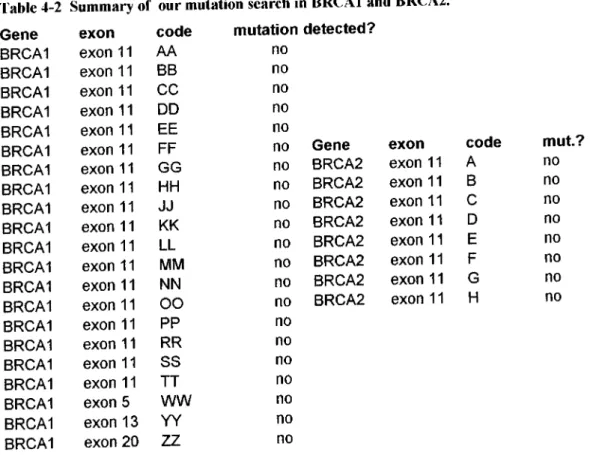
4. R E S U L T S ... 49
4.1. PCR...49
4.2. EFFICIENCY OF THE TECHNIQUES...60
4.3. MUTATION SEARCH OF THE PA TIEN TS... .62
4.3.1. BRCAl...52
4.3.2. BRCA2... .5.3 5. D ISC U SSIO N ... 55
LIST OF TABLES
Table 2-1 Important oncogenes in human cancers 5 Table 2-2 Important tumor suppressor genes in human cancers 6
Table 2-3 Some familial forms o f cancer 9
Table 2-4 BRCA1 and BRCA2 mutations o f familial breast cancer families 16
Table 3-1 The mutation carrying DNA 31
Table 3-2 List o f the Turkish familial breast cancer patients 32
Table 3-3 Breast cancer families 34
Table 3-4 PCR ingredients 40
Table 4-1 Mutation carrying DNA. SO
Table 4-2 Summary of our mutation search in BRCA 1 and BRCA2 54
LIST OF FIGURES
Figure 2-1 Mutations of BRCAI exons.____ Figure 2-2Mutations o f BRCA2 exons..___ Figure 2-3 Mutation distribution o f BRCAl. Figure 2-4 Mutation distribution o f BRCA1. Figure 2-5 BRCA } exon 11 mutations. ____ Figure 2-6 BRCA2 exon 11 mutations..____ Figure 2-7 Heteroduplex analysis.
Figure 2-8 Anatomy o f a heteroduplex gel. Figure 2-9 Anatomy of an SSCP g e l.____ Figure 2-10 SSCP analysis.___________ Figure 2-11 PIT.
Figure 3-1A PCR reaction._____________ Figure 3-2 The amplified regions o f BRCA 1. Figure 3-3 The amplified regions o f BRCA 2. Figure 4-1 Test ge!
Figure 4-2 Verification of the heteroduplex analysis trechnique.^ Figure 4-3 Heteroduplex analysis o f control DNA.___________ Figure 4-4 Heteroduplex analysis o f BRCA 1 fragments. ______ Figure 4-5 Heteroduplex analysis ofpatients for BRCA2._____
J O J O J i J 3 J 4 J 5 JO J 8 _44 JO _49 J i J 1 52 ^53 XII
ABBREVIATIONS
APS be b/o C-terminal del DNA dNTP ETBR HA HNPCC INS min. N-terminal PCR P I T rpm SSC SSCP TBE u v ammonium persulfate breast cancer breast/ovarian cancer carboxy-terminal deletion deoxyribonucleic aciddeoxy nucleotide triophosphates ethidium bromide
heteroduplex analysis
hereditary nonpolyposis colorectal cancer insertion
minutes
nitrogen-terminal
polymerase chain reaction protein truncation test revolutions per minute sodium citrate
single strand conformational polymorphisms Tris-boric acid-EDTA
ultraviolet
1.
INTRODUCTION
Ankara, year 2004, a woman at the age o f 34 enters the building on the wall o f which is written “Genetic Counseling” . She wants to know her risk o f developing breast cancer. Because o f breast cancer her mother has lost one o f her breasts, her aunt died at an early age and her grandfather bereaved them due to male breast cancer. She is aware o f the inherited forms o f cancer and wants to know the risk o f not only herself but also her two little children. She tells her story to the genetic counselor. “Either B R C A l, BRCA2 or BRCA3 ” he thinks, “ it is evidently a breast cancer family. Presence o f a male breast cancer patient increases the probability that this is a BRCA2 family, however we have to be sure o f the situation ”, “Would you please have these tests done?” requests the genetic counselor handling the prescription to the young woman, “ at the upper floor we have a genetic testing unit.” . At the unit, rapid mutation detection techniques are routinely being done. With reliable mutation screening techniques, a patient’s DNA is isolated from the blood and analyzed for the mutations in the genes B R C A l, BRCA2 and BRCA3 which gives information about the presence o f familial breast cancer.
The aim o f my thesis is establishment o f a rapid mutation detection technique which is the first step o f realizing a scene like the one described above. The discovery o f the cancer predisposition genes lead to pretty accurate estimation o f cancer risk in individuals. The incidence o f the inherited form o f breast cancer is 1 in every 200 individuals. This increases the importance o f the mutation detection techniques which are currently the only way to reveal the people at risk.
After giving general information about cancer and its causes, the genes predisposing to breast cancer, BRCAl and BRCA2 will be focused on. Widely utilized mutation detection techniques will be elucidated, proceeding the results o f the survey done on 15 breast cancer patients using heteroduplex analysis.
THEORETICAL BACKGROUND
2.1.
OF CELLS AND HUMAN BEINGS
The relation between each o f the 30 trillion cells o f a human body is similar to that o f a human being and his six billion counterparts. To live properly, the cells intercommunicate with each other by sending signals and receiving them They regulate their functions, size and maintain their location according to these signals[l]. In addition, they have a certain life cycle starting from each cell division. Decision to divide or stay at a certain point o f the cell cycle to differentiate is given by the inherent mechanisms and regulated upon communicating with the cellular and environmental signals[2]. They stop growing at a certain size and stay at a particular location, respecting every other cell. Some may even commit suicide for the sake o f not harming the other cells[3]. The entire knowledge leading to the delicate machinery o f a cell is saved in its nucleus in the form o f DNA, whose functional units are named as genes. There are around 100,000 genes dispersed in the chromosomes lying inside the nucleus o f a human being and each encode for a protein that will perform a certain function. Some o f them carry out structural functions, some specialized functions and some are in charge o f maintaining the
integrity o f the cell[4]. Another group o f genes encode for proteins that mediate the communications; sending and receiving signals, thereby regulating the cell behavior and cell cycle according to these signals. These genes, which play a major part in developing cancer are collectively termed as proto-oncogenes and tumor suppressor genes[l]. These genes will be the topics o f the following sections.
2.2.
WHAT IS CANCER?
Environment, even though it donates a cell its living conditions, may also pose problems by creating agents that can damage DNA. These agents vary from ultraviolet radiation coming from the sun, to the free radicals being formed from the food intaken. When a damage comes to the genes, a mutation might occur, which is a permanent change in the nucleotide sequence o f a gene. A mutation may occur in the genes called the proto-oncogenes and/or the tum or suppressor. Unless they are repaired by the mechanisms inside the nucleus[5], the cell or cells may begin to change their life-style and begin to follow their own agenda for reproduction disregarding the usual controls exerted from the cells in the vicinity. These cells may even migrate from the site they began, invading nearby tissues and forming masses at different sites o f the body. These cells called cancer cells may form tumors which become more and more aggressive over time, and they become lethal when they disrupt the tissues and organs needed for the survival o f the organism as hole[l]. Since any cell in the body might be transformed into a cancer cell, cancer is a collection o f about 100 diseases.
The first group o f genes that are discovered leading to cancer are the proto oncogenes. When mutated, proto-oncogenes can become carcinogenic oncogenes that drive excessive multiplication. The mutations may cause the proto-oncogene to yield too much o f its encoded growth-stimulatory protein or any overly active form o f it[6]. A mutation in one o f the alleles o f a proto-oncogene is enough to initiate carcinogenesis. Table 2-1 lists some o f the important proto-oncogenes and their functions:
2.2.1. ONCOGENES
I'able 2-1 Important oncogenes in human cancers (Adapted form reference 1).
ONCOGENES
denes fo r growth factors or receptors
PDGF Codes for platelet derived growtli factor. Involved in glioma. erb-B Codes for the receptor of the epidermal growth factor. Involved in
glioblastoma and breast cancer.
erb-B2 Codes for a growth factor receptor. Involved in thyroid cancer.
Genes fo r cytoplasmic relays in stimulatory signaling pathways
Ki-ras Involved in lung, ovarian, colon and pancreatic cancers. N-ras Involved in leukemias.
Genes fo r transcription factors that activate growth-promoting genes.
c-mvc Involved in leukemias and breast, stomach and lung cancers. N-mvc Involved in neuroblastoma and glioblastoma.
L-mvc Involved in lung cancer.
Genes fo r other kinds o f molecules
Bcl-2 Codes for a protein that normally blocks cell suicide. Involved in follicular B-cell lymphoma.
Bcl-1 Codes for cyclin D l, a stimulatory component of tlie cell cycle clock. Involved in breast, neck and head cancers.
MDM2 Codes for an antagonist of tlie p53 tumor-suppressor protein. Involved in sarcomas and other cancers.
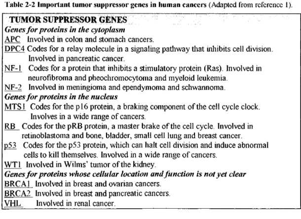
The second group discovered as a cause o f cancer is the tumor-suppressor genes. These genes usually encode for proteins that suppress the excessive growth o f a cell. To become malignant, cells must do more than overstimulate their growth- promoting machinery. They must also devise ways to evade or ignore braking signals issued by their normal neighbors in the tissue. Those braking signals are encoded by the tumor suppressor genes which are absent or inactive in many types o f cancer cells. For cancer to be initiated, both o f the alleles o f a tumor suppressor gene have to be impaired, since only one copy may carry out the adequate supressive function. Hereditary cancers occur due to the inheritance o f a mutated copy o f a tumor suppressor gene. For example, Li-Fraumeni syndrome occurs upon inheriting a gemline mutation o f the p53 gene, similarly, familial melanoma occurs as a result o f carrying a p i 6 gene mutation in the germline. The only documented hereditary neoplasm that is not due to a tum or suppressor gene mutation is multiple endocrine neoplasia[ 1 ], which arises from germline mutations o f the RET oncogene. Very recently, Vogelstein and Kinzler[7] classified the tumor-suppressor genes depending on their function and their relative risk o f leading to cancer when mutated. The two classes are named as gatekeepers and caretakers. They will be discussed in the following section. Table 2-2 is a list o f important tumor-suppressor
2.2.2. TUMOR SUPPRESSOR GENES
Table 2-2 Important tumor suppressor genes in human cancers (Adapted from reference 1).
Genes fo r proteins in the cytoplasm
A PC Involved in colon and stom ach cancers.
D P C 4 C odes for a relay m olecule in a signaling pathw ay that inliibits cell division. Involved in pancreatic cancer.
NF-1 C odes for a protein tliat inhibits a stim ulatory protein (Ras). Involved in neurofibrom a and pheochrom ocytom a and m yeloid leukemia.
N F -2 Involved in m eningiom a and ependym om a and schwannom a.
Genes for proteins in the nucleus
M T S l C odes for tlie p l 6 protein, a braking com ponent o f the cell cy cle clock. Involves in a w id e range o f cancers.
R B C odes for tlie pR B protein, a m aster brake o f the cell cycle. Involved in retinoblastom a and bone, bladder, sm all cell lung and breast cancer.
p53 C odes for the p53 protein, w hich can halt cell division and induce abnormal cells to kill them selves. Involved in a w id e range o f cancers.
W T l Involved in W ilm s’ tum or o f the kidney.
Genes for proteins whose cellular location and function is not yet clear
B R C A l Involved in breast and ovarian cancers. B R C A 2 Involved in breast and pancreatic cancers. V H L Involved in renal cancer.
2 2 3 GATEKEEPERS AND CARETAKERS
Genes that control the cellular proliferation directly are named as gatekeepers, while the genes that maintain the integrity o f the genome are called the caretakers. Retinoblastoma(Rb) and p53 can be regarded as gatekeeper genes. Germline mutations o f gatekeepers lead directly to the development o f hereditary cancers. For example Li-Fraumeni Syndrome is due to p53 gene and familial retinoblastoma is due to Rb gene mutations. For tum or development both the maternal and the paternal copies must be altered[8]. Predisposed individuals have one copy mutated, so they need one additional somatic mutation to initiate neoplasi, which is called “loss o f heterozygosity” . A hereditary mutation in a gatekeeper gene increases the
risk o f an individual around 1000 times more than the general population. However, inactivation o f a caretaker doesn’t lead directly to the development o f cancer. Instead, inactivation leads to genetic instabilities in the genome resulting in increased mutation o f all genes, including the gatekeepers. An accelerated rate o f mutation is induced by the mutation o f a caretaker. Therefore, in dominantly inherited cancer-predisposition syndromes o f the caretaker type, somatic mutations that involve the gatekeepers are required to initiate cancer. So the risk becomes 5- 50 fold greater than in the general population, which is quite low when compared to the inherited gatekeeper mutations. In addition, mutations in caretakers will not be expected to lead to sporadic cancers very often since both alleles o f the caretaker and in addition both alleles o f a gatekeeper should be inactivated for the initiation o f neoplasia. The known caretaker genes are the NER (Nucleotide Excision Repair) genes[5], mismatch repair genes(HNPCC)[9] and ATM gene[5], which all play roles in DNA repair.
2.3.
WHAT CAUSES CANCER?
Now comes the question, what triggers carcinogenesis ? The so-called carcinogens are either chemicals, physical effects or biological agents that are able to damage DNA. The top carcinogen seems to be the tobacco smoke which brings about 30% o f cancer deaths[l]. The only rival to tobacco as a cause o f cancer is diet. Animal fat in general, and red meat are associated with several cancers. Radiation, especially the ultraviolet B rays originating from the sun, cause more than 90% o f
skin cancers. Some o f the chemicals that are used in the workplaces for special purposes are proven to be causes o f cancer. Among these chemicals; asbestos, benzene, formaldehyde and soot are examples. In addition to the chemicals, biological agents can also damage DNA. The most common cancer causing pathogens are the DNA viruses. O f these viruses papilloma viruses types 16 and 18, and the hepatitis B virus are the most important. The former viruses can lead to cancer o f the cervix, while the latter can cause liver cancer. The insult on the DNA caused by these agents targets generally the somatic cells and rarely the germline. In the latter cases when the gatekeeper and the caretaker genes are affected, the result becomes familal forms o f cancers.
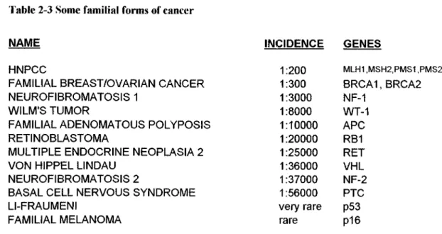
2.4. FAMILIAL FORMS OF CANCER
2.4.1. GENERAL VIEW
As stated earlier, some people are one step closer to cancer than the others. People who have inherited one mutant copy o f a caretaker or a gatekeeper gene have higher probablity o f lifetime risk o f cancer[10]. Some o f the important familal
cancers are hereditary non-polyposis colon cancer (HNPCC), familial
breast/ovarían cancer, neurofibromatosis type 1, familal adenomatous polyposis (APC) and familial retinoblastoma. Examples o f the genes that lead to familial cancers and their incidence is shown in Table 2-3.
NAME HNPCC
FAMILIAL BREAST/OVARIAN CANCER NEUROFIBROMATOSIS 1
WILM’S TUMOR
FAMILIAL ADENOMATOUS POLYPOSIS RETINOBLASTOMA
MULTIPLE ENDOCRINE NEOPLASIA 2 VON HIPPEL LINDAU
NEUROFIBROMATOSIS 2
BASAL CELL NERVOUS SYNDROME LI-FRAUMENI
FAMILIAL MELANOMA
Table 2-3 Some familial forms of cancer
INCIDENCE GENES 1:200 MLH1,MSH2,PMS1,PMS2 1:300 BRCA1, BRCA2 1:3000 NF-1 1:8000 WT-1 1:10000 APC 1:20000 RBI 1:25000 RET 1:36000 VHL 1:37000 NF-2 1:56000 PTC very rare p53 rare pi 6
Among these familial cancers, breast/ovarian cancer is o f special importance for us and therefore more information will be given in the following section.
2.5. FAMILIAL BREAST CANCER
2.5.1. CLINICAL CRITERIA
Breast cancer is the third most common tumor in the world after lung and colon cancers[l]. It is estimated that about one in ten women in the Western world develop cancer o f the breast by the age o f 70, and between 5% to 10% o f these cases are thought to result from a hereditary predisposition to the disease[10]. This number roughly represents 3 million breast cancer cases only this year in Turkey and about 100,000 o f these women develop the disease because o f having inherited either o f the two recently discovered breast cancer genes in a mutated form. The first o f these genes was found by positional cloning in 1994[11]. It was given the
name B R C A l, abbreviated form o f Breast Cancer Associated 1. The second gene, conceivably named as BRCA2, was cloned one year later[12]. These two genes account for nearly 90% o f the hereditary breast cancer cases[10].
2.5.2. THE GENES PREDISPOSING TO BREAST CANCER
2.5.2.1.BRCA1
The first gene that appeared in the familial breast cancer scene was BRCA l. It is linked to chromosome 17q21[13], and spans around 70kb o f genomic DNA. It has 24 exons, where exons 1 and 4 are not transcribed. A protein o f 1863 amino acids is brought about upon the translation.
The DNA sequence o f BRCAl includes, an N-terminal ring domain, a negatively charged region in its C-terminus, and C-terminal sequences are partially homologous to yeast RAD9 and to a cloned p53 binding protein[10]. Mutations in the BRCAl gene represent a predisposing factor in nearly 45% o f hereditary breast cancer cases and more than 80% o f hereditary ovarian cases[14, 15]. There is also an increased risk for BRCAl carriers to develop prostate cancer (3%) in males and colon cancer (4%) in males and females[16]. The average risk for developing breast or ovarian cancer for BRCAl carriers by age o f 70 years is 85% and 63%, respectively[17].
2.5.2.2. BRCA2
Following linkage o f BRCAl to chromosome 17, it soon became clear that many breast cancer families failed to show linkage to this locus. Whereas the majority o f breast and ovarian cancer families were linked to B R C A l, less than 50% o f breast site-specific cancer families showed linkage. So, non-BRCAl-linked families brought the gene BRCA2 to the scene, which is linked to chromosome 13ql2-13[12]. It has 27 exons, making up a 10,348 bp mRNA in length, spanning over 80kb gnomic DNA, encoding a protein o f 3418 aa[10].
Germline mutations in the BRCA2 gene are also associated with approximately 45% o f hereditary breast cancer families, but carriers have only a moderately increased risk for ovarian cancer [10]. There is an increased risk for developing a number o f other cancers, including pancreatic cancer [18, 19], prostate cancer [20], and breast cancer in males [21]. Among women who carry germ-line mutations in either o f BRCAl or BRCA2 genes, the cumulative risk o f breast cancer is estimated to range from 40% to 80%, and for ovarian cancer from 5% to 60%, depending on the population from which the data were derived [17, 22].
2.5.2.3. FINDINGS ON BRCAl AND BRCA2 FUNCTION
Both BRCAl and BRCA2 contain a region that can act as a transcriptional activation domain when it is fused to a DNA binding domain from another gene[23.
24, 25]. Naturally occurring mutations found in both o f the genes can compromise this transcriptional activation.
BRCAl can inhibit the growth o f cells in which it is overexpressed and there is a link between an inhibitor o f cell-cycle dependent protein kinases and the BRCAl protein[26]. Both BRCAl and BRCA2 bind to RadSl which functions in maintaining the integrity o f the genome[27, 28]. BRCA2 knock-out mice show early embryonic lethality and hypersensitivity to irradiation, similar to that observed in the RAD51 knock-out mice[28]. BRCAl knockout mice also show embryonic lethality[26, 29]. BRCAl and RadSl proteins share a striking colocalization along synaptonemal complexes (junctions between meiotic chromosomes necessary for homologous recombination)[28]. It is observed that embryonic and trophoblast cells form both BRCA2 and RadSl knockout mice show hypersensitivity to y- irradiation[27]. Based on these data it can be inferred that disruption o f a possible BRCA/RadS 1 pathway might be expected to lead to genetic instability.
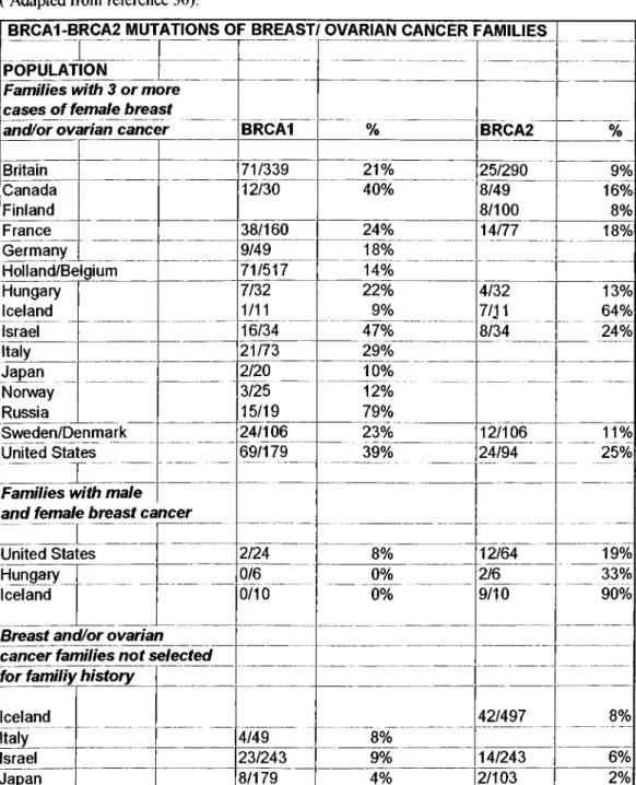
2.5.2.4. POPULATION GENETICS OF BRCAl AND BRCA2
Population geneticists raise some important questions on BRCAl and
BRCA2[30]. Some o f these are: W hat proportion o f high-risk families have mutations in B R C A l? In BRCA2? How do shared mutations reflect population structure ? How frequent are ancient mutations in populations in which they arose? W hat risks are associated with shared mutations ? The answers to these questions begin coming
upon the arrival o f mutation analysis data. The most recent statistics on BRCAl and BRCA2 mutations is shown in Table 2-4.
Here are some facts observed upon recent experimental data:
BRCA l and BRCA2 explain 6%-10% o f breast and ovarian cancer cases unselected for family history[10]. In Israel this number is higher (15%). In 53 site- specific breast cancer families ascertained in United States the frequency o f BRCAl mutations was twice those o f BRCA2 m utations[31, 32, 33].
The preponderance o f a single BRCA2 mutation in Iceland represents one o f the most dramatic examples o f a founder mutation in isolated populations where mutation rate (low), endogamy and isolation (high), and secular trend in penetrance all have contributed to the current population structure[34].
In families with male breast cancer, BRCA2 mutations are more common than BRCAl mutations. In United States, BRCA2 is responsible for 19% o f familial male breast cancers, but for a considerably lower fraction o f male breast cancer in the general population[21].
In all regions other than Iceland, the frequency o f BRCAl mutations is 1.5-2.0 fold higher than the frequency o f BRCA2 mutations. Lower prevalence o f BRCA2
mutations in families and patients could be due to fewer mutations, to lower penetrance, and/or to later age at onset o f BRCA2 breast cancer[30].
Lifetime risks o f breast cancer associated with BRCAl and BRCA2 are approximately equal but that, the age o f onset is later among BRCA2 mutation carriers[31].
In the Ashkenazim, two ancestral mutations BRCAl 185delAG and BRCA2 6174delT each appear in the general population at ~1% frequency and a third mutation; BRCAl 5382insC occurs at a population frequency o f 0.11% [35, 36]. On the other hand, 52% o f the Ashkenazi high-risk breast/ovarian cancer families do not carry any o f the recurrent mutations[35, 37, 38]. W hether these families carry other novel BRCAl or BRCA2 alleles, have mutations in other, as-yet- unidentified susceptibililty loci, or are high risk for nongenetic reasons remains to be determined.
The appearance o f the 185delAG mutation embedded in the same haplotype among Iranian, Iraqi, and Ashkenazi Jewish families suggest that this mutation predates the separation o f these communities from each other at the time o f the destruction o f the Second Temple in year 70 o f the common era. This mutation is more than 2,000 years old[37, 38]. The 5382insC mutation was observed in northern and eastern families, as well as in Russian , Hungarian and Ashkenazi Jewish
families[30]. Its high frequency in eastern European populations are consistent with a Baltic origin during the medieval period (~38 generations ago)[39],
BRCA2 mutations observed in Japan are unique to that country[40]. The distribution o f mutations in other Asian populations is not reported yet.
Table 2-4 B R C A l and BRCA2 mutations of fam ilial breast cancer fam ilies.
( Adapted from reference 30).
BRCA1-BRCA2 MUTATIONS OF BREAST/ OVARIAN CANCER FAMILIES POPULATION
Families with 3 or more cases o f female breast
and/or ovarian cancer BRCAl % BRCA2 %
Britain 71/339 21% 25/290 9% Canada 12/30 40% 8/49 16% Finland 8/100 8% France 38/160 24% 14/77 18% Germany 9/49 18% Holland/Belgium 71/517 14% Hungary 7/32 22% 4/32 13% Iceland 1/11 9% 7/J1 64% Israel 16/34 ‘ 47% 8/34 24% Italy 21/73 29% Japan 2/20 10% Nonway 3/25 12% Russia 15/19 79% Sweden/Denmark 24/106 23% 12/106 11%
United States i^/179__ 39% 24/94____ _25%
r . ...
Families with male and female breast cancer
I
United States 2/24 8% 12/64 19%
Hungary 0/6 0% 2/6 33%
Iceland b/ib 0% 9/10 90%
Breast and/or ovarian cancer families not selected for familiy history
Iceland 42/497 __ 8%
Italy 4/49 8%
Israel 23/243 9% 14/243 6%
Japan 8/179 4% 2/103 2%
The following figures shows the mutation distribution o f BRCAl and BRCA2 genes with respect to their exons The data is obtained from the Breast Cancer Information Core on the Intemet[41] The y-axis gives the number of mutations encountered up-to-date.
2.S.2.5. MUTATION STATUS OF BRCAl AM) BRCA2
# MUTATIONS
EXON NUMBERS
I'icurc 2-1 Mutations of lllW ’Al exons. In all the exons except for t and 4 which arc not transcribed harbor gcrmlinc mutations which lead to hereditars breast cancer. Due to its large size, the most frequently mutated exon is 11.
y //A # M UTATIO NS
f-igurc 2-2 IVluta(ion<i of 11R(,’A2 exons. Also in BRCA2 exon ) 1 harbors majorits (43 9%) of Ihc gcrnilinc mulahons. In addition exons 9. 10,23 and 24 are frequently nnitaleci
Figures 2-.'^ and 2-4 show the distribution o f mutations o f BRCAl and BRCA2 respectively, according to the exons and indicates which exon harbors whal percentage o f the total mutations
exon 1 1 4 7 .0 %
re s t o f exons 1 7 .0 %
Figure 2-3 Mutation distribution of BR i.’A l. Exons 11. 2. .S. 16 and 20 harbor 8.3% o f the BRCAl gcm ilinc mutations These exons altogether make up approximately 60'K, o f the total transcribed region
3.6%
l igurc 2-4 Mutation distribution of I{R(!A1. Similar to BRCAl. four c.xons -namely 11.0. 10 and 23- harbor majority (~f>2%) o f the BRCA2 mutations.
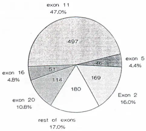
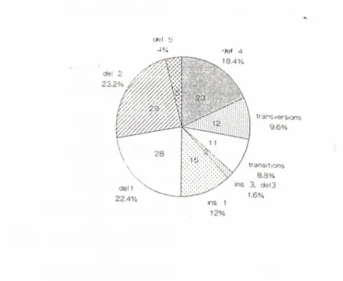
Figures 2-5 and 2-6 show the types o f mutations seen on exon 11 o f BRCA l and BRCA2 respectively. These data are important, since our mutation search is done on these exons.
transversion
Figure 2-5 B R C A l exon 11 mutations. Point mutations make up 41% o f the stnictural aberrations on the exon 11 of BRCAl gene. The rest o f the mutations are small deletions and rarely, small insertions.
d e l 5
4% del 4
Figure 2-6 BR C A 2 exon 11 mutations. The spectnim o f BR CAl exon 11 mutations are different than the BRCAl c.xon 11 mutations. Transitions and transversions make up 18% o f the mutations. The rest o f the mutations are deletions o f a single or a few base pairs.
From the data accumulated at the Breast Cancer Informatioii Core on the Internet [41], it can be observed that the mutations are spread evenly throughout the genome. Approximately 95% o f the mutations are predicted to give rise to truncating protein products o f variable size. Because o f its larger size exon 11 o f both o f the genes seem to accumulate the mutations; 47% and 44%, for BRCAl and BRCA2 respectively. Founder mutations increase the number o f mutations per exon. Exons 2, 5 and 20 harbor such mutations. One o f the most prevalent mutations in Ashkenazi Jews, 185delAG mutation, resides in exon 2 o f B R C A l.
If we look at the types o f mutations, single nucleotide substitutions (transversions and transitions) prevail in B R C A l; 44% o f all the mutations, while this number is slightly lower for BRCA2[41].
2.6.
THEORETICAL PRINCIPLES OF THE MUTATION DETECTION
TECHNIQUES
2.6.1. HETERODUPLEX ANALYSIS
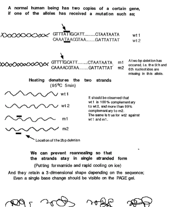
Heteroduplex analysis [HA] is one o f the cheapest, easiest and most reliable techniques in mutation detection [42J. The PCR amplified fragment is denatured by heating and then reannealed by slowly cooling down. So, two complementary strands, derived from alleles that differ in sequence, will include mismatched positions when base-paired. These mismatched positions will lead to migration differences when run on a polyacrylamide gel. Figures 2-7 explains the theory o f heteroduplex analysis and figure 2-8 shows an analysis performed in our laboratory.
HETERODUPLEX ANALYSIS
A normal human being has two copies of a certain gene, if one of the alleles has received a mutat ion such as;
GrTTATTGCATT... CTAATAATA CAAATAACGTAA... .G ATTATTAT
GTTTTG CATT... CT A AT A AT A m 1 CAAAACGTTAA...GATTATTAT m2
Heating denatures the two strands (95®C 5min)
wt1
wt2
A two bp deletion has occured, i.e. th e 5t h and 6th nucleot Ides are missing In this allele.
It should be observed that w tl Is 100% complementary to wt2, and more than 99% complementary to m2.
The same Is tr ue for wt2 against wt1 and m 1.
Locatl on of t he 2b p delet Ion
So cooling down slowly leads to reannealing of the fragments, and what is being formed is:
h o w t l GTTTATIGCATT... CTAATAATA „ CAAATAAOGTAA... GATTATTAT d U ^ ^ ^ ___GTTTTGCATT... CTAATAATA i* y>C >0000<i:>O 0O Q )i^ ^ 2 CAAAACGTAA... GATTATTAT e
X
e
wt 1 GTTTATtgCATT...CTAATAATA
m2 CAAA ACGTAA...GATTATTAT
w t2 CAAaTAacGTAA... ....GATTATTAT ‘ m1 GTTT TGCATT...,....CTAATAATA The site where the mismatch occurs
preventing compiete annelaiing
t e r 0 d u P 1 e X e s
Figure 2-7 H eteroduplex analysis. Here is a schematic view o f how heteroduplex formation is acliieved.
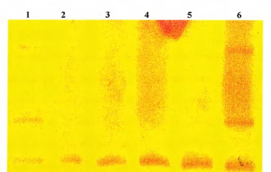
B
} ' ^ [ i<· '■, , '’'■*' > ' '* 1 >■, '· ' \' I'l v'i' S'· I '■' (^ , /" 'h· · i > V" ¡'’■f ' '"'‘/'hiH , W^''· - V i If' ,'' 'V,',!'·' C;' 'V',;^^‘■/i'A //F igure 2-8 A natom y o f a heteroduplex gel. A; Schematic representation o f the basis o f heteroduplex analysis is shown. B; The bands form during heteroduplex analysis appear as three bands i f there is a mutation (lane 1), one band if tlie two alleles are completely the same(lane 2) in the PAGE gel stained with silver nitrate.
2.6.2. SINGLE STRAND CONFORMATIONAL POLYMORPHISM
ANALYSIS (SSCP)
Single strand conformational polymorphism (SSCP) analysis, along with HA , is one o f the m ost straightforward, easiest and the cheapest methods o f mutation detection [43]. The PC R amplified target sequence is separated as single stranded molecules by application o f a denaturing chemical and loaded on a non-denaturing gel. This technique relies on the alteration o f the migration pattern o f the strands that have any sequence difference. This mobility shift is believed to be caused by mutation-induced changes o f tertiary structure o f the single-stranded DNA. The technique is schematically explained in figures 2-9 and 2-10.
SSCP
F igure 2-9 A natom y o f an SSC P gel. Due to the differential migration o f single strands, a mutation bearing D N A can be differentiated from that o f a non-mutation carrying homozygous D N A
SINGLE STRAND CONFORMATIONAL POLYMORPHISMS rSSCPI
A normal human being has two copies of a certain gene, if one of the alleles has received a mutation such as;
X y y O > O O C O iX x r GnTATlGCATT... CTAATAATA wt 1
CAAATAACGTAA...GATTATTAT wt 2
...Y Y T i- Iiili- " I
C A A A A C G T A A ...G A T T A T T A T m2 6th nucleotides are missing in this allele.
Heating denatures the two strands
(95^C 5min) wt 1
It should be observed that wt 1 Is 100 % complement ary W t 2 to wt2, and more than 99%
complementary to m2.
The same Is tr ue for wt2 against
m1 wt1 andm1.
m2
Locatl on of t he 2bp delet ion
We can prevent reannealing so that the strands stay in single stranded form
(Putting formamide and rapid cooling on ice)
And they retain a 3-dimensional shape depending on the sequence; Even a single base change should be visible on the PAGE gel.
They may migrate differently.
Figure 2-10 SSC P analysis. Here is a schematic view o f single strand conformational polymorpliisms are induced.
2.6.3. PROTEIN TRUNCATION TEST
The protein truncation test (PTT) detects mutations that interrupt the reading frames o f genes. The technique is based on coupled transcription and translation o f PCR products in vitro. The templates for PTT are generated by PCR either from reverse transcribed RNA or directly from genomic DNA. During PCR, a 36-base pair extension, encoding the bacteriophage T7 prom oter sequences as well as an eukaryotic translation initiation signal, is added to that end o f the PCR-product which correspods to the amino terminus. Simultaneous transcription and translation o f amplification products is performed in a rabbit reticulocyte lysate system using radiolabeled aminoacids. Translation products are subsequently resolved by SDS- PAGE and detected by autoradiography[43, 44]. Figure 2-11 depicts the basis o f a PTT reaction.
■■pnoTQfi •muwiATittN· Test'
A0GGfiC>.;i:iltAA,v.;GbtrAA6^^ C ' „ C UI
T7 promoter tagged primwif) , ,1
J
p rlm erfr). : f=CR amplification using modlfiad primars
i Z J
tn vitro rran;^;;rtptK>n1
noRMA Normal Sooation 0^ ofthBstopcoefon1
In Vitro translation IJWV Stcpoodonformacl <tLi0 lo t hn mutat ioni:
Figure 2-11 PTT. Tills is a schematic view o f how mutations leading to truncated products are revealed.
2.6.4. CHEMICAL MISMATCH CLEAVAGE
The chemical mismatch cleavage is a technique for detecting and localizing mismatches in heteroduplex DNA molecules[42, 45]. It relies on the chemical reactivity o f mismatched C and T bases to hydroxylamine and osmium tetroxide, respectively. Once reacted, the DNA strands are cleaved at the reacted mismatched base by piperidine and the molecules are separated by size to identify the location o f the mismatched positions.
2.6.5. DIRECT SEQUENCING
In today’s technology, it is the only technique that is capable o f finding out 100% o f the mutations. However, its high cost and arduous labor-requirement brings it among the rarely utilized techniques for mutation detection. On the other hand, it is an obligation to sequence, when a mutation is encountered by the indirect mutation detection techniques. It is the only means to identify the mutation and locate it precisely.
The aim o f this thesis is establishment o f a rapid mutation detection technique for the investigation o f the two breast cancer susceptibility genes BRCAl and BRCA2. Heteroduplex analysis is chosen to be the most suitable technique, which is accurate, rapid and cheap. Upon establishment o f this technique, 15 Turkish familial breast cancer patients are screened for BRCA l and BRCA2 mutations. Since the genes are too large (BRCAl requires 40 pairs, BRCA2 requires 44 pairs o f primers for entire screening.), we first chose the exons which harbor most o f the docum ented mutations to establish the technique. To screen B R C A l, primers for exon 11, as well as primers flanking exons 5, 13 and 20 have been synthesized. One o f the most frequently seen mutations 5382insC resides in exon 20. To screen BRC A2, primers spanning the first half o f exon 11 have been synthesized (16 pairs span the whole exon, while we have synthesized the first 8).
The results o f this study will help us to develop a non-readioactive indirect mutation screening technique, which can be used to characterize the mutations in BRCAl and BRCA2 genes in the Turkish population. Subsequently, genetic counselling for the high risk individuals can be offered and insight into the population genetics o f hereditary breast cancer can be obtained.
2.7. AIM
3.
MATERIALS AND METHODS
3.1.
MATERIALS
3.1.1. THE BIOLOGICAL MATERIAL
3.1.
LLMUTATION CARRYING DNA
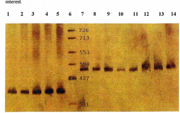
Four mutation carrying DNA samples which were characterized previously[46], belonging to French familial breast cancer patients on B RCA l that have been donated us kindly by Dr. Gilbert Lenoir, was used for the establishment o f the heteroduplex analysis technique in our laboratory. These DNA samples serve as positive controls to check the efficiency o f the mutation detection techniques.
Table 3-1 The mutation carrying DNA.
THE MUTATION CARRYING DNAs
No.
2638
Mutation
nt279J
Gene Prim er pairs
893del4 B BRCA1 GA 23-24I
2944 908G-T nt2841 BRCA1 GA 23-24
3300 1160del11 nt3599 BRCAl GA 31-32
2651 Ï234del5 nt3819 BRCA1 GA 33-34
3.I.I.2. DNA OF TURKISH BREAST CANCER PATIENTS
Blood samples have been obtained from 22 breast cancer patients in collaboration with Hacettepe University, Istanbul University and Ankara Oncology Hospital. The list o f the Turkish breast cancer patients that have been studied is shown in
Table 3-2.:
Table 3-2 List of the Turkish familial breast cancer patients. PATIENT LIST FAMILY CODE DNA CODE AGE OF DIAGN. DEPT. OBTAINED DR.
BRC-1 96-1 45 Capa Medical Fac. Dr.Mahmut Muslumanoglu BRC-2 96-2 25 Capa Medical Fac. Dr.Mahmut Muslumanoglu BRC-3 96-3 50 Capa Medical Fac. Dr.Mahmut Muslumanoglu BRC-4 96-4 50 Capa Medical Fac. Dr.Mahmut Muslumanoglu BRC-5 96-5 33 Capa Medical Fac. Dr.Mahmut Muslumanoglu BRC-6 96-6 51 Capa Medical Fac. Dr.Mahmut Muslumanoglu BRC-6 96-7 33 Capa Medical Fac. Dr.Mahmut Muslumanoglu BRC-7 96-8 37 Capa Medical Fac. Dr.Mahmat Muslumanoglu BRC-7 96-9' Capa Medical Fac. Dr.Mahmut Muslumanoglu BRC-8 96-10 4S "У
0/ Capa Medical Fac. Dr.Mahmut Muslumanoglu BRC-9 97-114 34 Hacettepe Med. Fac. Dr. Mesut Tez
BRC-10 97-137 32 Oncology Hospital Dr.Oguz Tarcan BRC-11 97-270 29 Oncology Hospital Dr.Oguz Tarcan BRC-12 97-274 40 Hacettepe Med. Fac. Dr. Mesut Tez BRC-13 97-343 35 Oncology Hospital Dr.Oguz Tarcan
' This patient is not a breast cancer patient, but a member seeking genetic counseling.
The families are grouped according to the criteria for the familial breast cancer cases[46]. Group 1 is the ‘breast cancer-only’ cases (be) where there is at least two breast cancer cases with one o f them either bilateral breast cancer or early onset (before age 40). The families are B R C l, BRC2, BRC3, BRC4, BRC6, BRC7, BRCIO. Group 2 is the familial breast/ovarían cancer cases (b/o), where at least three breast or ovarian cancer cases are present among the first or second degree relatives.The family is BRC8. Group 3 consists o f cases that are classified as sporadic. The families are BRC5, BRC9, B R C ll and B R C l2. Finally, group 4, which has only one family (BRC13), denotes the cases whose pedigrees could not be obtained. Family BRC13. Table 3-3 is the list o f the families we have obtained and grouped as mentioned. The pedigrees are given in the appendix.
3.1.2. PEDIGREES
Family Patient(s) t! patients
in family
Indication Group
BRC-1 96-1 6 3 be Patients 1
BRC-2 96-2 2 2 be cases, in first degree
relatives
1
BRC-3 96-3 2 2 be cases, one is early onset. 1
BRC-4 96-4 2 2 be cases, one is early onset. 1
BRC-5 96-5 2 2 be cases, one early onset,
but in second degree relatives
BRC-6 96-6/96-7 2 be cases, one is early onset. 1
BRC-7 96-8/96-9- 2 2 be cases, one early onset,
one is a male breast cancer case. 1
BRC-8 96-10 4 One be case with 2 b/o and
one be case in second degree relatives
2
BRC-9 97-114 1 One be case o f early onset
BRC-IO 97-137 4 3 be cases in first degree,
one be case in second degree relatives
1
BRC-Il 97-270 2 One early onset be case with
one be case in second degree relatives.
BRC-12 97-274 1 One early onset be case.
BRC-13 97-343 7 No data available 4
J ahic 3-3 Hrcflsl cancer lamilics.
3.1.3. CHEMICALS
Absolute ethanol (JD El-D elta) Acetic acid (A6283-Sigma) Acrylamide (A9099-Sigma) Agarose (A9539-Sigma) APS (420627-Carlo Erba) Bisacrylamide(M2022-Sigma) Boric Acid (B6768-Sigma)
Bromophenol blue (B5525-Sigma) Chloroform (438603-Carlo Erba) dNTP mix (A4916-Sigma)
EDTA (SE5134-Sigma)
Ethidium bromide (SE7637-Sigma) Ficoll (F4345-Sigma)
Formaldehyde (F8775-Sigma) Formamide (F7508-Sigma)
Isoamyl alcohol (413836Carlo Erba) NaOH (S0899-Sigma)
Parafilm (Can-Sigma) Phenol (P1037-Sigma)
Silvernitrate-A gN 03-(S8157-Sigma)
Sodium acetate (366207-Carlo Erba) Sodium Citrate (368007-Carlo Erba)
Size markers :
lOObp DNA ladder (G2101-Prome) OX174D N A/Hinf I (G1751-Prome) ADNA/EcoRI+HindIII(G 1731 Pro) TEM ED (ST8133-Sigma)
Tris Hcl (ST3253-Sigma) Xylene cyanol (X4126-Sigma)
5xPCR buffer ( Supplied with Taq polym erase)
10 X TBE buffer ( lit - 108gr Tris, 55gr Boric Acid, 0.3gr EDTA-pH 8.3 ) Non-denaturing polyacrylamide gel
( 50ml - 7.5 ml from 40% 39; 1 Acrylamide;bisacrylamide solution, 5ml lOXTBE, 350ul 10%stock o f APS, 3 7 .15ml ddH20 and just before pouring 40ul TEMED.)
Silver staining solutions:
* All the solutions should be prepared with deionized H2O.
Fixing solution
(600ml-Aqueous solution o f 10%(v/v) ethanol, 0.5%(v/v) acetic acid.)Staining solution
(300ml -0.1% aqueous solution o f silver nitrate (A gN O s)).Developer solution
(500ml -Aqueous solution o f J.5 % (w/v) NaOH, 0. l% (v/v) formaldehyde. Must be prepared immediately before use.)Protecting solution
(300ml - 7.5% (v/v) aqueous solution o f acetic acid.)3.1.4. SOLUTIONS
3.1.5. ENZYMES
Taq polymerases:AmpliTaq Gold (NN808-0241 Perkin Elmer) Taq Polymerase ( 1 146173-Boehringer Manheim)
Proteinase K (161519- Boehringer Manheim)
3.1.6.
PRIMERS
3.I.6.I. PRI.VIERS FOR BRCAl
CODE
r n m
From 5· to 3·
BRCAl exon 11 AA GA 1 GGAATTAAATGAAAGAGTATGAG BRCAl exon 11 AA GA 2 CTAAGCCAGGCTGTTTGCTT BRCAl exon 11 BB GA 3 ACAGCATGAGAACAGCAGTTT BRCAl exon 11 BB GA 4 CTCTAGGATTCTCTGAGCATTG
BRCAl exon 11 CC GAS GGTAGATCTCGAATGCTGATCCC
BRCAl exon 11 CC GA6 GCCTCATGAGGATCACTGG
BRCAl exon 11 DD GA 7 GCCAAAGTAGCTGATGTATTGG
BRCAl exon 11 DD GAS CGCTTTAATTTATTTGTGAGGG
BRCAl exon 11 EE GA 9 CCCAACTTAAGCCATGTAACTG BRCAl exon 11 EE GA 10 ATTCATCACTTGACCATTCTGA
BRCAl exon 11 FF GA11 ATTTGGCAGTTCAAAAGACTCC
BRCAl exon 11 FF GA 12 TTTAGGTGCTTTTGAATTGTGG
BRCAl exon 11 GG GA 13 AAAGCTGAACCTATAAGCAGCA
BRCAl exon 11 GG GA 14 GTTTCTGCTGTGCCTGACTG
BRCAl exon 11 HH GA 15 GCCCACCTAATTGTACTGAATT
BRCAl exon 11 HH GA 16 CTTGGAAGGCTAGGATTGACA
BRCAl exon 11 JJ GA17 TGAAGTTAACAAATGCACCTGG
BRCAl exon 11 JJ GA18 ACGAGATACTTTCCTGAGTGCC
BRCAl exon 11 KK GA 19 GGGTTTTGCAAACTGAAAGA BRCAl exon 11 KK GA 20 CTTGTTTCCCGACTGTGGTT BRCAl exon 11 LL GA 21 GGGACTAATTCATGGTTGTTCC
BRCAl exon 11 LL GA 22 TTTCTTTAAGGACCCAGAGTGG BRCAl exon 11 MM GA 23 TTTGCTCCGTTTTCAAATCC BRCAl exon 11 MM GA 24 TCGTTGCCTCTGAACTGAGA BRCAl exon 11 NN GA 25 GATAAGCCAGTTGATAATGCCA BRCAl exon 11 NN GA 26 CGGCTAATTGTGCTCACTGT
BRCAl exon 11 OO GA27 TTGAGGAACATTCAATGTCACC
BRCAl exon 11 0 0 GA28 ACCTCAGGTTGCAAAACCC BRCAl exon 11 PP GA 29 CCAGTGATGAAAACATTCAAGC BRCAl exon 11 PP GA 30 TTCACCATCATCTAACAGGTCA BRCAl exon 11 RR GA 31 AACTTAGAACAGCCTATGGGTT
BRCAl exon 11 RR GA 32 AACAAGTGTTGGAAGCAGGG
BRCAl exon 11 SS GA 33 AGGGGCCAAGAAATTAGAGTC BRCAl exon 11 SS GA 34 CTTCCAATTCACTGCACTGTG
BRCAl exon 11 TT GA35 AAAGGCATCTCAGGAACATCA
BRCAl exon 11 TT GA 36 CAAAAACCTGGTTCCAAGAC
BRCAl exon 2 QQ GA 47 AAACCTTCCAAATCTTCAAA
BRCAl exon 2 QQ GA 48 GTCTTTTCTTCCCTAGTATGT
BRCAl exon 5 WW GA 50 GCTCTTAAGGGCAGTTGTGA
BRCA1 exon 5 WW GA 52 ATAGCGTTCCTATAAAACCATT BRCAl exon 13 YY GA 53 AATGGAAAGCTTCTCAAAGTA
BRCAl exon 13 YY GA 54 TGTTGGAGCTAGGTCCTTAC
BRCAl exon 20 ZZ GA 55 ATATGACGTGTCTGCTCCAC