Dergi web sayfası:
www.agri.ankara.edu.tr/dergi www.agri.ankara.edu.tr/journalJournal homepage:
TARIM BİLİMLERİ DERGİSİ
—
JOURNAL OF AGRICUL
TURAL SCIENCES
20 (2014) 269-279
Morphological and Anatomical Investigations on in Vitro Micrografts
of OHxF 333 / Pyrus elaeagrifolia Interstock / Rootstock Combination
in Pears
Hatice DUMANOĞLUa, Aysun ÇELİKa, H. Nurhan BÜYÜKKARTALb, Saeed DOUSTİa
a Ankara University, Faculty of Agriculture, Department of Horticulture, 06110, Ankara, TURKEY b Ankara University, Faculty of Sciences, Department of Biology, 06100, Ankara, TURKEY
ARTICAL INFO
Research Article
Corresponding Author: Hatice DUMANOĞLU, E-mail: dmanoglu@agri.ankara.edu.tr, Tel: +90 (312) 596 12 99 Received: 03 December 2013, Received in Revised Form: 02 January 2014, Accepted: 13 January 2014
ABSTRACT
In this study, possibility of creating a specific interstock / rootstock combination to obtain a clonal, semi dwarf, composite pear rootstock tolerant to various stresses by micrografting was investigated. In vitro shoots of ‘Old Home x Farmingdale 333’ (Pyrus communis L.) which is a clonal semi dwarf pear rootstock resistant to fireblight and pear decline was used as interstock, and in vitro Pyrus elaeagrifolia Pallas seedlings known as tolerant to Fe-chlorosis, salinity and drought stresses was used as rootstock. Cleft grafting was applied in micrografts. Grafted seedlings were cultured on Murashige and Skoog basal medium with ½ strength of macronutrients for 8 weeks under white fluorescent light for 16 h day-1.
Cultures, except the control, received complete darkness either 1 or 2 weeks at the beginning of incubation. Graft take success in the control treatment was significantly higher (97.9%) than darkness treatments of 1 or 2 weeks (90.5% and 82.5%, respectively). Ultrastructural observations with transmission electron microscope revealed that dividing cambial initials reached to 2-3 layers, and new xylem and phloem elements distinctly differentiated in transverse sections of the graft union 8 weeks after micrografting in the control and darkness treatments. The results indicated a successful graft union formation.
Keywords: Micrografting; Graft union; Interstock; Darkness treatment
Armutlarda OHxF 333 / Pyrus elaeagrifolia Ara Anaç / Anaç
Kombinasyonunun in vitro Mikroaşılarında Morfolojik ve Anatomik
İncelemeler
ESER BİLGİSİ
Araştırma Makalesi
Sorumlu Yazar: Hatice DUMANOĞLU, E-posta: dmanoglu@agri.ankara.edu.tr, Tel: +90 (312) 596 12 99 Geliş Tarihi: 03 Aralık 2013, Düzeltmelerin Gelişi: 02 Ocak 2014, Kabul: 13 Ocak 2014
1. Introduction
Native Mediterranean Pyrus species P. elaeagrifolia Pallas which has habitat ranging from Turkey to Southeastern Europe and Ukraine (Bell 1990), is a potential pear rootstock in areas where lime, salt and drought are limiting factors for growing many Pyrus species (Lombard & Westwood 1987; Matsumoto et al 2006). Scions of cultivars (Pyrus communis L.) grafted on P. elaeagrifolia grow vigorously similar to pear seedlings causing a long juvenile period. This problem could be overcome by using a dwarf or semi dwarf clonal Pyrus interstocks such as ‘OHxF 333’ on P. elaeagrifolia. It is known that interstocks may reduce vegetative growth and enhance reproductive growth of the tree. Such double-worked plants have two unions, one between the rootstock and interstock and one between the interstock and the scion. The interstock piece is budded or grafted or bench grafted onto the rootstock prior to combine with the scion cultivar (Hartmann et al 2011). However, production of grafted-rootstock plants using conventional techniques takes time for at least more than one year considering the growth time of rootstock lines (Elivar & Dumanoğlu 1999).
In vitro grafting is an original and skillful technique which deserves greater consideration for overcoming the limitations of other vegetative
propagation methods, and also for studying more in depth the relationships between genetically different tissues and cells (Monteuuis 2012). In initial studies, the objective of micrografting was elimination of some virus diseases in tree fruit species (Jonard 1986). Murashige et al (1972) and Navarro et al (1975) were first to consider the use of micrografting technique in Citrus species in order to eliminate virus diseases. Micrografting has been successfully applied in Citrus (Edriss & Burger 1984; Sharma et al 2008), Prunus (Barba et al 1995; Jarausch et al 1999; Conejero et al 2013), Malus (Huang & Millican 1980; Bisognin et al 2008) and Pyrus (Faggioli et al 1997) species to get plants free from virus or virus like organisms. Besides, micrografting has been used for micropropagation, rejuvenation of mature tissues, determination of graft incompatibility and root to shoot communication, transport or cryopreservation in Citrus (Obeidy & Smith 1991; Parthasarathy et al 1997; Volk et al 2012), Prunus and Amygdalus (Ozzambak & Schmidt 1991; Ghorbel et al 1999; Amiri 2006; Yıldırım et al 2010; Isikalan et al 2011), Malus (Lane et al 2003; Nunes et al 2005; Dobranszki & Silva 2010), Pyrus (Musacchi et al 2004; Espen et al 2005; Hassanen 2013), Pistacia (Abousalim & Mantell 1992; Onay et al 2003; 2004;
ÖZET
Bu çalışmada, mikroaşılama yoluyla çeşitli streslere tolerant, klonal, yarı bodur, birleşik bir armut anacı elde etmek için spesifik bir ara anaç / anaç kombinasyonu oluşturmanın olabilirliği araştırılmıştır. Ateş yanıklığı ve armut göçüren hastalıklarına dayanıklı, yarı-bodur armut klon anacı ‘Old Home x Farmingdale 333’ (Pyrus communis L.)’ün in vitro sürgünleri ara anaç ve demir klorozu, tuzluluk ve kuraklık streslerine tolerant olarak bilinen Pyrus elaeagrifolia Pallas’ın in vitro çöğürleri anaç olarak kullanılmıştır. Mikroaşılarda yarma aşı tekniği uygulanmıştır. Aşılanmış çöğürler, makro element düzeyi ½ olan Murashige ve Skoog temel besin ortamı üzerinde, 8 hafta süreyle, 16 h gün-1
süreyle beyaz floresan ışık altında inkübe edilmiştir. Kontrol dışındaki kültürler, inkübasyonun başlangıcında, 1 ya da 2 hafta süreyle tamamen karanlık koşullara alınmıştır. Aşı başarısı kontrol uygulamasında (% 97.9), 1 ya da 2 hafta karanlık uygulamalarından (sırasıyla, % 90.5 ve % 82.5) önemli düzeyde daha yüksek olmuştur. Mikroaşılamadan 8 hafta sonra aşı kaynaşma yerinden alınan enine kesitlerde, transmisyon elektron mikroskopu ile yapılan ultrastrüktürel gözlemler, kontrolde ve karanlık uygulamalarında kambiyal inisyallerin bölünerek 2-3 sıraya ulaştığını ve yeni ksilem ve floem elemanlarının belirgin biçimde farklılaştığını ortaya koymuştur. Bulgular başarılı bir aşı kaynaşmasının meydana geldiğini göstermiştir.
Anahtar Kelimeler: Mikroaşılama; Aşı kaynaşması; Ara anaç; Karanlık uygulaması
Can et al 2006; Ozden-Tokatlı 2010), Castanea and Corylus (Nas & Read 2003), Actinidia (Ke et al 1993), Olea (Toroncoso et al 1999), Morus (Ma et al 1996), Anacardium (Thimmappaiah et al 2002), Opuntia (Estrada-Luna et al 2002) and Carica (Nava et al 2011) species. Creation of rootstocks by interstock/rootstock combination is possible by in vitro micrografting which is very fast, using in vitro rooted young rootstock plantlets and in vitro grown interstock scions. Thus, micrografting technique offers new possibilities for mass production of grafted rootstocks which might be later grafted in
vitro or in vivo with the scion cultivar.Up to date,
we are unaware of micrografting studies which have been done for micropropagation of rootstocks grafted with interstock.
The objective of this study was to develop a quick in vitro micrografting technique to create a specific pear rootstock combination using semi dwarf clonal interstock (‘Old Home x Farmingdale 333’, (OHxF 333)) and the vigorous seedling rootstock tolerant to different stress conditions (P. elaeagrifolia), and to study graft union formation on in vitro micrografted and complate darkness treated plantlets, since complete darkness may prevent internal auxin degradation, by morphological and anatomical (Transmission Electron Microscope) investigations.
2. Material and Methods
2.1. Rootstock and scion (interstock) sources for micrografting experiments
In vitro germinated seedlings of P. elaeagrifolia Pallas were used as the rootstock. The seeds were processed before culturing in aseptic conditions as follows; (1) the seeds were scarified in sulfuric acid (Merck, 95–98% H2SO4) for 2.5 min followed by rinsing in running water for 5 min, (2) the seeds were dipped in 85% ethanol for 3 min, and then sterilized in a solution of sodium hypochlorite (2.5% active chlorine) for 30 min followed by 3 rinses for 5 min with sterile distilled water. Then, the seeds were placed on germination medium in petri dishes by positioning vertically so that the
radical of the seed was in the medium. Five seeds were placed in each petri dish (70 x 10 mm). The seeds were stratified in vitro at 4 °C in dark for 8 weeks in order to break dormancy. Chilled seeds were germinated in a growth chamber at 25 ± 1 °C under 16/8 h day-1 (light/dark) photoperiod (cool white fluorescent light, 35 µmol m-2 s-1) for 10-14 days. MS basal medium (Murashige & Skoog 1962) with half-strength of macronutrients, containing 3% (w v-1) sucrose and 1% (w v-1) Difco Bacto agar was used in seed stratification and germination studies. The pH was adjusted to 5.8 before adding agar and autoclaving at 121 °C for 20 min.
In vitro shoots obtained from shoot-tip culture of ‘OHxF 333’ were used as scion (interstock). Shoot-tip cultures were established from actively growing shoots of plants. Shoot tips approximately 20 mm long were collected, washed in running tap water for 30 min, and sterilized by immersion in a solution of sodium hypochlorite (1% active chlorine) for 20 min followed by 3 rinses with sterile distilled water for 5 min. Approximately 10 mm long explants were cultured in glass tubes (120 x 25 mm) containing 10 mL of medium under aseptic conditions. The shoots (>10 mm long) from initial cultures were subcultured four times by four week intervals in glass flasks (250 mL) containing 50 mL of medium.
For initial culture and subcultures, MS basal
medium containing 3% (w v-1) sucrose and 0.7% (w
v-1) agar was used. MS medium was supplemented
with 4.4 µM BA and 0.9 µM GA3 which were were
added to media before autoclaving at 121 °C for 20 min. The pH was adjusted to 5.8 before adding agar. All cultures were incubated at the same conditions as seed germination.
2.2. In vitro micrografting experiments
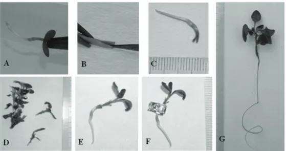
Cleft grafting was applied in micrografts under aseptic conditions. About 10-14 day-old seedlings with horizontal cotyledonary leaves, without true leaves, with roots about 15-30 mm in length were used as rootsock in grafts (Figure 1A).
The roots were cut to ~20 mm length. The tip of hypocotyl was decapitated just below the cotyledons. The stem was cut longitudinally 2-3 mm deep (Figure 1B & C). The scion, prepared from in vitro shoots of ‘OHxF 333’ was about 10 mm long and contained the apical meristem with 2-3 leaves. The basal end of the scion was cut to form a wedge about 2-3 mm long (Figure 1D). The scion was inserted into the cleft and the graft junction was wrapped with a sterile 5 x 20 mm piece of aluminum foil (12 µ thick) which was folded 4 times (Figure 1E & F). The basal end of grafted seedlings was placed in the medium with the graft junction above the level of
medium in glass tubes (120 x 25 mm) containing 10 mL of half-strength MS basal medium without
growth regulators containing 3% (w v-1) sucrose and
0.7% (w v-1) agar. The pH was adjusted to 5.8 before
adding agar and autoclaving.
For the micrografting experiments, grafted seedlings received either complete darkness for 1 or
2 weeks in a growth chamber or 16/8 h day-1 (light
/ night) photoperiod (control) at 25 ± 1 °C. At the end of the dark treatments, cultures were incubated under the photoperiod as the control received. The micrografting experiments were a completely randomized design with four replications with
Figure 1- Micrografting procedure of interstock ‘OHxF 333’ on Pyrus elaeagrifolia seedling under aseptic conditions. An in vitro germinated P. elaeagrifolia seedling (A); forming a cleft at about 2-3 mm in length on the top of hypocotyle of seedling which was cut just below the cotyledon leaves (B); an in vitro grown seedling ready for micrografting (C); a scion prepared from an in vitro grown interstock ‘OHxF 333’ micro shoots ready to be inserted into the cleft (D); an inserted scion into the cleft (E); a graft union wrapped around with sterile aluminum foil (F); a micrografted plantlet taken out from in vitro conditions after 8 weeks of grafting (G)
Şekil 1- Pyrus elaeagrifolia çöğürleri üzerine ‘OHxF 333’ ara anacının, aseptik koşullarda mikroaşılama işlemi. İn vitro koşullarda çimlendirilmiş P. elaeagrifolia çöğürleri (A); kotiledon yaprakların hemen altından tepesi vurulmuş fidelerin hipokotilinin üst kısmında yaklaşık 2-3 mm uzunluğunda bir yarık oluşturulması (B); mikroaşılama için hazır bir in vitro çöğür (C); in vitro koşullarda geliştirilmiş ara anaç ‘OHxF 333’den kalem olarak hazırlanmış, yarık içerisine yerleştirilmeye hazır mikro sürgünler (D); yarık içerisine kalemin yerleştirilmesi (E); aşı yerinin steril alüminyum folyo ile sarılması (F); aşılamadan 8 hafta sonra in vitro koşullardan çıkartılmış bir mikroaşılı bitkicik (G)
25 micrografts (tubes) in each replication. The experiment was repeated at least twice to ensure the accuracy of results.
2.3. Morphological investigations of micrografted plantlets
Grafted plantlets were removed from in vitro conditions 8 weeks after micrografting and the aluminum band was removed (Figure 1G). Percent graft take, graft junction rating on a scale of 1 to 3 (1 = weak, 2 = medium, 3 = good) by visual inspection, shoot length and number of leaves on the shoots were recorded. Morphological data were subjected to analyses of variance (ANOVA) at P<0.05, 0.01 or 0.001 using MINITAB statistical software (MINITAB Inc., UK). Means were separated by Duncan’s multiple range test at P = 0.05. The percent data were transformed into angle values prior to analyses.
2.4. Preparation of epon blocks and anatomic investigations
Tissue samples in size of 2 mm lenght were taken from the graft union 8 weeks after the micrografts were made. The samples were fixed in 3% glutaraldehyde buffered with 0.1 M phosphate (pH 7.2) for 3 h and then fixed in 1% osmium tetraoxide (in 0.1 M Na-P buffer) for 3 h at room temperature. Specimens were dehydrated in an ethanol series,
transferred to 100% propylene oxide and embedded in Epon 812 (Luft 1961). Ten blocks were prepared for each treatment. Ultrathin sections were stained with uranyl acetate (Stempak & Ward 1964) and lead citrate (Sato 1967). Ultrastructural observations were made under Jeol CXII Transmission Electron Microscope at 80 Kv.
3. Results and Discussion
In this study, very high graft take success was obtained in the micrografts of ‘OHxF 333’ / P. elaeagrifolia seedlings. Based on morphological observations, the highest graft take and graft junction rating was 97.9% and 2.70%, respectively (Table 1).
Hassanen (2013) found that the highest percentage of successful grafts was 83% in the Le-Cont pear / Pyrus betulaefolia combination. Nunes et al (2005) reported that in vitro micrografting techniques offer the potential for effective propagation of high quality genetic material in a short time under controlled and aseptic conditions. Grafting techniques and initial treatments applied prior to in vitro micrograftings are subjects of concern. Cleft grafting was used in this study because it has been reported as a successful micrografting technique in different fruit species such as cherry, pistachio, olive (Ozzambak & Schmidt 1991; Abousalim & Mantell 1992; Toroncoso et al 1999; Onay et al 2004).
Table 1- The effect of complete darkness treatments applied at the beginning of incubation, on graft take success and shoot development in ‘OHxF 333’ / Pyrus elaeagrifolia seedling interstock/ rootstock combinations in vitro (after 8 weeks of micro-cleft graftings)
Çizelge 1- İn vitro koşullarda inkübasyon başlangıcında uygulanan tamamen karanlık uygulamalarının ‘OHxF 333’ / Pyrus elaeagrifolia çöğürü ara anaç / anaç kombinasyonunda aşı başarısı ve sürgün gelişimi üzerine etkileri (mikro-yarma aşılamadan 8 hafta sonra)
Treatments Graft take
(%)
Graft junction rating (1-3)
Shoot length (mm)
Number of leaves
Control 97.9a 2.70 ± 0.06a 13.9 ± 0.8a 6.8 ± 0.5a
Complete darkness for a week 90.5b 2.33 ± 0.13b 10.6 ± 0.4b 3.9 ± 0.0b
Complete darkness for two weeks 82.5b 2.09 ± 0.13b 10.9 ± 0.6b 4.5 ± 0.3b
P 0.001 0.011 0.011 0.000
Graft junction rating 1, weak; 2, medium; 3, good. Means in each column followed by the same letter were not significantly different at P = 0.05, according to Duncan’s new multiple range test
We applied complete dark treatment for 1 or 2 weeks as an initial procedure with an expectation of increasing graft take success by promoting callus formation due to its role in internal auxin biosynthesis (Yin et al 2012). Endogenous growth regulators have been proposed to play a key role in grafting, with special reference to auxin which is known to be synthesized in shoot apices and degraded by light. Our results showed that although graft take success, graft junction rating, leaf number and shoot development were satisfactory in all treatments, the control treatment performed significantly better than the dark treatments (Table 1). The control treatment had the highest graft take among the treatments (P = 0.001) which was followed by 1 week (90.5%) and 2 weeks (82.5%) darkness treatments although the difference between last two was not statistically significant. The grade for level of graft junction was also high on grafts and the differences were significant. The control treatment had the highest grade (2.70 ± 0.06) followed by 1 week (2.33 ± 0.13) and 2 weeks of complete darkness (2.09 ± 0.13) treatments. (Table 1). The results are in agreement with the report of Monteuuis (1996) that the application of dark treatment for 2 weeks in in vitro micrografts did not improve the results in Acacia mangium. On the other hand, Monteuuis (1994) observed beneficial effects of 2 weeks to 3 weeks in darkness immediately after grafting in vitro seedlings of Picea abies which resulted in 52.4% success as compared to 32.6% for the control. Similarly, 5 days of dark treatment shortened the time required for grafting and increased the graft union rate (90.6%) compared with the control (80.7%) in the plug seedling of rose rootstock (Han et al 1998).
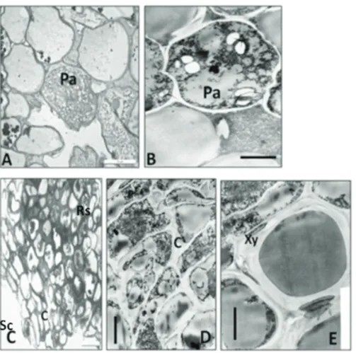
Ultrastructural observations with transmission electron microscope in ‘OHxF 333’ / P. elaeagrifolia micrografts revealed the successful graft union formation, and new xylem and phloem elements differentiated in all of the treatments. In the control, observation of callus tissue between graft elements at the graft union revealed that the cells at the graft union were generally oval in shape and scattered (Figure 2A).
Transverse sections of a region near graft elements showed that parenchymatous cells forming callus tissue were large and had larger intercellular spaces. In some of these cells the cytoplasm was less dense and contained vacuoles and a few organelles (Figure 2B). Some enlarged cells and occasionally divided cambial initials were observed at the graft union. Cambial cells were formed by couple of cell layers (Figure 2C), and cytoplasm of some of the cells were dense (Figure 2D). Xylem elements and phloem cells were formed by couple of cell layers and they were starting to differentiate. Tracheal elements which were in the process of differentiation had reached their normal size and the lumen of the cell was dense with electrons (Figure 2E). Phloem cells were found to be round, isodiametric or hexagonal in shape, thin walled and dispersed.
In one week complete darkness treatment, parenchymatous cells with rough walls forming the callus tissue between the graft elements were plenty and the cytoplasm stained dark (Figure 3A). At this stage, the cambial relation was present between graft elements. Cambium cells were formed with cells of different sizes and occasionally formed 7 or 8 layers. These cells had thin walls, were rectangular in shape, and formed radial rows. The cytoplasm stained very dark in some cells. There were few newly formed xylem and phloem elements (Figure 3B).
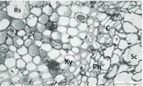
In two weeks of complete darkness treatment, callus tissue between the graft elements at the graft union was dense in samples treated. The cells at the union tissues were generally flattened and dispersed. However, parenchymatous cells producing the callus tissue were formed by large cells with large inter-cellular spaces. The cytoplasm in some cells was dense. Cambium was formed of couple of cell rows and the cytoplasm in some cells was dense. At this stage, the established cambial relation between graft elements was very clear. New xylem and phloem elements were few in number and they started to differentiate (Figure 4).
Espen et al (2005) indicated that the incompatible heterograft (Pyrus communis L. cv. ‘Bosc’ (B) /
Cydonia oblonga Mill. East Malling clone C (EMC)) showed a marked delay in internode cohesion compared with the autografts of B / B, and Pyrus communis L. cv. ‘Butirra Hardy’ (BH /
BH) and the compatible heterograft (BH / EMC). Thus we believe that, ‘OHxF 333’ / P. elaeagrifolia combination would form a compatible heterograft.
Figure 2- Transversal sections of the graft union in the control treatment after 8 weeks of micrografts. Rs, rootstock (P. elaeagrifolia seedling); Sc, scion (OHxF 333 interstock); Xy, xylem; C, cambium; Pa, parenchymatous cell. Semithin section showing parenchymatous cells at the graft union Bar= 20 µm (A); electron micrograph showing detail of parenchymatous cell cytoplasm Bar= 3 µm (B); semithin section showing cambial cells and newly differentiated xylem and phloem cells Bar= 50 µm (C); the cytoplasm of the active cambial cells Bar= 3 µm (D); detail of the newly differentiated xylem cells Bar= 3 µm (E)
Şekil 2- Mikroaşılamadan 8 hafta sonra kontrol uygulamasında aşı kaynaşma yerinden alınan enine kesitler. Rs, anaç (P. elaeagrifolia çöğürü); Sc, ara anaç (OHxF 333); Xy, ksilem; C, kambiyum; Pa, parankimatik hücre. Aşı kaynaşma yerinde parankimatik hücreleri gösteren yarı ince kesit Bar= 20 µm (A); parankimatik hücre sitoplazmasının ince yapısını gösteren elektron mikrografisi Bar= 3 µm (B); kambiyum hücreleri ve yeni farklılaşmış ksilem ve floem hücrelerini gösteren yarıince kesit Bar= 50 µm (C); aktif kambiyum hücrelerinin sitoplazması Bar= 3 µm (D); yeni farklılaşmış ksilem hücrelerinin ince yapısı Bar= 3 µm (E)
Figure 3- Transversal semithin sections of the graft union in 1 week complete darkness treatment after 8 weeks of micrografts. Rs, rootstock (P. elaeagrifolia seedling); Sc, scion (OHxF 333 interstock); Xy, xylem; Ph, phloem; C, cambium; Pa, parenchymatous cell. Parenchymatous cells at the graft union Bar= 20 µm (A), cambial cells, newly differentiated xylem and phloem cells Bar= 20 µm (B)
Şekil 3- Mikroaşılamadan 8 hafta sonra 1 hafta tamamen karanlık uygulamasında aşı kaynaşma yerinden alınan enine kesitler. Rs, anaç (P. elaeagrifolia çöğürü); Sc, ara anaç (OHxF 333); Xy, ksilem; Ph, floem; C, kambiyum; Pa, parankimatik hücre. Aşı kaynaşma yerinde parankimatik hücreler Bar= 20 µm (A), kambiyum hücreleri, yeni farklılaşmış ksilem ve floem hücreleri Bar= 20µm (B)
Figure 4- Transversal sections of the graft union in 2 weeks complete darkness treatment after 8 weeks of micrografts. Cambial ring and newly differentiated xylem and phloem cells Bar= 20 µm Rs, rootstock (P. elaeagrifolia seedling); Sc, scion (OHxF 333 interstock); Xy, xylem; Ph, phloem; C, cambium
Şekil 4- Mikroaşılamadan 8 hafta sonra 2 hafta tamamen karanlık uygulamasında aşı kaynaşma yerinden alınan enine kesitler. Kambiyum halkası ve yeni farklılaşmış ksilem ve floem hücreleri Bar= 20µm Rs, anaç (P. elaeagrifolia çöğürü); Sc, ara anaç (OHxF 333); Xy, ksilem; Ph, floem; C, kambiyum
4. Conclusions
In vitro micrografting technique was successfully applied to create ‘OHxF 333’ / P. elaeagrifolia seedling combination. Thus, with mass micropropagation it is possible to shorten the required time for production of a pear rootstock consisted of rootstock and interstock which is clonal, semi dwarf, tolerant to Fe-chlorosis, salinity and drought stresses, and resistant to fireblight and pear decline less than a year. This is the first report of in vitro micrografting on Pyrus elaeagrifolia Pallas wild pear, and micropropagation of rootstock plants which are micrografted with interstock in vitro.
Abbreviations and Symbols
C cambium
MS murashige & skoog
Pa parenchymatous
Ph phloem
Rs rootstock (P. elaeagrifolia seedling) Sc scion (OHxF 333 interstock)
Xy xylem
References
Abousalim A & Mantell S H (1992). Micrografting of pistachio (Pistacia vera L. cv. Mateur). Plant Cell,
Tissue and Organ Culture 29: 231-234
Amiri M A (2006). In vitro techniques to study the shoot-tip grafting of Prunus avium L. (cherry) var. Seeyahe Mashad. Journal of Food Agriculture and
Environment 4: 151-154
Barba M, Cupidi A, Loreti S, Faggioli F & Martino L (1995). In vitro micrografting: a technique to eliminate peach latent mosaic viroid from peach. Acta
Horticulturae 386: 531-535
Bell R L (1990). Pears (Pyrus). In: Moore J N & Ballington J R (Eds), Genetic Resources of Temperate Fruit and
Nut Crops, International Society for Horticultural
Science, Wageningen, pp. 655-696
Bisognin C, Ciccotti A, Salvadori A, Moser M, Grando M S & Jarausch W (2008). In vitro screening for resistance to apple proliferation in Malus species.
Plant Pathology 57: 1163-1171
Can C, Ozaslan M, Toremen H, Sarpkaya K & Iskender E (2006). In vitro micrografting of pistachio, Pistacia
vera L. var. Siirt, on wild pistachio rootstocks. Journal of Molecular Cell Biology 5: 25-31
Conejero A, Romero C, Cunill M, Mestre M A, Martínez-Calvo J, Badenes M L & Llácer G (2013). In vitro shoot-tip grafting for safe Prunus budwood exchange.
Scientia Horticulturae 150: 365-370
Dobranszki J & Silva J A T (2010). Micropropagation of apple - A review. Biotechnology Advances 28: 462-488
Edriss M H & Burger D W (1984). Micro-grafting shoot-tip culture of Citrus on three trifoliolate rootstocks.
Scientia Horticulturae 23: 255-259
Elivar D E & Dumanoğlu H (1999). Ayaş (Ankara) koşullarında elma, armut ve ayvada bir yaşlı fidan üretiminde ilkbahar sürgün ve sonbahar durgun göz aşılarının karşılaştırılması. Tarım Bilimleri Dergisi
5(2): 58-64
Espen L, Cocucci M & Sacchi G A (2005). Differentiation and functional connection of vascular elements in compatible and incompatible pear / quince internode micrografts. Tree Physiology 25: 1419-1425
Estrada-Luna A A, Lopez-Peralta C & Cardenas-Soriano E (2002). In vitro micrografting and the histology of graft union formation of selected species of pricly pear cactus (Opuntia spp.). Scientia Horticulturae 92: 317-327
Faggioli F, Martino L & Barba M (1997). In vitro micrografting of Pyrus communis shoot tips. Advances
in Horticultural Science 11: 25-29
Ghorbel A, Chatibi A, Thaminy, S & Kchouk M L (1999). Micrografting of almond Prunus dulcis (Miller) D.A. Webb.) cv. Achak. Acta Horticulturae 470: 429-433 Han Y Y, Woo J H, Sim Y G, Choi K B, Choi B S &
Yeo S K (1998). Studies on the plug seedling of rose rootstock (Rosa multiflora ‘Hort No. 1’). RDA
Journal of Horticulture Science 40: 82-88
Hartmann H T, Kester D E, Davies F T & Geneve R L (2011). Plant Propagation: Principle and Practices. Prentice-Hall, London
Hassanen S A (2013). In vitro grafting of pear (Pyrus spp.). World Applied Sciences Journal 21: 705-709 Huang S C & Millican D F (1980). In vitro micrografting
of apple shoot tips. HortScience 15: 741-743
Isıkalan C, Namli S, Akbas F & Ak B E (2011). Micrografting of almond (Amygdalus communis) cultivar ‘Nonpareil’. Australian Journal of Crop
Jarausch W, Lansac M, Bliot C & Dosbat F (1999). Phytoplasma transmission by in vitro graft inoculation as a basis for a preliminary screening method for resistance in fruit trees. Plant Pathology 48: 283-287 Jonard R (1986). Micrografting and its applications to tree
improvement. In: Bajaj Y P S (Ed). Biotechnology in
Agriculture and Forestry, Springer, Berlin, pp. 31-48
Ke S, Cai Q & Skirvin R M (1993). Micrografting speeds growth and fruiting of protoplast-derived clones of kiwifruit (Actinidia deliciosa). Journal of
Horticultural Science 68: 837-840
Lane W D, Bhagwat B, Armstrong J D & Wahlgren S (2003). Apple micrografting protocol to establish transgenic clones on field ready rootstock.
HortTechnology 13: 641-646
Lombard P B & Westwood M N (1987). Pear rootstocks. In: Rom R C & Carlson R F (Eds). Rootstocks for
Fruit Crops, A Wiley-Interscience Publication, John
Wiley and Sons, Inc., New York, pp. 145-183 Luft J H (1961). Improvements in epoxyresin embedding
methods. The Journal of Biophysical and Biochemical
Cytology 9: 409-414
Ma F, Guo C, Liu Y, Li M, Ma T, Mei L & Hsiao A (1996).
In vitro shoot-apex grafting of mulberry (Morus alba
L.). HortScience 31: 460-462
Matsumoto K, Tamura F, Chun J P & Tanabe K (2006). Native Mediterranean Pyrus rootstock,
P. amygdaliformis and P. elaeagrifolia, present
higher tolerance to salinity stress compared with Asian Natives. Journal of the Japanese Society for
Horticultural Science 75: 450-457
Monteuuis O (1994). Effect of technique and darkness on the success of meristem micrografting of Picea abies.
Silvae Genetica 43: 91-95
Monteuuis O (1996). In vitro shoot apex micrografting of mature Acacia mangium. Agroforestry Systems 34: 231-217
Monteuuis O (2012). In vitro grafting of woody species.
Propagation of Ornamental Plants 12: 11-24
Murashige T & Skoog F (1962). A revised medium for rapid growth and bioassays with tobacco tissue cultures. Physiologia Plantarum 15: 473-497 Murashige T, Bitters W P, Rangan T S, Nauer E M,
Roistacher C N & Holliday B P (1972). A technique of shoot apex grafting and its utilization towards recovering virus free Citrus clones. HortScience 7: 118-119
Musacchi S, Ganino T, Grassi S & Fabbri A (2004). Grafting in vitro shoots on acclimating plants: morphological and anatomical aspects of compatible and incompatible pear / quince combinations. Acta
Horticulturae 658: 559-563
Nas M N & Read P E (2003). Simultaneous micrografting, rooting and acclimatization of micropropagated American chestnut, grapevine and hybrid hazelnut.
European Journal of Horticultural Science 68:
234-237
Nava R, Garcia A V, Marin C & Villegas Z (2011). Clonal propagation of elite Carica papaya L. plants using in
vitro and in vivo micrografting. Interciencia 36:
517-523
Navarro L, Roistacher C N & Murashige T (1975). Improvment of shoot tip grafting in vitro for virus free Citrus. Journal of the American Society for
Horticultural Science 100: 471-479
Nunes J C O, Abreu M F, Dantas A C M, Pereira A J & Pedrotti E L (2005). Morphological characterization in apple micrografts. Revista Brasileira de Fruticultura
27: 80-83
Obeidy A A & Smith M A L (1991). A versatile new tactic for fruit tree micrografting. HortTechnology 1: 91-95 Onay A, Pirinc V, Adıyaman F, Isıkalan C, Tilkat E &
Basaran D (2003). In vivo and in vitro micrografting of pistachio, Pistacia vera L. cv. “Siirt”. Turkish
Journal of Biology 27: 95-100
Onay A, Pirinc V, Yıldırım H & Basaran D (2004). In vitro micrografting of mature pistachio (Pistacia vera var. Siirt). Plant Cell, Tissue and Organ Culture 77: 215-219
Ozden-Tokatlı Y, Akdemir H, Tilkat E & Onay A (2010). Current status and conservation of Pistacia germplasm. Biotechnology Advances 28: 130-141 Ozzambak E & Schmidt H (1991). In vitro and in
vivo micrografting of cherry (Prunus avium L.). Gartenbauwissenschaft 56: 221-223
Parthasarathy V A, Nagaraju V & Rahman S A S (1997).
In vitro grafting of Citrus reticulata Blanco. Folia Horticulturae 9: 87-90
Sato J E (1967). A modifred method for load staining of thin sections. Journal of Electron Microscopy 16: 133 Sharma S, Singh B, Rani G, Zaidi A A, Halan V K,
Nagpal A K & Virk G S (2008). In vitro production of Indian citrus ringspot virus (ICRSV) free Kinnow plants employing thermotherapy coupled with shoot
tip grafting. Plant Cell, Tissue and Organ Culture 92: 85-92
Stempak J C & Ward W P (1964). An improved staining method for electron microscopy. The Journal of Cell
Biology 22: 697-701
Thimmappaiah G T, Puthra S & Anil S R (2002). In vitro grafting of cashew (Anacardium occidentale L.).
Scientia Horticulturae 92: 177-182
Toroncoso A, Linan J, Cantos M, Acebedo M M & Rapoport H F (1999). Feasibility and anatomical development of an in vitro olive cleft-graft. The
Journal of Horticultural Science & Biotechnology 74:
584-587
Volk G M, Bonnart R, Krueger R & Lee R (2012). Cryopreservation of citrus shoot tips using micrografting for recovery. CryoLetters 33: 418-426 Yıldırım H, Onay A & Suzerer V (2010). Micrografting
of almond (Prunus dulcis Mill.) cultivars “Ferragnes” and “Ferraduel”. Scientia Horticulturae 125: 361-367
Yin H, Yan B, Sun J, Jia P, Zhang Z, Yan X, Chai J, Ren Z, Zheng G & Liu H 2012. Graft-union development: a delicate process that involves cell– cell communication between scion and stock for local auxin accumulation. Journal of Experimental


