Yazışma Adresi/Address for Correspondence: Dr. Murat Özkale, Baskent University Faculty of Medicine, Department of Pediatrics, Adana Teaching and Medical Research Center, Adana, Turkey, E-mail:drmuratozkale@gmail.com, drkaramurat@gmail.com
Geliş tarihi/Received: 01.05.2016 Kabul tarihi/Accepted: 06.06.2016
ARAŞTIRMA/RESEARCH
Association between platelet indices and febrile seizures in children
Çocuklarda trombosit belirteçleri ve febril konvülziyon arasındaki ilişki
Murat Özkale
1, İlknur Erol
2, Yasemin Özkale
1, Çağla Sarıtürk
31Baskent University Faculty of Medicine
,
AdanaTeaching and Medical Research Center, Department of Pediatrics, 2Department of Pediatrics, Neurology Division, 3Department of Biostatistics, Adana, TurkeyCukurova Medical Journal 2016;41(4):695-701.
Abstract Öz
Purpose: Febrile seizures (FS) are the most common type
of seizures in children.The aim of this study was to evaluate the relationship between platelet indices and FS in children.
Materials and Methods: This prospective study included
40 children who presented with FS and 30 controls who presented with febrile illnesses without seizures. Complete blood counts, including platelet count (PC), mean platelet volume (MPV), and platelet distribution width (PDW) from both groups within 1 hour of FS and 1 month later were obtained.
Results: We found that the MPV and PDW within 1 hour
of seizure in children with complex FS group was higher than simple FS group while there was no significant difference in MPV and PDW between patients in the simple and complex FS groups at 1 month. The mean PC was not significantly different between simple and complex FS groups; but, we found that the mean PC in the complex FS group was slightly lower than simple FS
group.There was a moderate significant positive correlation between MPV and PDW in children with FS
while there was a moderate significant negative correlation between PC and MPV, PDW for FS .
Conclusion: Our findings suggest that the increasing
platelet turnover in complex FS group causes a slightly decrease in the PC, an significantly increase of MPV and PDW values indicating that these parameters may play an important role in predicting the severity of FS in children at diagnosis.
Amaç: Febril konvülziyon çocuklarda en sık görülen
nöbet tipidir. Bu çalışmanın amacı çocuklarda trombosit belirteçleri ile febril konvülziyon arasındaki ilişkiyi incelemektir.
Gereç ve Yöntem: Bu prospektif çalışmada febril
konvülziyon ile başvuran 40 hasta ve kontrol grubu olarak nöbetsiz ateşli hastalık ile başvuran 30 hasta alındı. Febril konvülziyon ve ateşli hastalıkla başvuran her iki gruptada 1 saat ve 1 ay sonra trombosit sayısı, ortalama trombosit hacmi(MPV) ve trombosit dağılım genişliğini( PDW) içeren tam kan sayımına bakıldı.
Bulgular: Bu çalışmada 1. saatin sonunda bakılan MPV ve
PDW değerlerinin kompleks febril konvülziyon geçiren grupta basit febril konvülziyon geçiren gruba göre daha yüksek olduğunu bulurken 1. ayın sonunda iki grup arasında fark olmadığını bulduk. Ortalama trombosit sayısında basit ve kompleks febril konvülziyon geçiren gruplar arasında belirgin fark yoktu ama kompleks febril konvülziyon geçiren grupta daha düşük olarak bulduk. MPV ve PDW ile febril konvülziyon geçiren grupta orta derecede pozitif korelasyon varken, trombosit sayısı MPV ve PDW ile febril konvülziyon arasında orta derecede negatif korelasyon vardı.
Sonuç: Bulgularımız kompleks febril konvülziyon hasta
grubunda artmış trombosit döngüsünün trombosit sayısında düşüklüğe yol açtığını, belirgin olarak artan MPV ve PDW değerlerinin çocuklarda febril konvülziyonun şiddetini tahmin etmede önemli bir rol oynayabileceğini göstermektedir.
Key words: Febrile seizure, mean platelet volume,
INTRODUCTION
Febrile seizures which occur in association with fevers in children, are the most common type of seizures in young children1. The International
League Against Epilepsy (ILAE) has defined febrile seizures as seizures occurring in childhood after 1 month of age, (usually between 3 months and 6 years of age), associated with a febrile illness, not caused by an infection of the central nervous system, without previous neonatal or unprovoked seizures, and not meeting the criteria for other acute symptomatic seizures2. Although most FS are
benign, one-third are complex with a prolonged duration, a partial onset or showing focal features during the seizure or recurrent episodes, and associated with a risk of subsequent epilepsy3,4.
The mean platelet volume (MPV) reflects platelet size and the rate of platelet production in the bone marrow, and it may be used as an indicator of platelet activation and severity of inflammation5-9.
Another marker of platelet activation is the platelet distribution width (PDW), which reflects the variability in platelet size10. There is a growing body
of clinical evidence suggesting that platelets play an important role in the inflammatory response11.
Some studies have reported that a high MPV is associated with a variety of established risk factors, cardio-and cerebro-vascular disorders, and low-grade inflammatory conditions prone to arterial and venous thromboses12. In contrast, other studies
reported that high-grade inflammatory diseases, such as active rheumatoid arthritis or attacks of familial Mediterranean fever (FMF), present with a low MPV13,14. Some clinical and experimental
studies suggest that inflammation, which is intrinsic to the fever response, is involved in the generation of FS; however, the association between MPV and FS has previously been reported in only one study involving children8,15.
The aim of this study was to evaluate the association between platelet indices and FS. This is the fırst study to investigate PC, MPV and PDW together in patients with FS.
MATERIALS AND METHODS
The study was approved by the Baskent University Institutional Review Board and Ethics Committee (Project no: KA13/28O). Written informed consent was obtained from the parents of all participants. All
consecutive febrile children <6 years of age, who were admitted to Pediatric Emergency Department at Baskent University Hospital, Adana Teaching and Medical Research Center, Adana, Turkey, between February 2014 and July 2014 with or without seizures, were eligible to participate. All children were diagnosed using reliable clinical, laboratory, and neuroimaging techniques according to ILAE criteria. Febrile seizures were classified as simple FS, complex FS, and febrile status epilepticus. The exclusion criteria were as follows: infection of the CNS or any confirmed neurological illness; regular blood transfusion; chronic disease; malignancy; hypercholesterolemia; and aspirin, non-steroidal anti-inflammatory drugs, oral anticoagulant, steroid therapy, and children with or without seizure who were admitted to the emergency department 1 hour after the onset of fever. A total of 3002 patients were admitted with febrile illness between these dates. One hundred thirty-one of these patients had febrile seizure. Eighty-four of these 131 patients with FS was admitted within 1 hour of seizure. However, 40 of 84 patients with FS enrolled in study due to the exclusion criteria and refusion of familes. Approximately 28 of families with FS refused to participate. Many of the families of the 2871 febrile children without seizure did not know the time of the onset of the fever. Beside this, 1255 of them was not admitted within 1 hour of fever onset. Therefore, only thirty of 2871 febrile children without seizure enrolled in study due to the exclusion criteria and refusion of families. All of the patients with and without seizure who enrolled in study came to the follow up.
All 40 patients in the FS group underwent a comprehensive clinical evaluation.The febrile illness, duration of seizures, medical histories of the patient and parents, and time of admission to the hospital after the FS or onset of fever were recorded. Hemoglobin (Hb) concentration, white blood cell (WBC) and C-reactive protein (CRP) levels within 1 hour of seizure onset of fever. Platelet count, MPV and PDW were measured from both groups within 1 hour of seizure onset of fever, and 1 month after a seizure or febrile illness.
For the 30 control children, we recorded body temperature at presentation and the underlying illness at the time of presentation. Hemoglobin concentration, WBC, PC, MPV, and PDW were measured within 1 hour of the fever onset and 1 month after the febrile illness.
Testing
Blood samples were obtained from the antecubital vein from all participants with minimal stasis and collected into tubes containing ethylene-diamine-tetra-aceticacid for determination of a CBC within the first hour and 1 month after a seizure attack or febrile period. All blood specimens were analyzed on an Cell-Dyn 3700 autoanalyser.
The PC, MPV and PDW are simple and useful parameters detected in routine CBC parameters. The normal ranges for the PC, MPV and PDW parameters are 150-400 x 103/uL, 7–11 fl and 0– 25%, respectively.
The mean MPV, PDW and PC were compared between the FS and control groups within 1 hour and at 1 month after FS or the febrile illness. In the FS group, we compared the mean PC, MPV and PDW within 1 hour of the FS and at 1 month after the FS attack. Similarly, the mean PC, MPV and PDW within 1 hour and at 1 month after the febrile illness were compared in the control group. The mean MPV, PDW and PC between patients in the simple FS and complex FS groups were also compared.
Statistical analysis
Data were analyzed using the Statistical Package for the Social Sciences (SPSS, version 15.0; IBM, USA). Descriptive statistics were calculated for demographic data. The results were reported as the mean ± standard deviation or as the median and range. For each continuous variable, the normality of the distribution was confirmed using the Kolmogorov-Smirnov test. Results for categorical variables were compared using a chi-square test. The means for Hb, WBC, PC, CRP level, MPV, and PDW were compared using the Mann-Whitney U-test, Student’s t-U-test, or ANOVA. Spearman or Pearson correlation analysis was used to assess the relationships between FS, and PC, MPV, PDW. P-values < 0.05 were considered statistically significant.
RESULTS
Based on these criteria, 40 febrile children with seizures (FS group) and 30 febrile children without seizures who had a similar age distribution (control group) were enrolled. The FS group was comprised of 23 boys and 17 girls with a mean age of 2.1±1.1
years (range, 0.5 – 5.0 years). The control group was comprised of 18 boys and 12 girls with a mean age of 2.4 ± 1.4 years (range, 0.5–6 years). There were no significant differences between the two groups with respect to age or gender (p > 0.05). Of the 40 FS patients, 32 (80%) were diagnosed with simple FS, 8 (20%) with complex FS. Mean temperatures of children in the FS group and control groups were 38.6 ± 0.7°C and 38.4 ± 0.8 °C, respectively ( p > 0.05) (Table 1).
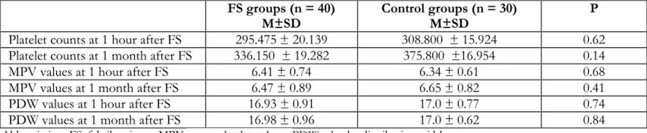
Table 2 summarizes the median Hb, WBC count, and CRP level in the FS and control groups. There were no significant differences between the group means for Hb concentration, WBC count, and CRP level ( p > 0.05). Table 3 summarizes the mean values and standard deviations of PC, MPVs and PDWs in then FS and control groups. There were no significant differences in the group means for PC, MPV and PDW values between the FS and control groups within 1 hour and at 1 month after FS or febrile illness. In the FS group there were also no significant differences between the group means for MPV and PDW values within 1 hour of FS and 1 month after a FS attack (p > 0.05). In the control group, we detected no significant differences between the group means for MPVs and PDWs within 1 hour of the fever onset and 1 month after the febrile illness (p > 0.05).
Table 4 summarizes the mean ± standard deviation for PC, MPVs and PDWs with the FS patients according to type of FS. MPVs and PDWs within 1 hour of seizure in children with complex FS significantly higher than simple FS (p = 0.04 , p = 0.001 respectively). But there was no significant difference in PC between simple and complex FS within 1 hour and at 1 month after a FS. There was also no significant difference between the simple and complex FS groups for MPVs and PDWs at 1 month after seizure.
Table 5 summarizes the correlation between mean PC, MPV and PDW value in children with FS. The mean PC count in the simple FS and complex FS groups were not significantly different; however, the mean PC in the complex FS group was lower than in the simple FS group. There was a moderate significant positive correlation (p < 0.05) between MPV and PDW in children with FS while there was a moderate significant negative correlation between mean PC and MPV (r = - 0.40), PDW (r = -0.29) for FS (p < 0.05).
Table 1. Demographic and clinical characteristics of the study groups
FS group (n=40) Control group (n=30)
Age in years (range) 2.2±1.1 (0.5–5.5) 2.4±1.4 (0.5–6.0)
Gender (F/M) 23/17 18/12
Body temperature at time of admission (°C) 38.6±0.7 38.4±0.8
FS: febrile seizure; (age and body temperature are reported as the mean ± standard deviation)
Table 2.Comparison of Hb concentration, CRP level and WBC count, between the groups
FS group(n = 40) Control group(n = 30)
Median Min Max Median Min Max p
WBC (×10) 9650 4500 25400 10850 4500 21800 0.87
Hb (mg/dl) 11.3 7.2 13.6 11.5 8.6 12.9 0.73
CRP((mg/dl) 10.3 1.0 146 12.2 3 197 0.19
Abbreviation: FS, febrile seizure; MPV, mean platelet volume; PDW, platelet distribution width; WBC, white blood cell; PLT, platelet; Hb, hemoglobin; CRP, C-reactive protein
Table 3.. Results (mean ± standard deviation = M ± SD) for Platelet, MPV and PDW at 1 hour and 1 month of admission in the FS patients and controls
FS groups (n = 40)
M±SD Control groups (n = 30) M±SD P
Platelet counts at 1 hour after FS 295.475 ± 20.139 308.800 ± 15.924 0.62 Platelet counts at 1 month after FS 336.150 ± 19.282 375.800 ±16.954 0.14
MPV values at 1 hour after FS 6.41 ± 0.74 6.34 ± 0.61 0.68
MPV values at 1 month after FS 6.47 ± 0.89 6.65 ± 0.82 0.41
PDW values at 1 hour after FS 16.93 ± 0.91 17.0 ± 0.77 0.74
PDW values at 1 month after FS 16.98 ± 0.96 17.0 ± 0.62 0.84
Abbreviation: FS, febrile seizure; MPV, mean platelet volume;PDW, platelet distribution width
Table 4. Results for Platelet, MPV and PDW at 1 hour and 1 month of admission in the FS patients Simple FS group (n = 32)
X ± SS Complex FS group (X ± SS n = 8) P
Platelet counts at 1 hour after FS 308.218 ± 23.979 244.500 ± 25463 0.21 Platelet counts at 1 month after FS 339.281 ± 22.563 323.625 ± 36219 0.75
MPV values at 1 hour after FS 6.30 ± 0.67 6.97 ± 0.88 0.04
MPV values at 1 month after FS 6.30 ± 0.79 6.87 ± 1.14 0.08
PDW values at 1 hour after FS 16.7 ± 0.63 17.8 ± 1.3 0.001
PDW values at 1 month after FS 16.89 ± 0.99 17.35 ± 0.73 0.23
Abbreviation: FS, febrile seizure; MPV, mean platelet volume; PDW, platelet distribution width
Table 5. Correlation between mean platelet values, MPV and PDW in children with FS
r p
Platelet count-MPV -0.40 0.0001
Platelet count-PDW -0.29 0.01
MPV-PDW 0.43 0.0001
Abbreviation: FS, febrile seizure; MPV, mean platelet volume; PDW, platelet distribution width
DISCUSSION
The occurrence of FS in only a sub-group of children with febrile illnesses indicates the importance of host factors and genetic susceptibility.
There is a growing body of evidence to suggest that inflammatory cells and pro-inflammatory cytokines play significant roles in the generation of FS16,17. It is
known that brain inflammation promotes increased neuronal excitability, decreases the seizure
threshold, and is likely to be involved in the molecular, structural, and synaptic changes which characterize epileptogenesis18. Previous studies have
also revealed increased levels of inflammatory cytokines in the plasma and cerebrospinal fluid of patients with FS19.
Platelet indices have been shown to be an important component of the inflammatory response and the size of platelet is associated with the intensity of inflammation12 . The role of MPV as an indicator of
platelet function has been investigated in association with several inflammatory disorders, such as cystic fibrosis, ulcerative colitis, rheumatoid arthritis, FMF, and infections6,10,12,20,21. In acute
exacerbations of chronic obstructive pulmonary disease in which the intensity of inflammation increases, the MPV has been shown to be significantly lower, while the serum leukocyte count and percentage of neutrophils were higher than during the stable period22. In support of this
observation, some studies have suggested that cytokines, such as IL-3 and IL-6, which play a role in the inflammatory process, may influence megakaryocyte ploidy and lead to the production of larger and more reactive platelets, thus affecting the MPV22. The relationship between MPV and
infection, especially sepsis, has also been evaluated in a number of studies23-25. Robbins et al reported
that at least two patterns of platelet size changes are possible in response to infection (an early rise in MPV in severe infections, such as septicemia (may be associated with thrombocytopenia); and a later decrease in MPV with a sustained, chronic, or persistent bacterial infection (may be associated with thrombocytosis)26. As reported in the literature, a
number of studies have shown that low or high MPV values are associated with acute or chronic inflammatory disorders8-14,20-23,25,26. In contrast, the
clinical validity and utility of PDW have not been established; however, some authors argue for use of the PDW in inflammatory disorders. Herve et al reported that because the PDW represents the variability in PLT size, the PDW may potentially provide more information than MPV concerning the activity of the number of PLT10.
MPV values were previously evaluated in only one study involving children with FS; however, the relationship between FS and MPV and PDW has not been evaluated previously8. Ozaydin et al
speculated that because epilepsy is a brain inflammatory disorder and MPV decreases in
inflammatory conditions, MPVs in complex FS should be lower than simple FS8. Therefore,
Ozaydin et al retrospectively compared MPVs between simple and complex FS and found that MPV was significantly lower in children with complex FS than in the simple FS group. Although the sample size was large, it was a retrospective study. In contrast to Ozaydin et al we found that MPVs and PDWs within 1 hour of seizure in children with complex FS significantly higher than simple FS. However there was no significant difference in MPVs and PDWs between patients in the simple FS and complex FS groups at 1 month after seizure. This finding shows that more inflammatory changes occur in the acute phase of disease activity in the brain in the complex FS. Arıca et al compared the MPVs levels in children diagnosed with FMF, during attack and attack-free periods and they reported that during acute attacks, the MPV values in children with FMF were rising as the disease severity score increased27.
Some studies showed that MPVs levels were increased due to decreased platelet count during acute infection. Therefore platelets play an important role during infection and inflammation28,29. Some other studies suggests that
inflammation, which is intrinsic to the fever response, is involved in the generation of FS15. It is
also reported that inflammatory cells and pro-inflammatory cytokines play significant roles in the generation of FS16,17. In our study, we also found
that lower PLT count and increased MPV and PDW values in complex FS group and we thought that since new produced platelets are larger and more reactive because of secreted chemokines, cytokines, and other inflammatory mediators, MPV and PDW values are higher in complex FS. Therefore we speculated that the increased MPVand PDW values show the increased intensity of inflammation process in complex FS group.
It is known that chronic epilepsy syndromes, including temporal lop epilepsy with hypocampaol sclerosis associated with complex FS15. However the
mechanisms is not yet understood, some studies suggest that inflammation might contribute to the development of temporal lop epilepsy following FS30. Although the sample size was small in our
study, increased intensity of inflammation process in complex FS group may supports the idea which claims the association of temporal lop epilepsy and hypocampaol sclerosis with complex FS in children.
In conclusion, this is a prospective study which has evaluated the association between MPV, PDW and FS in the literature. The present study demonstrated that there was no difference in MPVs and PDWs between febrile patients with and without seizures. But this study also revealed that there was a significant difference in MPVs and PDWs between patients in the simple and complex FS groups. The increased MPV and PDW values show the increased intensity of inflammation process in complex FS group which supports the idea that claims the association of temporal lop epilepsy and hypocampaol sclerosis with complex FS. A limitation of the study was the relatively small sample size. For this reason, these findings cannot be generalized based on this study alone. Further prospective studies involving a larger patient population are needed to establish the association between MPV and PDW in patients with FS.
This research was supported by the Baskent University Research Fund (Project no: KA13/28O).
REFERENCES
1. Hauser WA. The prevalence and incidence of convulsive disorders in children. Epilepsia. 1994;35:1-6.
2. Engel J. Report of the ILAE Classification Core Group. Epilepsia. 2006;47:1558–68.
3. French JA, Williamson PD, Thadani VM, Darcey TM, Mattson RH, Spencer SS et al. Characteristics of medial temporal lobe epilepsy: I. Results of history and physical examination. Ann Neurol. 1993;34:774– 80.
4. Sagar HJ, Oxbury JM. Hippocampal neuron loss in temporal lobe epilepsy: correlation with early childhood convulsions. Ann Neurol. 1987;22:334– 40.
5. Renshaw AA, Drago B, Toraya N, Gould EW. Respiratory syncytial virus infection is strongly correlated with decreased mean platelet volume. Int J Infect Dis. 2013;17:678-80.
6. Kapsoritakis AN, Koukourakis MI, Sfiridaki A,
Potamianos SP, Kosmadaki MG, Koutroubakis IE et al. Mean platelet volume: a useful marker of inflammatory bowel disease activity. Am J Gastroenterol. 2001;96:776–81.
7. Bath PM, Butterworth RJ. Platelet size: measurement, physiology and vascular disease. Blood Coagul Fibrinolysis. 1996;7:157–61.
8. Ozaydin E, Arhan E, Cetinkaya B, Ozdel S,
Değerliyurt A, Güven A et al.Differences in iron deficiency anemia and mean platelet volume between children with simple and complex febril seizures. Seizure. 2012;21:211–14.
9. Yavuz S, Ece A. Mean platelet volume as an indicator of disease activity in juvenile SLE. Clin Rheumatol. 2014;33:637–41.
10. Herve P, Humbert M, Sitbon O, Parent F, Nunes H,
Legal C et al. Pathobiology of pulmonary hypertension: the role of platelets and thrombosis. Clin Chest Med. 2001;22:451–58.
11. Sakallı H, Kal O. Mean platelet volume as a potential predictor of proteinuria and amyloidosis in familial Mediterranean fever. Clin Rheumatol. 2013;32:1185– 190.
12. Gasparyan AY, Ayvazyan L, Mikhailidis DP, Kitas GD. Mean platelet volume: a link between thrombosis and inflammation? Curr Pharm Des. 2011;17:47–58.
13. Makay B, Türkyilmaz Z, Unsal E. Mean platelet volume in children with familial Mediterranean fever. Clin Rheumatol. 2009;28:975–78.
14. Kisacik B, Tufan A, Kalyoncu U, Karadag O,
Akdogan A, Ozturk MA et al. Mean platelet volume (MPV) as an inflammatory marker in ankylosing spondylitis and rheumatoid arthritis. Joint Bone Spine. 2008;75:291–94.
15. Choy M, Dubé CM, Ehrengruber M, Baram TZ. Inflammatory processes, febrile seizures, and subsequent epileptogenesis. Epilepsy Curr. 2014;14:15–22.
16. De Simoni MG, Perego C, Ravizza T, Moneta D,
Conti M, Marchesi F et al. Inflammatory cytokines and related genes are induced in the rat hippocampus by limbic status epilepticus. Eur J Neurosci. 2000;12:2623–633.
17. Vezzani A, Conti M, De Luigi A, Ravizza T, Moneta
D, Marchesi F et al. Interleukin-1 beta immunoreactivity and microglia are enhanced in the rat hippocampus by focal kainate application: functional evidence for enhancement of electrographic seizures. J Neurosci. 1999;19:5054– 065.
18. Ravizza T, Balosso S, Vezzani A. Inflammation and prevention of epileptogenesis. Neurosci Lett. 2011;27;497:223–30.
19. Nur BG, Kahramaner Z, Duman O, Dundar NO,
Sallakcı N, Yavuzer U et al. Interleukin-6 gene polymorphism in febrile seizures. Pediatr Neurol. 2012;46:36–38.
20. Uysal P, Tuncel T, Olmez D, Babayigit A, Karaman O, Uzuner N. The role of mean platelet volume predicting acute exacerbations of cystic fibrosis in children. Ann Thorac Med. 2011;6:227–30.
21. Yazici S, Yazici M, Erer B, Calik Y, Ozhan H, Ataoglu S. The platelet indices in patients with rheumatoid arthritis: mean platelet volume reflects disease activity. Platelets. 2010;21:122–25.
22. Ulasli SS, Ozyurek BA, Yilmaz EB, Ulubay G. Mean platelet volume as an inflammatory marker in acute exacerbation of chronic obstructive pulmonary disease. Pol Arch Med Wewn. 2012;122:284–90. 700
23. Becchi C, Al Malyan M, Fabbri LP, Marsili M, Boddi V, Boncinelli S. Mean platelet volume trend in sepsis: is it a useful parameter? Minerva Anestesiol. 2006;72:749–56.
24. Catal F, Bavbek N, Bayrak O, Uz E, Isik B, Karabel M et al. Platelet parameters in children with upper urinary tract infection: is there a specific response? Ren Fail. 2008;30:377–81.
25. Van der Lelie J, Von dem Borne AEG. Increased mean platelet volume in septicaemia. J Clin Pathol. 1983;36:693–96.
26. Robbins G, Barnard DL. Mean platelet volume changes in infection. J Clin Pathol. 1983;36:1320. 27. Catal F, Bavbek N, Bayrak O, Uz E, Isik B, Karabel
M et al. Platelet parameters in children with upper urinary tract infection: is there a specific response? Ren Fail. 2008;3:377-81.
28. Van der Lelie J, Von dem Borne AK. Increased mean platelet volume in septicaemia. J Clin Pathol. 1983;36:693-96.
29. Emsley HC, Appleton RE,Whitmore CL, Jury F, Lamb JA, Martin JE et al. Variations in inflammation related genes may be associated with childhood
febrile seizure susceptibility. Seizure. 2014;23:457-61.
30. Sibson NR, Blamire AM, Bernades-Silva M, Laurent S, Boutry S, Muller RN et al. MRI detection of early endothelial activation in brain inflammation. Magn Reson Med. 2004;51:248–52.