I
NTRODUCTIONPulmonary hamartomas are the most frequent benign tumors of the lungs, usually occurring in the parenchyma, with less than 10% located endobronchially.1
Endobronchial hamartoma may cause recurrent obstructive pneumonia, bronchiectasis, atelectasis, and eventually destruction of parenchyma distal to the tumor occlusion.2 Therefore, endobronchial hamartomas
may be misdiagnosed as a bronchial carcinoma or an adenoma of the bronchus, since these lesions may show the same radiological appearances. Reported is a case of endobronchial hamartoma developing from multiple foci, with a brief discussion of tumor genesis and surgical management of the tumor.
C
ASER
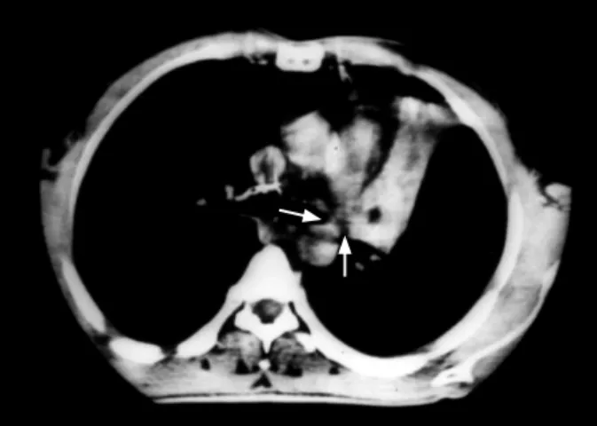
EPORTA 64-year-old male was admitted with complaints of gradually increasing cough and dyspnea during the previous year. Posteroanterior X-Ray showed consolidation of the left upper lobe, whereas computed tomographic (CT) scan of the chest revealed atelectasis of the anterior segment and lingular segments of the left upper lobe (Figure 1). Fiberoptic bronchoscopy demonstrated a large endobronchial polypoid mass, occluding the left main bronchus, which extended to the lingula. Irregular mucosal areas were also noted around the mass. Initially, benign endobronchial tumor
was suspected, but histopathologic evaluation of bronchoscopic biopsy material showed dysplasia and metaplasia at the squamous epithelium. A sleeve left upper lobectomy was thus performed. Macroscopic examination of the resected material demonstrated a yellowish endobronchial mass of 1 cm diameter. The histopathological examination of the tissue showed bronchial glandular tissue, smooth muscle, and early connective tissue distributed throughout the mature adipose tissue. Multiple growths of the hamartomatous foci, constituting adipose and glandular tissues, were detected in the biopsy material taken from the irregular areas that were located at the inner surface of the left upper lobe bronchus.
The patient’s postoperative recovery was uneventful without evidence of recurrence for 3 years after the operation.
D
ISCUSSIONHamartomas are the most common benign lung neoplasms, consisting of cartilage, connective tissue, fat, smooth muscle and respiratory epithelium that locate either in the parenchyma or in the bronchi.1
Although the majority are located in the periphery of the lung, approximately 10% are found endobronchialy,1–3
and are usually only diagnosed incidentally on chest
C
ASE
S
TUDY
Endobronchial Lipomatous Hamartoma
Ibrahim C Kurkcuoglu, MD, Sedat Demircan, MD, Ismail C Kurul, MD,
Funda Demirag, MD
1Department of Thoracic Surgery School of Medicine
Gazi University
1Department of Pathology
Ataturk Center for Chest Disease and Thoracic Surgery Ankara, Turkey
For reprint information contact:
Ibrahim C Kurkcuoglu, MD Tel: 90 312 214 1000 ext. 5635 Fax: 90 312 202 5635 Email: [email protected] Department of Thoracic Surgery, Medical Faculty, Gazi University, Ankara 06500, Turkey.
A
BSTRACTPulmonary hamartomas are the most common benign neoplasm of the lung, occurring in the parenchyma or sometimes within the bronchi. Reported is a case of a 64-year-old male patient with an endobronchial tumor. Sleeve left upper lobectomy was performed and histhopathological examination revealed multiple growths of endobronchial hamartomatous foci. This report demonstrates that endobronchial hamartomas may develop from multiple foci.
(Asian Cardiovasc Thorac Ann 2005;13:372–3)
Kurkcuoglu Endobronchial Hamartoma
2005, VOL. 13, NO. 4 373 ASIAN CARDIOVASCULAR & THORACIC ANNALS
initial excision.6 Incomplete removal of the hamartoma
was thought to be the etiologic factor, but we believe that multiple developmental foci could explain apparent recurrences.6–7 Treatment of endobronchial hamartomas
usually involves open surgery, but sometimes endobronchial resection via bronchoscopy is considered to be a safe and effective treatment. Thus, we believe that effective and appropriate operations require clear bronchial resection margins in open surgery, as can be controlled with frozen section studies. Consequently, in order to maximize possibility of a cure, long-term follow up is needed, particularly for patients undergoing endoscopic excision.
In conclusion, we believe that lung hamartomas are benign fi brous tissue neoplasms which can result from multiple development foci. Therefore, resection borders must be controlled for multiple growths, especially in endobronchial hamartomas.
R
EFERENCES1. Keller MS, Katariya K. Primary Lung Tumors other than Bronchogenic Carcinoma: Benign and Malignant. In: Fishman AP, ed. Fishman’s Pulmonary Diseases and Disorders. New York: McGraw-Hill 1998;1833–40.
2. Shields TW, Robinson PG. Benign Tumors of the Lung. In: Shields TW, LoCicero III J, Ponn RB, eds. General Thoracic Surgery. Philadelphia: Lippincott Williams & Wilkins 2000;1515–32.
3. Borro JM, Moya J, Botella JA, Padilla JD, Canto A, Paris F. Endobronchial Hamartoma. Scand J Thorac Cardiovasc Surg 1988;23:285–7.
4. Bateson EM. Relationship between intrapulmonary and endobronchial cartilage-containing tumors (so-called hamartoma). Thorax 1965;20:447–61.
5. Albrecht E. Ueber Hamartoma. Verh Dtsch Ges Pathol 1904;7:153–7.
6. Van den Bosch JM, Wagenaar SS, Corrin B, Elbers JR, Knaepen PJ, Westermann CJ. Mesenchymoma of the lung (so called hamartoma): a review of 154 parenchymal and endobronchial cases. Thorax 1987;42:790–3.
7. Andersen MN. Multicentric hamartomas of the lung. Report of a case with two additional lesions nine years after primary resection. Ann Thorac Surg 1968;6:469–72.
radiographic examination.1–3 Endobronchial hamartomas
can cause irreversible pulmonary destruction due to bronchial obstruction, so early diagnosis and proper treatment is very important.
The majority of hamartomas are seen in adults aged 50–60 years and are more common in men than in women.2,4 The origin of pulmonary hamartoma is a subject
of controversy. The term hamartoma, was fi rst described by Albrecht, being a combination from the Greek term ‘hamartia’ (defect) and ‘oma’ (tumor).5 In 1922, Feller fi rst
reported a case of pulmonary hamartoma and concluded hamartomas were a developmental malformation of the lung, however, Bateson suggested that hamartomas were true neoplasms originating from the undifferentiated mesenchymal tissues in the submucosa of the bronchi.3–4 Indeed, the onset of the disease in older
age and the demonstration of multiple developmental foci on microscopic evaluation in our patient supports Bateson’s hypothesis. Van den Bosch and colleagues reported hamartomas that recurred in the same pulmonary segment that were detected 10 and 12 years after
Figure 1. CT scan at the level of the left upper lobe bronchus shows an endobronchial mass with distal atelectasis (Arrows).