Oc 2009 Federation of European Microbiological Societies Published by Blackwell Publishing Ltd. All rights reserved
FEMS Microbiol Lett 296 (2009) 198–202 R E S E A R CH L E T T E R
Description of
Oxalicibacterium horti
sp. nov. and
Oxalicibacterium
faecigallinarum
sp. nov., new aerobic, yellow-pigmented,
oxalotrophic bacteria
Nurettin Sahin1, Maria C. Portillo2, Yuko Kato3 & Peter Schumann4
1Mugla University, Egitim Fakultesi, Mugla, Turkey; 2Instituto de Recursos Naturales y Agrobiologia, CSIC, Sevilla, Spain; 3Microbiological and Analytical
Group, Food Research Laboratories, Mitsui Norin Co. Ltd, Shizuoka, Japan; and 4DSMZ – Deutsche Sammlung von Mikroorganismen und Zellkulturen
GmbH, Braunschweig, Germany
Correspondence: Nurettin Sahin, Mugla University, Egitim Fakultesi, TR-48170 Kotekli,
Mugla, Turkey. Tel.: 190 252 211 1826; fax:
190 252 223 8491; e-mail: [email protected]
Received 7 April 2009; accepted 17 April 2009. Final version published online 15 May 2009.
DOI:10.1111/j.1574-6968.2009.01636.x
Editor: Aharon Oren
Keywords
oxalate-oxidizing bacteria; Oxalobacteraceae;
Oxalicibacterium horti; Oxalicibacterium faecigallinarum.
Abstract
Three strains of aerobic, Gram-negative, rod-shaped, non-spore-forming, yel- low-pigmented bacteria (OD1T, YOxT and NS13), which were isolated in
previous studies by enrichment in a mineral medium with potassium oxalate as the sole carbon source, were characterized. On the basis of 16S rRNA gene sequence similarity, strains OD1T, YOxT and NS13 belong to the Betaproteobac-
teria, most closely related to Oxalicibacterium flavum TA17T (97.2–99.7%
sequence similarity). The major whole-cell fatty acids were C16:0, C16:1o7c and
C17:0 cyclo. The results of DNA–DNA hybridization and physiological and
biochemical tests allowed genotypic and phenotypic differentiation of strains OD1T and YOxT from O. flavum TA17T and from each other. Therefore, it is
concluded that the strains OD1T and YOxT represent novel species within the
genus Oxalicibacterium, for which the names Oxalicibacterium horti sp. nov. (type strain OD1T = DSM 21640T = NBRC 13594T) and Oxalicibacterium
faecigallinarum sp. nov. (type strain YOxT = DSM 21641T = CCM 2767T) are
proposed.
Introduction
The genus Oxalicibacterium, with the type species
Oxalicibacterium flavum, was established by Tamer et al.
(2002) in the order Burkholderiales, class Betaproteobacteria, to accommodate an aerobic oxalic acid-utilizing yellow- pigmented bacterium isolated from a litter of oxalate- producing plants (Rumex sp. and Mesembryanthemum sp.).
Oxalicibacterium flavum was characterized by a relatively
high growth rate and yield in a basal mineral medium with oxalate as the sole carbon source, together with a limited substrate spectrum. Later, the genus has been assigned to the family Oxalobacteraceae according to Garrity
et al. (2005).
In the present article, we describe the physiological, chemotaxonomic and phylogenetic characteristics of three aerobic, yellow-pigmented, oxalate-utilizing strains sharing the highest 16S rRNA gene sequence similarity to O. flavum TA17T.
Materials and methods
Bacterial strains
Strain NS13 was isolated from a forest soil sample (Sahin
et al., 2002), strain OD1T was from a garden soil sample
(Jayasuriya, 1955) and strain YOxT was from a chicken dung
sample (Chandra & Shethna, 1975). All of the strains were isolated by enrichment in a mineral medium with potassium oxalate as the sole carbon source.
Phenotypic characterization
Physiological and biochemical tests were performed at 28 1C. Conventional biochemical tests were performed ac- cording to standard methods (Smibert & Krieg, 1994). API 20NE and API ZYM strips (BioMe´rieux) and Biolog GN plates were used according to the manufacturer’s instruc- tion, except that nitrate reduction and indole production
D ow n lo ad ed f rom ht tps :// ac a dem ic .ou p. c om /fem s le/ a rti c le/ 29 6/ 2 /1 98 /60 44 07 by M u gl a U ni v us er on 29 D ec em ber 2 02 0
Oc 2009 Federation of European Microbiological Societies Published by Blackwell Publishing Ltd. All rights reserved
FEMS Microbiol Lett 296 (2009) 198–202 199
Two new Oxalicibacterium species
from tryptophan were read after 2 days, while other reactions of the API 20NE strips were observed for 7 days. Utilization and assimilation of carbohydrates was determined on Biolog GN plates and incubated for 7 days before reading.
Tolerance to metals was tested by growing the isolates on nutrient lactate agar that contained 5 mM of one of the following compounds: Cd(NO3)2, NiCl2 · 6H2O and ZnCl.
Antimicrobial susceptibility testing was performed by the disk-diffusion method using antibiotic-impregnated disks (Oxoid). The following antibiotics were tested: chloramphe- nicol (30 mg), colistin sulfate (10 mg), erythromycin (15 mg), penicillin G (10 IU) and streptomycin (10 mg). An inhibition zone of 12 mm or more in diameter was scored as sensitive to that antibiotic.
Lipophilic pigments were extracted with acetone–metha- nol (3 : 1 v/v); spectra were determined in the same solvent using a UV/visible spectrophotometer (Shimadzu 1601). 16S rRNA gene sequencing and phylogenetic analysis
The 16S rRNA gene sequences were analyzed as described by Sahin et al. (2008). Evolutionary distances were calculated using Kimura’s two-parameter model (Kimura, 1980) with- out taking into account the alignment gaps and unidentified base positions. Phylogenetic trees were constructed from the distance data using the neighbor-joining method of Saitou & Nei (1987). The robustness for individual branches was estimated by bootstrapping with 1000 replicates (Felsen- stein, 1985).
Chemotaxonomic characterization
For analysis of fatty acids, cells were grown on tryptic soy broth agar (Difco) at 28 1C for 4 days. Cells were saponified, methylated to create fatty acid methyl esters and extracted as described previously (Ka¨mpfer & Kroppenstedt, 1996). Peaks were automatically integrated, and fatty acid names and percentages were determined using the MICROBIAL IDENTI-FICATION standard software package MIDI (Sasser, 1990).
Ribotyping, DNA base compositions and DNA--DNA hybridizations
Ribotyping of strains was performed with the automated RiboPrinter Microbial Characterization System (Qualicon, DuPont, Wilmington, DE). Riboprint analyses, using EcoRI, followed described methods (Bruce, 1996).
For determination of the G1C content, DNA was de- graded to nucleosides using P1 nuclease and bovine intest- inal mucosa alkaline phosphatase, as described by Mesbah
et al. (1989). The nucleosides were separated by reversed-
phase HPLC (Shimadzu Apparatus) according to the meth- od described by Tamaoka & Komagata (1984), and the G1C
content was calculated from the ratio of deoxyguanosine to thymidine.
The degree of DNA–DNA relatedness between the three isolates and previously described O. flavum type strain (TA17T) was determined by measuring the divergence
between the thermal denaturation midpoint of homoduplex DNA and heteroduplex DNA (DTm) as described by
Gonza´lez & Sa´iz-Jime´nez (2005).
Results and discussion
Phenotypic characteristics
The strains had a very limited substrate spectrum regarding the substrates in the API 20NE and Biolog GN plates. All strains utilized oxalate, formate, glycollate, lactate, pyruvate, succinate and malate. Other carboxylic acids, alcohols and all amino acids, except alanine, were not utilized. Differen- tial phenotypic characteristics between the type strains of the three species of the genus Oxalicibacterium are given in Table 1. All strains produced a yellow, water-insoluble intracellular pigment. The absorption spectrum of the
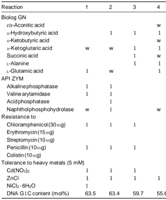
Table 1. Differential characters of Oxalicibacterium strains
Reaction 1 2 3 4 Biolog GN cis-Aconitic acid w a-Hydroxybutyric acid 1 1 1 a-Ketobutyric acid w a-Ketoglutaric acid w w 1 1 Succinic acid 1 w L-Alanine 1 1 L-Glutamic acid 1 w 1 API ZYM Alkaline phosphatase 1 1 Valine arylamidase 1 1 Acid phosphatase 1 Naphthol phosphohydrolase w 1 w Resistance to Chloramphenicol (30 mg) 1 1 1 Erythromycin (15 mg) Streptomycin (10 mg) Penicillin (10 mg) 1 1 1 Colistin (10 mg)
Tolerance to heavy metals (5 mM)
Cd(NO3)2 1 1 1
ZnCl 1 1 1 1
NiCl2 · 6H2O 1
DNA G1C content (mol%) 63.5 63.4 59.7 55.6
Strains: 1, Oxalicibacterium flavum TA17T; 2, O. flavum NS13; 3, OD1T; 4,
YOxT. Biolog GN and API 20NE reactions after 7-day incubation at 28 1C.
All strains were positive for: (API 20NE) malate, (API ZYM) esterase (C4), esterase lipase (C8), leucine arylamidase and (Biolog GN plates), formic
acid, DL-lactic acid, bromosuccinic acid, alanine amide and D-alanine, but
negative for the other substrates or reactions of the panels not men-
tioned. , negative; 1, positive; w, weakly positive.
D ow n lo ad ed f rom ht tps :// ac a dem ic .ou p. c om /fem s le/ a rti c le/ 29 6/ 2 /1 98 /60 44 07 by M u gl a U ni v us er on 29 D ec em ber 2 02 0
200 N. Sahin et al.
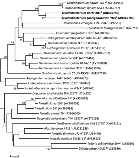
Fig. 1. Phylogenetic tree, based on
neighbor-joining method (Saitou & Nei, 1987), derived from an alignment comprising 16S rRNA gene partial region sequences (1367 bp). The data set was resampled 1000 times using the bootstrap option, and the percentage values are given at the nodes. The scale bar indicates the number of substitutions per nucleotide position. Bold type indicates two new species.
pigment is almost the same for all strains, which showed major peaks between 430–435 and 450–455 nm, with a shoulder at 480 nm. During the cultivation on tryptic soy agar or nutrient lactate agar plates, a variation in the colony appearance was observed, especially for strains OD1T and
YOxT. The variants are (1) circular, raised, more or less
translucent, with an entire margin and/or (2) a wrinkled surface with an undulate margin. All variants had similar protein and FT-IR patterns (data not shown). Strains OD1T,
YOxT and NS13 were able to grow in the presence of 25 g L 1
potassium oxalate. The optimal growth in mineral medium with potassium oxalate as the sole source of carbon and energy occurred as 8 g L 1.
Phylogenetic and chemotaxonomic characteristics
Phylogenetic analyses using the 16S rRNA gene sequences indicated that strains OD1T, YOxT and NS13 belong to the
family Oxalobacteraceae of the b-subclass of the Proteobac-
teria; O. flavum TA17T showed 97.17%, 97.44% and 99.73%
sequence similarity in the 16S rRNA gene with strains OD1T,
YOxT and NS13, respectively. 16S rRNA gene sequence
similarities were o 96.2% with all established type species of all the genera of the family Oxalobacteraceae (Fig. 1).
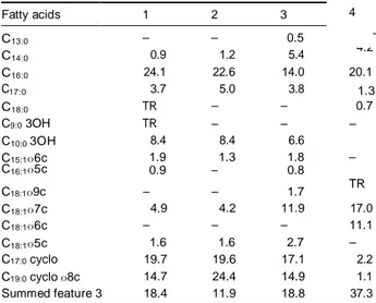
The fatty acid compositions of the genus Oxalicibacterium were not included in the species descriptions earlier. The major fatty acids were C16:0 (14–24%), summed feature 3
(C16:1o7c/C15:0 iso 2OH; 11.9–37.3%) and moderate amounts
of C18:1o7c (4.2–17%). In addition, C10:0 3OH was the only
hydroxylated fatty acid detected (Table 2). Furthermore, C18:1o7c (17%) and summed feature 3 (37.3%) seems to be
present in strain YOxT in significantly higher amounts than all
other Oxalicibacterium type species examined. The fatty acids C18:1o6c (11%) and C13:0 (0.5%) were detected only in strains
YOxT and OD1T, respectively, in contrast to others. In conclu-
sion, Oxalicibacterium type strains can be distinguished from each other based on their fatty acid profiles.
DNA--DNA hybridizations and ribotyping
The fragment patterns of strain O. flavum TA17T and of the
strains OD1T, YOxT and NS13 varied, resulting in a unique
pattern for each of the strains (Fig. 2). The lack of a high
D ow n lo ad ed f rom ht tps :// ac a dem ic .ou p. c om /fem s le/ a rti c le/ 29 6/ 2 /1 98 /60 44 07 by M u gl a U ni v us er on 29 D ec em ber 2 02 0
Oc 2009 Federation of European Microbiological Societies Published by Blackwell Publishing Ltd. All rights reserved
FEMS Microbiol Lett 296 (2009) 198–202 201
Two new Oxalicibacterium species
similarity of strains OD1T and YOxT to the reference type
strain O. flavum TA17T excludes the possibility of their
affiliation to the O. flavum genomospecies. As displayed in the dendrogram, the patterns of the type strains of O. flavum TA17T and the strain NS13 showed high similarities, and
most of the differences observed were within the target recognition sites of the restriction enzymes used in the analysis.
The DNA–DNA relatedness studies among strains OD1T,
YOxT and O. flavum TA17T, sharing a 16S rRNA gene
sequence similarity of 97.4–97.2%, showed that the DTm
values were 4 6 1C, which confirms that strains OD1T and
YOxT do not belong to the genospecies O. flavum. The DT m
value of O. flavum TA17T and strain NS13, sharing a 16S
rRNA gene sequence similarity value of 99.73%, is 4.3 1C. The reassociation values of strains TA17T-OD1T, TA17T-
YOxT, YOxT-NS13 and YOxT-OD1T were 9.7, 6, 14 and 8 1C,
respectively, which are well above the 5 1C cut-off point recommended for the delineation of species (Wayne et al., 1987; Rossello´-Mora & Amann, 2001).
Table 2. Whole-cell fatty acid composition of Oxalicibacterium strains
On the basis of the results described above, it can be concluded that the strains OD1T and YOxT represent novel
species within the genus Oxalicibacterium, for which the names Oxalicibacterium horti sp. nov. and Oxalicibacterium
faecigallinarum sp. nov. are proposed, respectively.
Description of Oxalicibacterium horti sp. nov.
Oxalicibacterium horti (hor’ti. L. gen. n. horti, of a garden,
isolated from a garden soil).
Gram-negative, small rods 0.5 x 1.5 mm, motile by polar flagella. No spores found. Colonies on nutrient agar (Oxoid CM3) with 0.2% lactate are yellow pigmented. Forms smooth, raised, opaque with entire edges; diameter is up to 1.5–2 mm after 3 days of incubation at 28 1C. Growth occurs weakly at 37 1C, but not at 42 1C. Optimum growth occurs at 25 1C and pH 7.0. Grows in media containing 3% NaCl. Oxidase and catalase positive. Nitrate is not reduced to nitrite. Poly-b-hydroxybutyrate is accumulated. Negative for indole production, arginine dihydrolase, urease, esculin, casein and gelatine hydrolysis and b-galactosidase. Oxalate,
DL-lactate, glycollate, DL-malate and succinate are utilized as
17:0 C18:1o7c 4.9 4.2 11.9 17.0 C18:1o6c – – – 11.1 C18:1o5c 1.6 1.6 2.7 – C17:0 cyclo 19.7 19.6 17.1 2.2 C19:0 cyclo o8c 14.7 24.4 14.9 1.1 Summed feature 3 18.4 11.9 18.8 37.3
Strains: 1, Oxalicibacterium flavum TA17T; 2, O. flavum NS13; 3, OD1T; 4,
YOxT. Cells were grown on tryptic soy broth (TSB) agar at 28 1C for 3
days. Data are percentages of total fatty acids. TR, fatty acid content o 0.5%. Summed feature 3 comprises 16:1o7c, 15 iso 2OH or any combination of these fatty acids.
in a mineral medium.
Description of Oxalicibacterium faecigallinarum sp. nov.
Oxalicibacterium faecigallinarum (L. n. faex faecis, the dregs,
feces; L. gen. pl. n. gallinarum, of hens; N.L. gen. pl. n.
faecigallinarum, isolated from feces of hens).
Gram-negative, small rods 0.75 x 1.5 mm, motile by polar flagella. No spores found. Colonies on nutrient agar (Oxoid
Fig. 2. Diversity of normalized ribotype patterns
found in type strains of Oxalicibacterium.
Cluster analysis was performed using the unweighted pair group method with the arithmetic averages method based on the Pearson correlation coefficient. VCA indicates a standard EcoRI batch.
D ow n lo ad ed f rom ht tps :// ac a dem ic .ou p. c om /fem s le/ a rti c le/ 29 6/ 2 /1 98 /60 44 07 by M u gl a U ni v us er on 29 D ec em ber 2 02 0
Fatty acids 1 2 3 4 the sole carbon and energy sources, but not other carbohy- drates or carboxylic acids, acetate, citrate, malonate, metha-
C13:0 – – 0.5 – nol or ethanol. Substrate utilization, enzyme production
C14:0 0.9 1.2 5.4
4.2
and other physiological characteristics are given in Table 2.
C16:0 24.1 22.6 14.0 20.1
C 3.7 5.0 3.8 1.3 The main fatty acids are summed feature 3 (C16:1o7c/C15:0 C18:0 TR – – 0.7 iso 2OH; 18.8%), C16:0 (14%), C17:0 cyclo (17.1%), C18:1o7c
C9:0 3OH TR – – – (11.9%) and C19:0 cyclo o8c (14.9%). Does not contain C18:0
C10:0 3OH 8.4 8.4 6.6 4.6 and C18:1o6c fatty acids. The G1C content of DNA is
C15:1o6c C16:1o5c 1.9 0.9 1.3 – 1.8 0.8
– 59.7 mol% (as determined by HPLC).
C18:1o9c – – 1.7
The type strain, OD1T ( = DSM 21640T = NBRC 13594T), TR
202 N. Sahin et al.
CM3) with 0.2% lactate are yellow pigmented. Forms smooth, glistening, raised, translucent with entire edges; the diameter is up to 1–1.5 mm after 3 days of incubation at 28 1C. Growth occurs at 37 1C, but not at 42 1C. Opti- mum growth occurs at a pH between 7.0 and 7.5. Grows in media containing 3% NaCl. Oxidase and catalase positive. Very limited substrate spectrum. Oxalate, DL-lactate and DL
-malate are utilized as the sole carbon and energy sources, but not other carbohydrates or carboxylic acids, acetate, citrate, malonate, methanol or ethanol. No acid produced from glucose, galactose, arabinose, sucrose and lactose. Nitrate is not reduced to nitrite. Indole and H2S are not
produced. Substrate utilization, enzyme production and other physiological characteristics are given in Table 2. The main fatty acids are summed feature 3 (C16:1o7c/C15:0 iso
2OH; 37.3%), C16:0 (20.1%), C18:1o7c (17%) and C18:1o6c
(11.1%). Does not contain C15:1o6c, C16:1o5c and C18:1o5c
fatty acids. The G1C content of DNA is 55.6 mol% (as determined by HPLC).
The type strain, YOxT ( = DSM 21641T = CCM 2767T),
was isolated from chicken dung after enrichment with oxalate in a mineral medium.
Acknowledgement
We are grateful to Dr Jean P. Euze´by for his help with the Latin nomenclature of the species epithets.
Statement
The GenBank/EMBL/DDBJ accession numbers for the 16S rRNA gene sequences of strains OD1T, NS13 and YOxT are
AB469786, AB469787 and AB469788, respectively.
References
Bruce J (1996) Automated system rapidly identifies and
characterized microorganisms in food. Food Technol 50: 77–81.
Chandra TS & Shethna YI (1975) Isolation and characterization
of some new oxalate-decomposing bacteria. Antonie Van
Leeuwenhoek 41: 101–111.
Felsenstein J (1985) Confidence limits on phylogenies – an
approach using the bootstrap. Evolution 39: 783–791.
Garrity GM, Bell JA & Lilburn T (2005) Family II. Oxalo-
bacteraceae fam. nov. Bergey’s Manual of Systematic
Bacteriology, Vol. 2 (Brenner DJ, Kreig NR, Staley JT & Garrity GM, eds), pp. 623–647.Springer, Heidelberg.
Gonza´lez JM & Sa´iz-Jime´nez C (2005) A simple fluorimetric method for the estimation of DNA–DNA relatedness between closely related microorganisms by thermal denaturation
temperatures. Extremophiles 9: 75–79.
Jayasuriya GCN (1955) The isolation and characteristics of
an oxalate-decomposing organism. J Gen Microbiol 12:
419–428.
Ka¨mpfer P & Kroppenstedt RM (1996) Numerical analysis of fatty acid patterns of coryneform bacteria and related taxa. Can J Microbiol 42: 989–1005.
Kimura M (1980) A simple method for estimating evolutionary rates of base substitutions through comparative studies of
nucleotide sequences. J Mol Evol 16: 111–120.
Mesbah M, Premachandran U & Whitman WB (1989) Precise
measurement of the G1C content of deoxyribonucleic acid by
high-performance liquid-chromatography. Int J Syst Bacteriol
39: 159–167.
Rossello´-Mora R & Amann R (2001) The species concept for
prokaryotes. FEMS Microbiol Rev 25: 39–67.
Sahin N, Gokler I & Tamer AU (2002) Isolation, characterization and numerical taxonomy of novel oxalate-oxidizing bacteria. J Microbiol 40: 109–118.
Sahin N, Kato Y & Yilmaz F (2008) Taxonomy of oxalo-
trophic Methylobacterium strains. Naturwissenschaften 95:
931–938.
Saitou N & Nei M (1987) The neighbor-joining method – a new
method for reconstructing phylogenetic trees. Mol Biol Evol 4:
406–425.
Sasser M (1990) Identification of bacteria by gas chromatography
of cellular fatty acids. USFCC Newsl 20: 1–6.
Smibert RM & Krieg NR (1994) Phenotypic characterization. Methods for General and Molecular Bacteriology (Gerhardt P, ed), pp. 607–654. American Society for Microbiology, Washington, DC.
Tamaoka J & Komagata K (1984) Determination of DNA-base composition by reversed-phase high-performance
liquid-chromatography. FEMS Microbiol Lett 25:
125–128.
Tamer AU, Aragno M & Sahin N (2002) Isolation and
characterization of a new type of aerobic, oxalic acid utilizing
bacteria, and proposal of Oxalicibacterium flavum gen. nov.,
sp. nov. Syst Appl Microbiol 25: 513–519.
Wayne LG, Brenner DJ, Colwell RR et al. (1987) Report of the ad
hoc-committee on reconciliation of approaches to bacterial
systematics. Int J Syst Bacteriol 37: 463–464.
D ow n lo ad ed f rom ht tps :// ac a dem ic .ou p. c om /fem s le/ a rti c le/ 29 6/ 2 /1 98 /60 44 07 by M u gl a U ni v us er on 29 D ec em ber 2 02 0