Sevim Çiftçi Yegin
The Effect of Lycopene Application on the Antioxidant Activity
in Liver and Kidney Tissues of Diabetic Rats
*Sevim ÇİFTÇİ YEGİN
1a, Fatmagül YUR
2b1. Giresun University, Health Service Vocational School of Higher Education, Giresun, TURKEY.
2. Mugla Sıtkı Kocman University, Fethiye Faculty of Health Science, Department of Nutrition and Dietetics, Mugla, TURKEY. ORCID: 0000-0003-3950-4399a, 0000-0002-5536-9169b
Abstract: The scope of the present study was to determine the effect of lycopene on the activity of malondialdehyde (MDA),
glutathione, vitamin A, vitamin E and catalase in liver and kidney tissues of diabetic rats. Wistar albino male rats were randomly allocated into four groups: control, diabetic, diabetic+lycopene and lycopene group (n=7). Rats in the respective groups were treated with intraperitoneal streptozotocin (45 mg/kg) to induce diabetes. Rats in the group lycopene and diabetic+lycopene were given orally 1 ml lycopene every day for 1 month. Catalase levels in the liver tissues of lycopene group was significantly lower than other groups (P<0.05). A significant decrease was observed at Vitamin A levels in the kidney tissues of the groups diabetic, diabetic+lycopene and lycopene compared to the control group (P<0.05). Other non-significant differences between the groups except catalase levels in liver tissue and vitamin A levels in kidney tissues may be result of a short period of diabetes mellitus. The results of the present study supported the positive correlation among metabolic control and oxidative stress in diabetes mellitus.
Keywords: Antioxidants, Experimental Diabetes Mellitus, Lycopene.
Diyabetik Ratların Karaciğer ve Böbrek Dokularındaki Antioksidan Aktivite
Üzerine Likopen Uygulamasının Etkileri
Öz: Bu çalışmanın amacı, diyabetik ratlarda likopen uygulamasının karaciğer ve böbrek dokusundaki malondialdehit,
glutatyon, vitamin A, vitamin E ve katalaz üzerine etkilerini araştırmaktır. Çalışmada kullanılan Wistar cinsi albino erkek ratlar, içlerinden rastgele seçilerek kontrol grubu, diyabet grubu, likopen grubu ve diyabet+likopen grubu olmak üzere 4 farklı gruba ayrıldı. Her grupta 7 adet rat bulunmaktadır. Deneysel diyabet oluşturmak için diyabet grubu ve diyabet+likopen grubundaki ratlara intraperitonel yoldan streptozotosin (45 mg/kg) uygulandı. Diyabet+likopen grubu ile likopen grubundaki ratlara 1 ay boyunca her gün oral yoldan 1 ml likopen (ayçiçeği yağında eritildi) uygulandı. Likopen grubunun karaciğer dokularındaki katalaz düzeyi diğer gruplardan önemli derecede düşük bulundu (P<0.05). Diyabet, diyabet+likopen ve likopen gruplarının böbrek dokularındaki vitamin A düzeyleri diğer gruplarla kıyaslandığında istatistiksel olarak önemli bir azalış göstermiştir (P<0.05). Böbrek dokularındaki vitamin A düzeyleri ve karaciğer dokularındaki katalaz düzeyleri hariç diğer gruplar arasındaki istatistiksel olarak önemli olmayan farklılıkların olması Diabetes Mellitusun kısa periyodunun sonuçları olabileceği kanaatindeyiz. Çalışmanın sonucu olarak, yaptığımız araştırma Diabetes Mellitusta oksidatif stres ile metabolik kontrol arasındaki pozitif korelasyonu destekler niteliktedir.
Anahtar Kelimeler: Antioksidanlar, Deneysel Diabetes Mellitus, Likopen. Geliş Tarihi/Received 22.11.2018 Kabul Tarihi/Accepted 13.03.2019 Yayın Tarihi/Published 25.10.2019
Bu makaleye atıfta bulunmak için/To cite this article:
Çiftçi Yeğin S, Yur F: The Effect of Lycopene Application on the Antioxidant Activity in Liver and Kidney Tissues of Diabetic Rats. Atatürk Üniversitesi Vet. Bil. Derg., 14(2): 119-128, 2019. DOI: 10.17094/ataunivbd.486629
INTRODUCTION
iabetes mellitus is a lifelong chronic metabolic disease with mortality, morbidity and economic burden. The deficiency or abnormality of insulin results in failures of target tissues at carbohydrate, lipid and protein metabolisms. Polyuria, polydipsia, polyphagia, weight loss, blurred vision and tendency to infections are important clinical findings of hyperglycemia. The risk for the development of long-term complication gets high, while the disease progresses (1).
Glutathione is a tripeptide, which has thiol group functions as a substrate for many enzymes like transferase and peroxidase, thus it prevents or decreases the destructive effects of free radicals. Thiol groups are cellular antioxidants that catch free radicals via enzymatic reactions. Glutathione is a water-soluble thiol and exists in many cells at high concentration protecting membranes against lipid peroxidation. This protection occurs enzymatically (2). Glutathione peroxidase (GSH-Px) needs selenium minerals for its activity and it converts the reduced form of GSH to the oxidized form as glutathione disulfide (GSSG). Glutathione reacts with very harmful oxidants like single oxygen (O2), superoxide
anion (·O2-), hydroxi radicals (·OH) without enzyme
catalysis (3).
Vitamin E prevents oxidation of unsaturated fatty acids in cellular membranes and also prevents membrane degradation. Unsaturated fatty acids in membrane phospholipids are oxidized by hydrogen peroxides which are formed by flavoproteinoxidase enzyme (4).
Vitamin A exists in vegetables in the form of β-carotene as a provitamin, which is a yellow pigment, and it consists of two retinal molecules linked with aldehyde points. Vitamin A is stored in lipocytes of liver in the form of ester, possibly as a lipoprotein complex. β-carotene is an antioxidant and it can restrain peroxy free radicals in tissues at low oxygen partial pressure. The antioxidant effect of β-carotene is dependent on its ability to get stabilized the organic peroxide free radicals in stable conjugated
alkyl structures. β-carotene is effective in lower oxygen concentrations, so that it completes the antioxidant features of vitamin E, which is effective in higher oxygen concentrations (5).
Malondialdehyde (MDA) is final products of lipid peroxidation. Free radicals with their autocatalytic characteristics, cause lipid oxidation and membrane damage (6). Lipid peroxidation leads to a harmful chain reaction. It directly damages membrane structure and indirectly damages other cell components because of reactive aldehydes production. It results in oxidative cellular membrane destruction and serious tissue damage. Peroxidation induced MDA causes cross linking of membrane components and their polymerization. In turn, it changes the intrinsic membrane property causing ion transport, deformation, enzyme activity and aggregation of cell surface components. Antioxidant defense systems have a great importance in preventing tissue damage connected with oxidation (7).
Catalase (CAT) is a hemoprotein consisting four heme groups and exists in all cell types at different concentrations. It catalyzes hydrogen peroxide to molecular oxygen and water. It is mostly localized in peroxisomes. CAT’s reducing activity is observed on small molecules like hydrogen peroxide, methyl and ethyl hydro peroxides. It has no effect effect on big molecule like lipid hydro peroxides. CAT can be found in blood, bone marrow, mucous membranes, liver and kidney at high amounts (8). Antioxidant enzymes like superoxide dismutase (SOD), catalase, GSH-Px, and neutralize reactive oxygen species (ROS) (9). Carotenoids and antioxidant vitamins scavenge effectively ROS and regulate the activities of antioxidant enzymes (10). Carotenoids behavior as strong antioxidants that protect cell membranes from lipid peroxidation and repair free radicals as well as other reactive molecules (11). Carotenes (α-carotene, lycopene, β-carotene) and xanthophylls (zeaxanthin, lutein) regard the class of carotenoids. Between the carotenoids, lycopene is an effective
free radical scavenger and has the highest antioxidant activity (12).
Lycopene is plenty in red fruits like tomato and water melon. The protective effects of lycopene against many different types of cancer inclusive prostate, skin cancer and breast have been reported in many studies (13,14). These anti-cancer effects seem to be generated by the antioxidant properties of lycopene. Lycopene helps to preserve cells from free radical attack via scavenging of ROS and detoxicate lipid peroxide (15).
In the present study, we have studied the renal and hepatic changes excited by acute exposure to STZ (streptozotocin) in rats, furthermore antioxidant enzymes, vitamin A, E levels were controlled. We evaluated also the possible preventive effect of lycopene in rats.
MATERIALS and METHODS Experimental Procedures
The ethics committee of the Giresun University (no 2013/5) approved the study.
In the present study, twenty-eight Wistar-albino male rats with weight among 250 – 300 g were used. Rats were provided by the Experimental Research Laboratory of Medical Faculty, University of Yuzuncu Yıl. Study rats were arbitrarily separeted into four groups with seven rats in groups: control group (C), diabetic rats not treated with lycopene (D), diabetic rats treated with lycopene (DL) and lycopene cleansed rat group (L).
Single dose of 45 mg/kg STZ (Sigma, USA) solved in citrate buffer with pH 4.5 was intraperitoneally injected to rats to induce diabetes in D and DL groups (16). The exact, same amount of physiological saline solution was injected using the same method to the control group. Blood samples were collected from rat tail veins 72 hours after the injection of STZ. Blood glucose meter (Plus Med Accuro Biosensor Blood) measurement device and its blood strips were used for the measurement of blood glucose levels. Rats with blood glucose levels above 270 mg/dl were admitted as patients. 1 ml/day lycopene (10% FS;
Redivivo TM; Code 7803; DSM Inc., Istanbul, Turkey) was solved in sunflower oil and the preparation was administered orally to rats in respective groups every day (17).
For four weeks, rats were hold in cages at room temperature (22±2°C) and for 12 hours in dark and light periods, also food and fresh water were provided at all times.
Biochemical Analysis
GSH Assay: In this method, 5 ml sulphosalicylic acid was added to 0.5 g of tissue, which was homogenized afterwards. The sample was centrifuged for 15 min at 4500 g. After removing 1 ml of supernatant, 5 ml phosphate buffer was added to the sample. The sample was brooded for 10 minutes at 60°C and then it was cooled down to room temperature, finally 1 ml 5.5’ Dithiobis-2-Nitrobenzoik Acid (DTNB) was added to the sample. Sample was tested at 412 nm towards blank (distilled water) and the calculation was performed (18).
Vitamin E Assay: In this method, 1 ml ethanol was added to 0.5 g of tissue and the sample was homogenized. After centrifugation of the sample, 1 ml of supernatant was distanted from and blended with 1 ml of xylene. This sample was strongly rinsed for 30 seconds, afterwards it was centrifuged for 5 min at 2500 g. 1 ml of supernatant was taken and mixed with 0.5 ml of 2,4,6-Tri(2-Pyridyl)-s-Triazine (TPTZ). First determination was performed at 410 nm, afterwards 0.1 ml of FeCl3 was added to the
sample and finally second determination was performed at 600 nm (19).
Vitamin A Assay: In this method, 0.5 g of tissue and 1 ml ethanol was blended and homogenized. After the centrifugation of the sample, 1 ml of supernatant was distanted from and mixed 1 ml ethanol and 3 ml hexane. The sample was stirred for 10 min followed by the centrifugation for 10 min at 2000 g. Finally, sample was tested at 453 nm and 325 nm (20).
MDA Assay: In this method, 0.5 g of tissue was blended with 1,5 ml of cold Tris-HCl buffer and
homogenized for 5 minutes. It was centrifuged for 30 min at 9500 g and 200 µl of supernatant was removed from the sample. This sample was blended with 0,8 ml phosphate buffer, 0,025 ml BHT and 0,5 ml 30% TCA and stirred. The sample was kept at -20° C for 2 hours and it was centrifuged for 15 min at 2000 g. 1 ml supernatant was taken from the sample
and mixed with 75 µl 0,1 M
Ethylenediaminetetraacedic acid (EDTA) and 250 µl Tiobarbutiric acid (TBA). This sample was kept at 90°C for 15 min. When it cooled down to room temperature, the sample was tested at 532 nm (21). In this assay, the level of MDA was determined via spechtrophotometric measurement. For this reason, the color created by the reaction of TBA with MDA. The level of MDA is expressed as nmol/gram protein by using standard calibration curve (R2=0.9974).
Catalase Assay: A commercial test kit from Cayman trademark was used for the catalase assay. Tissue homogenate: 1 gr tissue was homogenized in 5 ml buffer and centrifuged for 15 min at 10.000 g at 4°C. Supernatant was taken from the sample.
Standard preparation: In this study, 7 tubes were labeled from A to G. 100 µl sample buffer was placed into tube A, 990 µl sample buffer and 10 µl formaldehyde was placed into tube B, 970 µl sample buffer and 30 µl formaldehyde was placed into tube C, 940 µl sample buffer and 60 µl formaldehyde was placed into tube D, 910 µl sample buffer and 90 µl formaldehyde was placed into tube E, 880 µl sample buffer and 120 µl formaldehyde was placed into tube
F, 850 µl sample buffer and 150 µl formaldehyde was placed into tube G.
In measurement method, 100 µl diluted assay buffer, 30 µl methanol and 20 µl standard solution were added twice into standard wells of plate. 100 µl assay buffer, 30 µl methanol and 20 µl diluted CAT were added into positive control wells. 100 µl assay buffer, 30 µl methanol and 20 µl sample were added into sample wells. All wells on the plate were filled with 20 µl of H2O2. Plates were closed on and placed
on the shaker to incubate for 20 min. Finally, 30 µl KOH and 30 µl Purpald (chromogen) were added into all wells and the reaction was completed. Plates were again closed on and were stirred and brooded for 10 min at room temperature. All wells were filled with 10 µl of KIO4 and they were incubated for 5 min at
room temperature. Samples absorbance were determined at 540 nm (22).
Statistical Analysis
All data were statistically analyzed using ANOVA. DUNCAN multi comparison test was applied to determine any significant differences. Correlations between the tested biochemical parameters were calculated. MEANS, CORR and GLM procedures of SAS software package were used to evaluate the data.
RESULTS
The obtained results were summarized in Table 1 and 2.
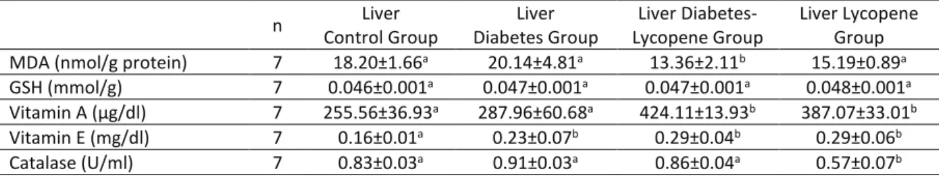
Table 1. Effects of lycopene on hepatic MDA, GSH, vitamin A, vitamin E levels and catalase activities in control
group (C), diabetic rats not given lycopene (D), diabetic rats given lycopene (DL) and lycopene given rats group (L).
Tablo 1. Kontrol grup (K), diyabetli olup likopen verilmeyen grup (D), diyabetli olup likopen verilen grup (DL) ve
likopen gruplarında (L) karaciğer MDA, GSH, vitamin A, vitamin E düzeyleri ve katalaz aktivitesi üzerine likopenin etkileri. n Liver Control Group Liver Diabetes Group Liver Diabetes-Lycopene Group Liver Lycopene Group
MDA (nmol/g protein) 7 18.20±1.66a 20.14±4.81a 13.36±2.11b 15.19±0.89a
GSH (mmol/g) 7 0.046±0.001a 0.047±0.001a 0.047±0.001a 0.048±0.001a
Vitamin A (µg/dl) 7 255.56±36.93a 287.96±60.68a 424.11±13.93b 387.07±33.01b
Vitamin E (mg/dl) 7 0.16±0.01a 0.23±0.07b 0.29±0.04b 0.29±0.06b
Catalase (U/ml) 7 0.83±0.03a 0.91±0.03a 0.86±0.04a 0.57±0.07b
MDA: Malondialdehyde, GSH: Glutathione
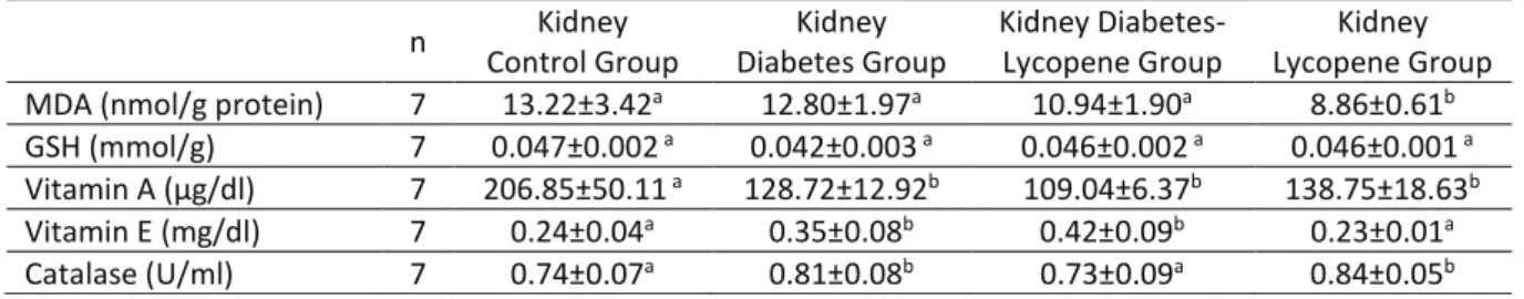
Table 2. Effects of lycopene on kidney MDA, GSH, vitamin A, vitamin E levels and catalase activities in control
group (C), diabetic rats not given lycopene (D), diabetic rats given lycopene (DL) and lycopene given rats group (L).
Tablo 2. Kontrol grup (K), diyabetli olup likopen verilmeyen grup (D), diyabetli olup likopen verilen grup (DL) ve
likopen gruplarında (L) böbrek MDA, GSH, vitamin A, vitamin E düzeyleri ve katalaz aktivitesi üzerine likopenin etkileri. n Kidney Control Group Kidney Diabetes Group Kidney Diabetes-Lycopene Group Kidney Lycopene Group
MDA (nmol/g protein) 7 13.22±3.42a 12.80±1.97a 10.94±1.90a 8.86±0.61b
GSH (mmol/g) 7 0.047±0.002 a 0.042±0.003 a 0.046±0.002 a 0.046±0.001 a
Vitamin A (µg/dl) 7 206.85±50.11 a 128.72±12.92b 109.04±6.37b 138.75±18.63b
Vitamin E (mg/dl) 7 0.24±0.04a 0.35±0.08b 0.42±0.09b 0.23±0.01a
Catalase (U/ml) 7 0.74±0.07a 0.81±0.08b 0.73±0.09a 0.84±0.05b
MDA: Malondialdehyde, GSH: Glutathione
Results were presented as median±standard error. [a-b: difference between groups assigned different letters in the same line is statistically significant (P<0.05)].
MDA levels in the liver of diabetic-lycopene group were significantly lower compared to the control group. No significant change was observed in GSH levels between groups. Vitamin A levels in the liver of diabetes-lycopene and lycopene groups were significantly increased compared to the control group. Liver vitamin E levels in all treatment groups were observed to be significantly higher than untreated control group. It was observed that Catalase levels in the liver of lycopene group was significantly decreased (Table 1).
Kidney MDA levels in diabetes+lycopene group and lycopene group were observed lower than other groups, but this decrease was not statistically significant in diabetes+lycope group. MDA levels was decreased notably (P<0.05) in the kidney tissue of lycopene group in comparison with the control group and diabetes group.
No significant difference was observed between the groups regarding the glutathione levels in the tissues of kidney and liver in this study. Vitamin A level in the kidney tissue was higher in the control group in proportion to other groups and this difference was statistically significant. Kidney vitamin A level in all therapy group was notably lower than control group. Vitamin E levels in the kidney tissues of diabetic group and diabetic+lycopene group were notably higher than the lycopene and control groups. Rats in the diabetes group and lycopene group had
higher catalase levels in the kidney tissue than control group and this was statistically significant (Table 2).
DISCUSSION and CONCLUSION
Diabetes is a chronic metabolic disease and also a state of increased oxidative stress. Increased free radicals interact with proteins, lipids and nucleic acids so that it causes loss of functional alterations, structural and membrane integrity of proteins and genetic mutations. Organisms has non enzymatic and enzymatic antioxidant advocacy systems to overcome these injurious effects of radicals (23).
Lipid peroxidation level (LPO), with structural and functional varieties at cell membrane, is a significant indicator of membrane damage. Free radicals induced lipid peroxidation is linked with degenerative diseases including diabetes (24). In accordance with the findings of a study (25), MDA-TBA levels were increased in kidney and liver tissues of diabetic rats. The increase in MDA-TBA levels is a significant indicator of peroxidative stress and advanced complications of diabetes (25). In the present study, the liver MDA level of diabetes group was observed to be higher than the other groups but that was not notably significant. Vitamin A and vitamin E levels of diabetes-lycopene and lycopene groups were found to be notably higher than the other groups. A statistically significant decrease was
observed in liver catalase activity in lycopene group compared to other groups.
Certain studies reported that if the duration of diabetes takes longer, the severity of oxidative stress is also increased. Singh et al. (26) determined a strong correlation between the duration of diabetes, metabolic control and oxidative stress (26). Another study determined that oxidative stress decreased as soon as the metabolic control was under the control in Type I and Type II diabetes patients (27).
The reason that there was no important difference in point of glutathione levels in the liver and kidney tissues of the groups could be a result of the diabetes created within a short time. Yılmaz et al. (28), reported high levels of LPO in the groups with diabetes longer than five years and low levels of antioxidant enzymes like GSH-Px and SOD. However, they did not report statistical significance (28). They attributed this finding to the fact that there was no difference between glucose regulation at both groups and they reported that the findings were supportive for the idea of positive correlation between metabolic control and oxidative stress in diabetes (26). Another study showed similar findings in parallel with our study (28). Consequently, they came to the conclusion that there was no important difference in terms of oxidative stress among the groups that was having diabetes treatment for five years or more and the group with shorter treatment period. There was also no difference in the glucose regulation among these two groups which may be a factor for this result (29).
The most important cellular defense unit against ROS is glutathione (GSH) exists in all mammalian cells. GSH as an electron donor in non-enzymatic reactions acting directly with radicals to eliminate those radicals. It was stated that the decrease of GSH level in the tissues is parallel with an increase in LPO (30). Study findings demonstrated that GSH levels decreased both in kidney, liver tissues in a diabetic group. Demir and Yılmaz (31) stated that increased both in liver and kidney tissues in the group treated with pine oil. It was predicted that
antioxidant molecules of pine oil were the reason for this increase (31). This present study showed that there was no significant difference among liver and kidney tissues with regard to glutathione.
Catalase activity is intensive in kidney, erythrocyte and liver. In another study, it was emphasized that patients with type II diabetic have enhanced serum catalase activity (32). Yet, that increase could be a compensative mechanism to protect itself from lipid peroxidation (33). In our study, a statistically important decrease was determined in the catalase activity of liver of lycopene group compared to the other groups. In other words, diabetes induced high catalase activity and lycopene induced low catalase activity was observed in the present study. It was concluded that the catalase activity decreased in lycopene group due to its supportive effect on lipid peroxidation and increased in diabetic groups due to increasing lipid peroxidation. Salvi et al. (34), reported that catalase in liver mitochondria has antioxidant defense properties and it is especially effective on increasing hydrogen peroxide. The most affected factor by peroxidation in liver was the catalase activity, which is also observed in the present study.
Vitamin A is a group of compounds required for vision, reproduction, growth and strength of epithelial tissue (35). Vitamin A (retinol) displays antioxidant activity against free radicals. It is accepted that free radicals have roles in many pathogenesiz of degenerative diseases including diabetes (36). Demir and Yılmaz (31), found that vitamin A level in liver and kidney tissues increased in rats with diabet, however it decreased in kidney tissues especially after pine oil application (31). Vitamin A level increased in kidney, liver tissues of diabetic rats, but as a result of pine oil treatment vitamin A decreased especially kidney tissue. Vitamin A level decreased in STZ induced diabetes of rat’s plasma but it increased in liver tissue. It was hypothesized that decreasing activity of retinol carrier protein (RBP) was responsible for that increase (31). Abnormalities in vitamin A metabolism
in diabetes were optimized by applying of insulin. In a study, it was determined that there weren’t any changes in retinol level of rat’s plasma with STZ induced diabetes but retinol level was continuously increased during the experiment (37). Similar findings were found in previous studies (38). In our study, it was observed that vitamin A level of kidney tissue was lower in all groups in proportion to the control group.
α-tocopherol is the most plenty form of vitamin E in the nature which has the highest biological activity. Demir and Yılmaz (31), have determined that α-tocopherol level was increased in diabetic group but as a result of pine oil treatment, α-tocopherol and δ-tocopherol levels were slightly decreased in kidney, liver tissues (31). α-tocopherol transfer protein (α-TTP) is responsible for the regulation and distribution of α-tocopherol in plasma and peripheral tissues. An increase of α-tocopherol in diabetic human plasma and rodent plasma and liver tissue was reported by others (30, 39). It was stated that α-tocopherol level was increased as a result of hyperglycemia or insulin resistance regulated α-TTP gene expression (39). The findings of the present study were consistent with previous studies. It can be speculated that the increase of α-TTP activity can be responsible for the α-tocopherol increase in liver and kidney tissues. The similar findings were reported in previous studies (40, 41). In the present study, vitamin E levels in kidney tissues of diabetic group and diabetes+lycopene group were determined higher than the control group and lycopene group, but that increase was not statistically significant.
Under normal conditions, free radicals are neutralized by natural antioxidants such as catalase, superoxide dismutase (SOD), glutathione peroxidase (GSH-Px). In the present study a significant difference between the catalase levels in kidney and liver tissues of groups were not observed, similar results were also reported by others (42).
Kidney MDA levels in diabetes+lycopene group and lycopene group were observed lower than the other groups, but this decrease was not statistically
significant. Important difference was not observed among the groups regarding glutathione levels of kidney and liver tissues in the present study. Vitamin A level in kidney tissues of the control group was higher than the other groups and this difference was found to be statistically important. Vitamin A level in kidney tissues was determined lower in lycopene group than the other groups and that difference was statistically significant. Vitamin E level in kidney tissues of diabetic group and diabetes+lycopene group were found to be higher than the lycopene and control groups, but that difference was not statistically important.
In this present study, there was not a important difference among the groups except the catalase levels of liver and kidney tissues. Therefore, lycopene might have activated antioxidant system by increasing antioxidant enzymes even additional oxidative stress has not been given. Lycopene protection against diabetes due to its antioxidant effect observed in the antioxidant enzymes activities could be linked to the changes in the antioxidant-prooxidant homeostasis. In accordance, lycopene therapy has been beforehand showed to modulate in vivo the activity of antioxidant enzymes (decrease CAT activity and increase SOD activity), even in the lack of a pro-oxidant therapy (43). The present results showed that Vitamin A levels of kidney tissues could be induced by short period of diabetes, which supports the idea of positive connection between metabolic control and oxidative stress during diabetes.
The study recommend that type 2 diabetes mellitus is associated with decreased plasma Pyridoxal 5’-phosphate concentrations and changes in vitamin B6 metabolism, especially in patients with
incoming nephropathy (44).
The study claim that concentrations of together with lycopene and lutein/zeaxanthin check against pro-vitamin A carotenoids were notably lower in the retinopathy group than non-retinopathy group (45).
Garcinia kola seed practice notably improved hyperglycemy intervene injury by decrescent the
glucose level, augmentation of the antioxidant system, inhibition of lipid peroxidation, development the architecture of the liver, testes and kidney in rats with diabet. In addition, G. kola seed agency repaired the liver, kidney function biomarkers, the sperm characteristics additionally the plasma levels of LH, FSH, testosterone, T3 and thyroxine to normal in rats
with diabet (46).
The study, its was explored the performance of rosmarinic acid (R.A.) in forbit the change of oxidative parameters in the liver, kidney of rats with diabet. Its results state that RA influentially decreased the oxidative stress excited by STZ, claim that RA is a potential factor for the prohibit and therapy of pathological conditions in Diabetes Mellitus (47).
As a result, liver MDA level of diabetes group was observed to be higher than the other groups. Therefore, lycopene might have initiated antioxidant system by expansioning anti-oxidant enzymes even tough supplementary oxidative stress was not given. In our study, any important difference was found among the groups except for the catalase levels of kidney liver, tissues. Catalase activities in kidney tissues of diabetic group and lycopene group were found to be higher than the diabetic + lycopene and control groups. A significant decrease in catalase activities was only observed in the lycopene group in liver tissues. It can be concluded that the failure of liver and kidney caused by diabetes can be prevented by the antioxidant effect of lycopene which increases the activity of antioxidant enzymes in the oxidant defense system.
This data claim that lycopene might be useful in the therapy of Diabetes Mellitus.
Conflict of interest
The authors explain that they have no conflict of interest.
REFERENCES
1. Onat T., Emerk K., Sözmen YE., 2006. İnsan Biyokimyası. In “Diabetes Mellitus”, Ed. T Onat, K Emerk, EY Sözmen, 2nd ed., 280-281, Palme
Yayıncılık, Ankara.
2. Di Mascio P., Murphy ME., Sies H., 1991. Antioxidant defense systems: the role of carotenoids, tocopherols, and thiols. Am J Clin Nutr, 53, 194-200.
3. Larson RA., 1988. The antioxidants of higher plants. Phytochemistry, 27, 969-978.
4. Kalaycıoğlu L., Serpek B., Nizamlıoğlu M., Başpınar N., Tiftik AM., 2000. Biyokimya, 265-305, 2nd ed., Nobel Yayın Dağıtım Ltd Şti, Ankara.
5. Murray RK., Granner DK., Mayes PA., Rodwell VW., 2004. Harper Biyokimya, 642-653, 25nd ed., Nobel Matbaacılık, Ankara.
6. Stringer MD., Gorog PG., Freeman A., Kaskar VV., 1989. Lipid peroxides and atherosclerosis. Br Med J, 298, 281-284.
7. Karabulut H., Gulay MŞ., 2016. Serbest radikaller. MAKÜ Sag Bil Enst Der, 4, 50-59. 8. Dikici İ., 1999. Akut viral hepatitlerle interferon
tedavisi görmüş kronik viral hepatitlerde oksidatif stresin araştırılması. Selçuk Üniv Tıp Fak Biyokimya Anabilim Dalı, Uzmanlık Tezi, Konya. 9. Blokhina O., Virolainen E., Fagerstedt KV., 2003.
Antioxidants, oxidative damage and oxygen deprivation stress: a review. Ann Bot Fennici, 91, 179-194.
10. Martinez A., Rodriguez-Girones MA., Barbosa A., Costas M., 2008. Donator acceptor map for carotenoids, melatonin and vitamins. J Phys Chem A, 112, 9037-9042.
11. Lauretani F., Semba RD., Dayhoff-Brannigan M., Corsi AM., Di Iorio A., Buiatti E., Bandinelli S., Guralnik JM., Ferrucci L., 2008. Low total plasma carotenoids are independent predictors of mortality among older persons: The In CHIANTI study. Eur J Nutr, 47, 335-340.
12. Omoni AO., Aluko RE., 2005. The anti-carcinogenic and anti-athero- genic effects of lycopene: a review. Trends Food Sci Tech, 16, 344-350.
13. Amin AR., Kucuk O., Khuri FR., Shin DM., 2009. Perspectives for cancer prevention with natural
compounds. J Clin Oncol, 27, 2712-2725. 14. Ellinger S., Ellinger J., Müller SC., Stehle P., 2009.
Tomatoes and lycopene in prevention and therapy-is there an evidence for prostate diseases? Aktuelle Urol, 40, 37-43.
15. Gupta SK., Trivedi D., Srivastava S., Joshi S., Halder N., Verma SD., 2003. Lycopene attenuates oxidative stress induced experimental cataract development: an in vitro and in vivo study. Nutrition, 19, 794-799. 16. Karabay G., Zagyapan R., Take G., 2006.
Streptozotosinle oluşturulan diabetin sıçan periferik sinirleri üzerine etkisinin elektron mikroskobik incelenmesi. Uludag Uni Tıp Fak Derg, 32, 77-81.
17. Rencuzogullari N., Erdogan S., 2007. Oral administration of lycopene reverses cadmium-suppressed body weight loss and lipid peroxidation in rats. Biol Trace Elem Res, 118, 175-183.
18. Beutler E., Duran O., Kelly B., 1963. Improved method for the determination of blood glutathione. J Lab Clin Med, 61, 882-888. 19. Martinek RG., 1964. Method for the
determination of Vitamin E (total tocopherols) in serum. Clin Chem, 10, 1078-1086.
20. Suzuki I., Katoh N., 1990. A simple and cheap methods for measuring serum vitamin A in cattle using only a spectrophotometer. Jpn J Vet Sci, 52, 1281-1283.
21. Gutteridge JM., 1995. Lipid peroxidation and antioxidants as biomarkers of tissue damage. Clin Chem, 41, 1819-1828.
22. Aebi H., 1984. Catalase in Vitro. Methods Enzymol, 105, 121-126.
23. Memisogullari R., Bakan E., 2004. Levels of ceruloplasmin, transferrin, and lipid peroxidation in the serum of patients with Type 2 diabetes mellitus. J Diabetes Complicat, 18, 193-197.
24. Ramachandran V., Saravanan R., 2013. Asiatic acid prevents lipid peroxidation and improves antioxidant status in rats with
streptozotocin-induced diabetes. J Funct Food, 5, 1077-1087. 25. Saravanan G., Ponmurugan P., 2011.
Ameliorative potential of S-allyl cysteine on oxidative stress in STZ induced diabetic rats. Chem-Biol Interact, 189, 100-106.
26. Singh S., Melkani GC., Rani C., Gaur SPS., Agrawal V., Agrawal CG., 1997. Oxidative stres and metabolic control in non-insulin dependent diabetes mellitus. Indian J Biochem Bio, 34, 512-517.
27. Wierusz-Wysocka B., Wysocki H., Byks H., Zozulinska D., Wykretowicz A., 1995. Metabolic control quality and free radical activity in diabetic patients. Diabetes Res Clin Pract, 27, 193-197.
28. Yılmaz N., Vural H., Eren Z., Ceylan C., Nazlıgül Y., 2000. Tip 2 diyabetik hastalarda diyabet süresinin oksidatif stres üzerine etkisi. Türk Tıp Derg, 7, 37-39.
29. Ciftci Yegin S., Mert N., 2013. Deneysel Olarak Diyabet Oluşturulmuş Sıçanlarda Hba1c, Mda, Gsh-Px ve Sod Miktarlarının Tayini. Van Vet J, 24, 51-54.
30. Maritim AC., Sanders RA., Watkins JB., 2003. Diabetes, oxidative stress, and antioxidants: a review. J Biochem Mol Toxic, 17, 24-38.
31. Demir E., Yılmaz Ö., 2014. Streptozotosinin neden olduğu tip-1 diyabette cam yağının karaciğer ve böbrek dokusundaki bazı biyokimyasal parametrelere etkisi. Karaelmas Fen Müh Derg, 4, 43-51.
32. Memisogullari R., Taysi S., Bakan E., Capoglu I., 2003. Antioxidant Status and Lipid Peroxidation Type II Diabetes Mellitus. Cell Biochem Funct, 21, 291-296.
33. Komosinska-Vassev K., Olczyk K., Olczyk P., Winsz-Szczotka K., 2005. Effects of metabolic control and vascular complications on indices of oxidative stress in type 2 diabetic patients. Diabetes Res Clin Pr, 68, 207-216.
34. Salvi J., Matabosch C., Fofi D., Forest J., 2007. A review of recent range image registration methods with accuracy evaluation. Image Vis
Comput, 25, 578-596.
35. Memisogulları R., 2005. Diyabette serbest radikallerin rolü ve antioksidanların etkisi. Düzce Tıp Fak Derg, 3, 30-39.
36. Cao U., Dc E., In U., Ac N., 2006. Effect of glycaemic control on serum retinol and beta carotene levels in Type 2 diabetics in Calabar, Nigeria. Malays J Nutr, 12, 55-65.
37. Tsin ATC., Griffin BW., Mata NL., Yu HS., Williams GW., Crider JY., Chandler ML., 1993. Vitamin A homeostasis in the diabetic rat. J Clin Biochem Nutr, 15, 23-31.
38. Cemek M., Kaga S., Simsek N., Buyukokuroglu ME., Konuk M., 2008. Antihyperglycemic and antioxidative potential of Matricaria chamomilla L. in streptozotocin induced diabetic rats. J Nat Med, 62, 284-293.
39. Miyazaki H., Takitani K., Koh M., Takaya R., Yoden A., Tamai H., 2013. α-tocopherol status and expression of α-tocopherol transfer protein in type 2 diabetic Goto-Kakizaki rats. J Nutr Sci Vitaminol, 59, 64-68.
40. Hozumi M., Murata T., Morinobu T., Manago M., Kuno T., Tokuda M., Konishi K., Mingci Z., Tamai H., 1998. Plasma beta-carotene, retinol, and alpha-tocopherol levels in relation to glycemic control of children with insulindependent diabetes mellitus. J Nutr Sci Vitaminol, 44, 1-9. 41. Arulselvan P., Subramanian SP., 2007. Beneficial
effects of Murraya koenigii leaves on antioxidant defense system and ultra structural changes of pancreatic β-cells in experimental diabetes in rats. Chem.-Biol. Interact, 165, 155-164. 42. Noyan T., Balahoroglu R., Komuroglu U., 2004.
Diyabetik sıçanlarda ünsülinle kombine edilmiş A, E ve C vitamini tedavisinin antioksidan enzimler üzerine Etkileri. Turkiye Klinikleri J Med Sci, 2, 113-119.
43. Moreira EAM., Fagundes RLM., Filho DW., Neves D., Sell F., Bellisle F., Kupek E., 2005. Effects of diet energy level and tomato powder comsumption on antioxidant status in rats. Clin Nutr, 24, 1038-1046.
44. Nix WA., Zirwes R., Bangert V., Kaiser RP., Schilling M., Hostalek U., Obeid R., 2015. Vitamin B status in patients with type 2 diabetes mellitus with and without incipient nephropathy. Diabetes Res Clin Pract, 107, 157-165.
45. Yan MKW., Khalil H., 2017. Vitamin supplements in type 2 diabetes mellitus management: A review. Diabetes Metab Syndr, 11, 589-595 46. Adedara IA., Awogbindin IO., Anamelechi JP.,
Farombi EO., 2015. Garcinia kola seed ameliorates renal, hepatic, and testicular oxidative damage in streptozotocin-induced diabetic rats. Pharma Biol, 53,695-704.
47. Mushtaq N., Schmatz R., Ahmed M., Pereira LB., Costa P., Reichert KP., Dalenogare D., Pelinson LP., Vieira JM., Stefanello N., Oliveira LS., Mulinacci N., Bellumori M., Morsch VM., Maria Rosa Schetinger MR., 2015. Protective effect of rosmarinic acid against oxidative stress biomarkers in liver and kidney of strepotozotocin-induced diabetic rats. J Physiol Biochem, 71, 743-751.