Functional Role of the Noncatalytic Domains of Elongation Factor Tu in the
Interactions with Ligands
†Rengu¨l Cetin,‡,⊥Pieter H. Anborgh,‡,§Robbert H. Cool,|
and Andrea Parmeggiani*,‡
Groupe de Biophysique-Equipe 2, Ecole Polytechnique, F-91128 Palaiseau Cedex, France, and Abteilung Strukturelle Biologie, Max-Planck-Institut fu¨r molekulare Physiologie, D-44026 Dortmund, Germany
ReceiVed February 26, 1997; ReVised Manuscript ReceiVed September 30, 1997X
ABSTRACT: Elongation factor (EF) Tu from Escherichia coli contains three domains, of which domain 1 (N-terminal domain) harbors the site for nucleotide binding and GTP hydrolysis. To analyze the function of domains 2 [middle (M) domain] and 3 [C-terminal (C) domain], EF-Tu(∆M) and EF-Tu(∆C) were engineered as GST-fused products and purified. Circular dichroism and thermostability showed that both constructs have conserved organized structures. Though inactive in poly(Phe) synthesis the two constructs could bind GDP and GTP with comparable micromolar affinities. Therefore, like the isolated N-terminal domain, they had lost a typical feature of EF-Tu, the >100 times stronger affinity for GDP than for GTP. EF-Tu(∆M) and EF-Tu(∆C) had an intrinsic GTPase activity comparable to that of wild-type EF-Tu. Ribosomes did not stimulate the GTPase activity of either factor, while kirromycin increased the GTPase activity of both constructs, particularly of EF-Tu(∆C), to a level, however, much lower than that of the intact molecule. The interaction with aa-tRNA of both mutants was >90% reduced. As a major result, their GDP-bound form could efficiently respond to EF-Ts. All four EF-Tu-specific antibiotics [kirromycin, pulvomycin, GE2270 A ()MDL 62 879), and enacyloxin IIa] retarded significantly the dissociation of EF-Tu(∆C)·GTP, showing the same kind of effect as on Tu‚GTP, but they were little active on EF-Tu(∆M)‚GTP. Like EF-Tu(∆C)‚GTP, EF-Tu(∆M)‚GTP was, however, able to bind efficiently kirromycin and enacyloxin IIa, as determined via competition with EF-Ts. Together, these results enlight selective functions of domains 2 and 3, particularly toward the interaction with EF-Ts and antibiotics, and emphasize their functional cooperativity for an efficient interaction of EF-Tu with ribosomes and aa-tRNA and for maintaining the differential affinity for GTP and GDP.
In bacterial protein biosynthesis, the GTP-bound elonga-tion factor Tu (EF-Tu)1 acts as the carrier of aa-tRNA to
the mRNA-programmed ribosome (Miller & Weissbach, 1977; Kaziro, 1978; Weijland et al., 1992). The recycling
of EF-Tu‚GTP requires two events: the hydrolysis of GTP, leading to the GDP-bound state with low affinity for aa-tRNA and ribosomes, and the exchange of bound GDP and free GTP, a reaction accelerated by EF-Ts. EF-Tu is the target of four families of antibiotics: kirromycin, pulvomy-cin, GE2270 A [for references, see Parmeggiani and Swart (1985) and Anborgh and Parmeggiani (1991)], and enacy-loxin IIa (Cetin et al., 1996). Kirromycin prevents the dissociation of EF-Tu‚GDP from the ribosome‚aa-tRNA complex; GE2270 A and pulvomycin inhibit the interaction between EF-Tu‚GTP and aa-tRNA, and enacyloxin IIa hinders the incorporation of aa-tRNA into polypeptidyl-tRNA by acting on both EF-Tu and the ribosome.
Recently, the progress of our knowledge of the tertiary structure of EF-Tu has helped in clarifying several aspects of the interactions between EF-Tu and its ligands. X-ray diffraction studies (Kjeldgaard & Nyborg, 1992; Berchtold et al., 1993; Kjeldgaard et al., 1993, Polekhina et al., 1996; Abel et al., 1996) have revealed marked differences in the orientations of the EF-Tu domainssparticularly of domains 2 and 3 toward domain 1sdepending on whether GDP or GTP is bound. Moreover, they have shown that the binding of aa-tRNA to EF-Tu‚GTP takes place in the interfaces between domains 1 and 2 and domains 2 and 3 (Nissen et al., 1995) and that the EF-Tu‚EF-Ts complex has the major contact on domain 1 (Kawashima et al., 1996).
†This work was carried out in the framework of Contract ERB-CHRXCT 940510 (Program Human Capital and Mobility of the E. C.) and was suported by grants from the Ligue Nationale Franc¸aise Contre le Cancer, Fe´de´ration National des Centres de Lutte Contre le Cancer, and Association pour la Recherche sur le Cancer (no. 6377). R.C. was the recipient of a Fellowship of the Ministe`re des Affaires Estrange`res and P.H.A. of the Fondation pour la Recherche Me´dicale. * Corresponding author. Tel: x33-169334180. Fax: x33-169334840. E-mail: [email protected].
‡Ecole Polytechnique.
⊥Present address: Faculty of Science, Department of Molecular
Biology and Genetics, Bilkent University, TR-06533 Bilkent, Ankara, Turkey.
§Present address: Laboratory of Cellular Oncology, National Cancer Institute, Bethesda, MD 20892.
|Max-Panck-Institut fu¨r molecular Physiologie, D-44026 Dortmund,
Germany.
XAbstract published in AdVance ACS Abstracts, December 1, 1997. 1Abbreviations: EF-Tu, GST-fused elongation factor Tu from Escherichia coli; EF-Ts and EF-G, elongation factors Ts and G; G domain, EF-Tu lacking residues 203-392; EF-Tu(∆C), GST-fused EF-Tu lacking residues 301-392; EF-EF-Tu(∆M), GST-fused EF-Tu lacking residues 205-294; GST, glutathione S-transferase; ME, 2-mercapto-ethanol; DDT, dithiothreitol; PMFS, phenylmethane sulfonyl fluoride; PK, pyruvate kinase; PEP, phosphoenolpyruvate; CD, circular dichro-ism; t50, temperature for half-maximum activity; 3D, three dimensional; IPTG, isopropylβ-D-thiogalactopyranoside.
S0006-2960(97)00443-1 CCC: $15.00 © 1998 American Chemical Society Published on Web 01/13/1998
observations encouraged us to apply the mutagenic approach to define the functions of the two noncatalytic domains. In this work, we report on the characterization of two E. coli EF-Tu constructs lacking either domain 2 [EF-Tu)∆M)] or domain 3 [EF-Tu(∆C)] and, in particular, on their role in the interaction with ligands.
MATERIALS AND METHODS
Materials. Restriction enzymes, T4 DNA ligase, and calf intestinal alkaline phosphate were from Boehringer, Mann-heim; glutathione-Sepharose 4B was from Pharmacia-LKB; human thrombin and reduced glutathione were from Sigma Chemical Co. (St. Louis, MO). For other biological materials see Parmeggiani and Sander (1981). Kirromycin, pulvomy-cin, and GE2270 A ()MDL 62 879) were used as described (Anborgh & Parmeggiani, 1991, 1993). Enacyloxin IIa, obtained from Dr. K. Izaki (Tohoku University, Sendai, Miyagi 982, Japan), was kept as a 10 mM stock solution in 100% methanol at -20°C (Cetin et al., 1996).
Construction, Production, and Purification of Deleted EF-Tu’s. Site-directed mutagenesis was carried out on the E. coli tufA gene cloned in pEMBL9+, using synthetic oligode-oxynucleotides (Parmeggiani et al., 1987). For constructing EF-Tu(∆C), 5′-C ATC AAG CCG4GGC TAA TTG CA-3′ was used as mutagenic primer. Its 5′-half hybridized to nucleotides 891-900 of tufA and its 3′-half to nucleotides 1177-1187, comprising the C-terminal Gly393, the termina-tion codon, and five nucleotides of the C-terminal flanking region. With this primer we could loop out the nucleotides encoding His301 to Leu392, a region of the molecule including the C-terminal domain of EF-Tu. For constructing EF-Tu(∆M), the 5′-half of the mutagenic primer 5′-A GAG CGT GCG4AAG CCG GGC A-3′was hybridized to nucle-otides 608-615 and its 3′-half to nucleotides 880-891, looping out the nucleotides encoding Ile206 to Ala293 and thus the middle domain. Both constructs were checked by DNA sequence. The Mr’s of the two constructs are 32 907
[EF-Tu(∆C) and 33 483 [EF-Tu(∆M)].
To prepare the GST fusions, the coding sequence of tufA (or its deleted mutants) was inserted into the unique BamHI site of pGEX-2T, under control of the IPTG-inducible tac promoter (Smith & Johnson, 1988). The BamHI site was engineered upstream to the 5′-end of the first codon of tufA, resulting in the in-frame fusion of the GST and tufA gene. Preserved at the fusion junction was the site for endopro-teolytic cleavage by thrombin (Leu-Val-Pro-ArgVGly-Ser). Since the Ser residue in this sequence corresponds to the N-terminal Ser of Tu, after cleavage the resulting EF-Tu contains an additional N-terminal Gly. SDS-PAGE of
with 20 mL of the same buffer, the elution was carried out with 10 mL of buffer A, containing 5 mM reduced glu-tathione. The next step was on an Ultrogel AcA44 column (100 cm× 3 cm), preequilibrated and eluted with buffer A. To cleave GST, thrombin (1µM/mL) in buffer A plus 2.5 mM CaCl2 was slowly passed on the GST-fused EF-Tu
bound to the affinity column at 4°C (flow rate: 0.5 mL/ min). The action of thrombin was stopped with 0.2 mM PMSF.
Circular Dichroism Measurements. The circular dichro-ism (CD) spectra of the various GST-fused products were measured with a Jasco 710 spectropolarimeter (Japan Spec-troscopic Co., Ltd., Tokyo) in a 0.1 cm cuvette at room temperature and at a concentration of 0.15 mg of protein/ mL in 20 mM potassium phosphate buffer, pH 7.0, and 2 mM MgCl2. Since the proteins were diluted at least
10-fold in the phosphate buffer, the remaining storage buffer components (<1 mM ME and <5% glycerol) did not interfere with CD measurements. Ten spectra were taken for each protein and subsequently averaged. The mean residue ellipticity [θ]mwas calculated from the ellipticity (θ)
by the equation [θ]m) (θ)/(nc), where n indicates the number
of peptide bonds in the protein and c the molar concentration of the protein.
Other Methods. For DNA sequencing by the dideoxy chain-termination method, Sequenase 2.0 (USB) was used. Plasmid purification, agarose gel electrophoresis, and DNA transformation were performed by standard methods (Sam-brook et al., 1990). Poly(Phe) synthesis (Jacquet & Parmeg-giani, 1988), GDP or GTP dissociation rates and dissociation constants (Fasano et al., 1978; Parmeggiani et al., 1987; Anborgh et al., 1993), spontaneous hydrolysis of Phe-tRNAPhe(Cetin et al., 1996), equilibrium dialysis (Cre´chet
& Parmeggiani, 1986), and EF-Tu-dependent binding to poly(U)‚ribosomes and native PAGE (Anborgh & Parmeg-giani, 1991) were carried out as described. Hydrolysis of [γ-32P]GTP was measured according to the charcoal method
(Ivell et al, 1981) and the complex between Phe-tRNAPhe
and the GTP-bound EF-Tu forms by filtration on Sephadex G25 columns (0.37 × 15 cm) (Cre´chet & Parmeggiani, 1986). In the experiments involving GTP, it was carefully controlled that the presence of trace contaminations of nucleotidases did not influence the reactions by affecting critically the concentration of GTP. When needed, an excess of ATP was added to damp down unspecific nucleotidase activities. The standard buffer we referred to in the legends to the figures was 50 mM Tris-HCl, pH 7.6, 60 mM NH4Cl,
10 mM MgCl2, and 1 mM DTT. The G-domain, the isolated
N-terminal domain of EF-Tu, was purified according to Parmeggiani et al. (1987) and Jensen et al. (1989). Protein
concentration was determined by the method of Bradford (1976), using bovine serum albumin as standard.
RESULTS
Production of EF-Tu(∆C) and EF-Tu(∆M). Maximum production of the recombinant protein was obtained after 3 h of induction at 30°C or 7 h at 24°C in the presence of 0.1-0.3 mM IPTG. In the cell extract, EF-Tu(∆C) and EF-Tu(∆M) were 30-40% soluble, while EF-Tu was >80% soluble. In the case of EF-Tu(∆M), it was important to keep the induction temperature at 24°C. After affinity chroma-tography, EF-Tu(∆C) or EF-Tu(∆M) were∼80% pure, as tested on SDS-PAGE. The yield was∼85% of the initial soluble product, i.e., 2-3.5 mg/g of induced cells at 2.0A600.
As shown in Figure 1, passage on Ultrogel AcA44 increased the purification of EF-Tu(∆C) (lane 3) and EF-Tu(∆M) (lane 4) to∼90% and of EF-Tu (lane 2) to ∼100%. GST could be cleaved by thrombin digestion; unfortunately, release of GST made the mutated proteins more prone to aggregation, reducing markedly the yield. Therefore, since the presence of GST did not appear to influence much the majority of the EF-Tu activities, the mutated forms were used in most experiments as GST fusions, unless otherwise stated, taking GST-fused GlyEF-Tu as reference.
Circular Dichroism and Stability to Heat Denaturation. In order to verify the influence of the deletion of domain 2 or 3 on the tertiary structure, the CD spectra of the two mutated forms fused with GST were compared to the spectra of GST-EF-Tu (Figure 2). As a first result, each product showed a CD spectrum of a protein with a defined structure. Furthermore, the percentage of R-helical stretches was larger in the two deletion mutants (208-222 nm), whereas the signal around 203 nm, indicative for the β-strand content, was smaller in the mutants, as compared to the full-length protein. These differences are to be expected since deletion of the middle or of the C-terminal domain removes parts of EF-Tu that contain only β-strands. The spectrum of EF-Tu(∆C) bears strong resemblance to that reported for the corresponding construct [EF-Tu(I, II)] from Thermus ther-mophilus (Nock et al., 1995).
Concerning the resistance to heat denaturation (Figure 3), inactivation of GDP-bound EF-Tu(∆C) and G domain
occurred at lower temperature than that of the full-length molecule (t50) 37 Vs 51°C). EF-Tu(∆M)‚GDP (t50) 51
°C) was less stable than full-length EF-Tu between 42 and 51 °C and more stable at >52 °C. The border course of inactivation of the mutated forms suggests a more pro-nounced conformational heterogeneity than in the case of EF-Tu.
EF-Tu(∆C) and EF-Tu(∆M) Are InactiVe in Poly(Phe) Synthesis. GST-fused EF-Tu, though much less active in poly(Phe) synthesis than unfused EF-Tu, showed a significant activity (∼10% that of native EF-Tu or of thrombin cleaved GlyEF-Tu, not shown). The presence of an additional glycine at the N-terminus of EF-Tu was irrelevant for the rate of poly(Phe) synthesis or any other activity of EF-Tu. EF-Tu(∆C) and EF-Tu(∆M) were totally inactive in poly-FIGURE1: SDS-PAGE patterns of unfused Tu, GST-fused
EF-Tu, EF-Tu(∆C), and EF-Tu(∆M) after purification by GST affinity and AcA44 gel filtration chromatography. Protein bands were visualized by staining with Coomassie Brilliant Blue. Of each protein, 1µg was applied. Lanes: (1) unfused EF-Tu; (2) fused EF-Tu; (3) EF-Tu(∆C); (4) EF-Tu(∆M).
FIGURE2: Circular dichroism of the various forms of EF-Tu: CD signal of 0.15 mg/mL GDP-bound GST-fused products [EF-Tu (O), EF-Tu(∆C) (2), and EF-Tu(∆M) (0)] in 20 mM potassium phosphate buffer, pH 7.0, and 2 mM MgCl2at room temperature after determination of the molar residual ellipticity, [θ]m.
FIGURE 3: Temperature-induced denaturation of EF-Tu, EF-Tu-(∆C), EF-Tu(∆M), and G domain. Twenty picomoles of GDP-bound EF-Tu (O), EF-Tu(∆C) (2), EF-Tu(∆M) (0), or G domain‚ GDP (b), preincubated for 15 min at 30°C in 20µL of standard buffer in the presence of 140 pmol of [3H]GDP (specific activity, 3100 dpm/pmol), was incubated for 8 min at the indicated temperatures, cooled on ice, and then spotted on nitrocellulose filters. The results are expressed as a percentage of the [3H]GDP binding obtained upon incubation for 8 min at 0°C.
(Phe) synthesis, even after cleavage of GST by thrombin (not shown).
The Affinity for GDP Is the Most Affected Parameter in the Guanine Nucleotide Interaction of Mutated EF-Tu’s. The presence of GST did not essentially modify the nucleotide interaction of full-length EF-Tu [Table 1; cf. also Fasano et al. (1978) and Scarano et al. (1995)]. A typical property of EF-Tu is its 2 orders of magnitude higher affinity for GDP than for GTP. An important modification of the two mutated forms is that the affinities for GDP and GTP both lie in the micromolar range, a property also observed with the G domain (Parmeggiani et al., 1987; Jensen et al., (1989). The Kdvalue of the GDP complex of EF-Tu(∆C) and
EF-Tu-(∆M) has thus increased 3 orders of magnitude Vs that of the full-length EF-Tu, whereas the Kdof the GTP complex
has only increased 1 order of magnitude. Noteworthy, the GDP off-rates of EF-Tu(∆C)·GDP and EF-Tu(∆M)‚GDP, though faster than the off-rate of EF-Tu·GDP, are severalfold slower than that of the G domain·GDP.
EF-Tu(∆C) and EF-Tu(∆M) Can Still Express an Intrinsic GTPase ActiVity. Effect of Kirromycin and Ribosomes. EF-Tu is endowed with a very low intrinsic GTPase activity that can be specifically enhanced by ribosomes or kirromycin (Fasano et al., 1982). In studying the GTPase activity of the two mutated EF-Tu’s, we observed the presence of trace contaminations of nucleotidases (GTPases/ATPases), even in our most purified preparations, especially with EF-Tu-(∆M). Considering the very low activity of the intrinsic GTP hydrolysis of EF-Tu (Fasano et al., 1982), it was important to neutralize this unspecific activity. Since the nucleotide binding to EF-Tu is strictly specific for the guanine base, as shown by 3D analysis (Jurnak, 1985, Kjeldgaard & Nyborg, 1992), we damped down the nucleotidase activity by adding to our assay (i) high concentrations of KCl that, besides increasing the intrinsic GTPase activity of EF-Tu (Fasano et al., 1978; Ivell et al., 1981) or G domain (Jensen et al., 1989), strongly inhibit nucleotidase activities (A. Parmeg-giani, unpublished results) and (ii) high concentrations of ATP. Moreover, we also tested the GTPase activity in the presence of kirromycin, whose action is known to enhance selectively the EF-Tu-dependent GTPase. The results show that the GTPase activity, determined at concentrations of GTP corresponding to a 50% saturation of wild-type EF-Tu and the two mutated constructs, yields comparable values, that of EF-Tu(∆C) being somewhat lower (Figure 4). The presence of 50µM kirromycin enhanced the activity of wild type ∼8 times, that of EF-Tu(∆C) ∼2 times, and that of EF-Tu(∆M)∼30%.
Unprogrammed ribosomes that efficiently stimulate the intrinsic GTPase activity of full-length EF-Tu, whether fused or unfused, and also slightly enhance the intrinsic GTPase of the G domain (Jensen et al., 1989) were inactive on both EF-Tu mutants (not shown).
The RemoVal of Either One of the Noncatalytic Domains Strongly Weakens the Interaction with aa-tRNA. The ability of the EF-Tu mutants to interact with aa-tRNA was determined by (i) formation of a stable complex, (ii) protection against spontaneous deacylation of aa-tRNA, and (iii) binding of aa-tRNA to poly(U)-programmed ribosomes. Since the half-life of the EF-Tu‚GTP complex is strongly increased (>100 times) by the presence of aa-tRNA, the stability of this interaction was determined by following the migration of [γ-32P]GTP on Sephadex G25 gel filtration
columns (Cre´chet & Parmeggiani, 1986). As shown in Figure 5A, in the presence of Val-tRNAVal the EF-Tu‚[
γ-aDetermined at 0°C. Association rate constants were determined as k
+1) k-1/Kd.bTaken from Jensen et al. (1989).
FIGURE 4: Intrinsic GTPase activity of EF-Tu, EF-Tu(∆C), and EF-Tu(∆M) and effect of kirromycin. Reaction mixtures (200 µL) containing 50 mM imidazolium acetate, pH 7.5, 2 M KCl, 10 mM MgCl2, 1 mM DTT, 2 mM PEP, 40µg/mL PK, 200 µM ATP, and
1 µM EF-Tu (circles), EF-Tu(∆C) (triangles), or EF-Tu(∆M)
(squares) were incubated for 20 min at 30°C. After a further 5 min incubation without (open symbols) or with (closed symbols) 50µM kirromycin at 30°C, the reaction preceded by addition of 0.9, 6, and 10 µM [γ-32P]GTP for EF-Tu, EF-Tu(∆C), and EF-Tu(∆M), respectively (specific activity, 1500 dpm/pmol). At the indicated times, aliquots (20µL) were withdrawn and the liberated [γ-32P]Piwas determined.
32P]GTP formed a discrete peak preceding the free [γ-32
P]-GTP, while in its absence EF-Tu-bound [γ-32P]GTP was not
detectable. With the mutant EF-Tu’s only a small amount of bound [γ-32P]GTP preceded the free [γ-32P]GTP,
corre-sponding to∼9% Tu(∆M), Figure 5B] and ∼5% [EF-Tu(∆C), Figure 5C] of that bound to full-length EF-Tu.
In experiments not illustrated, neither EF-Tu(∆M) nor EF-Tu(∆C) in complex with GTP could influence the rate of the spontaneous deacylation of Phe-tRNAPhe, differently from
EF-Tu. This shows that the interaction between the EF-Tu variants and the aminoacylated 3′-end of aa-tRNA is anomalous.
Furthermore, whereas EF-Tu‚GTP strongly enhanced the binding of Phe-tRNAPheto poly(U)‚ribosomes (from about
10% in its absence to >90%), only a very low EF-Tu(∆M)-or EF-Tu(∆C)-dependent stimulation of the Phe-tRNAPhe
binding could be detected, corresponding to 5-8% that induced by EF-Tu‚GTP (Figure 6). These results are in agreement with the reduced capability of the deleted EF-Tu’s to form a stable complex with aa-tRNA (see above). EF-Ts Interacts Efficiently with GDP-Bound EF-Tu(∆C) and EF-Tu(∆M). The action of Ts on the mutant Tu was determined by measuring the exchange rate of EF-Tu-bound [3H]GDP or [3H]GTP with the corresponding free
unlabeled nucleotide (Figure 7). The addition of EF-Ts (closed symbols with dashed lines) in a 1:100 molar ratio markedly decreased the half-life of EF-Tu(∆C)‚GDP (panel 7B) and EF-Tu(∆M)‚GDP (panel 7C) to values comparable to those obtained with EF-Tu. If the GDP release was estimated by taking into account the higher (∼2 times) intrinsic GDP/GDP exchange of the two mutants (Table 1), the effect of EF-Ts on EF-Tu(∆M) was about half and that on EF-Tu(∆C) one-third of the stimulation on the control Tu. Addition of more Ts to a 1:1 molar ratio to EF-Tu further increased the velocity of the dissociation rate without essentially affecting the difference between mutated forms and wild-type factor. EF-Ts did not enhance the dissociation rate of G domain‚GDP, which is intrinsically much faster than that of full-length EF-Tu and also of the two mutated forms.
Remarkably, we were unable to detect any effect of EF-Ts on the dissociation rate of EF-Tu(∆C)‚GTP and EF-Tu-(∆M)‚GTP even if EF-Ts was added to a 2:1 molar excess (not shown).
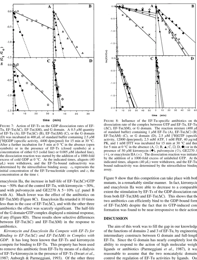
Tu-Specific Antibiotics Are ActiVe on GTP-Bound EF-Tu(∆C). A common effect of the action of the EF-Tu-specific antibiotic is the strong retardation of the EF-Tu‚ GTP dissociation rate (Parmeggiani & Swart, 1985; Anborgh & Parmeggiani, 1993; Cetin et al., 1996). Therefore, the dissociation rate of the GTP complex is the most representa-tive parameter for analyzing the response to antibiotics of the mutated EF-Tu’s. As shown in Figure 8, kirromycin, pulvomycin, GE2270 A, and enacyloxin IIa were able to retard consistently the dissociation rate of EF-Tu(∆C)‚GTP from 4- to 7-fold, depending on the kind of antibiotic. With FIGURE5: Formation of a stable complex between Val-tRNAValand EF-Tu, EF-Tu(∆C), or EF-Tu(∆M). The reaction mixtures contained, in 30µL of standard buffer, 1 µM EF-Tu(A), EF-Tu(∆M) (B), or EF-Tu(∆C) (C), 5 µM [γ-32P]GTP (specific activity, 3500 dpm/pmol), 1 mM PEP, and 40µg/mL PK incubated at 10 min at 30°C in the absence (open symbols) or presence (closed symbols) of 5µM Val-tRNAValand were subsequently passed onto a Sephadex G25 column (0.37× 15 cm) equilibrated with standard buffer. Fractions (100 µL) were collected and the radioactivity was determined.
FIGURE6: Enzymatic binding of [14C]Phe-tRNAPhe to the A-site of poly(U)-programmed ribosomes mediated by EF-Tu, EF-Tu-(∆C), or EF-Tu(∆M). The reaction mixture (20 µL) contained 25 mM Tris-HCl, pH 7.6, 86 mM NH4Cl, 10 mM MgCl2, 7 mM ME, 1 mM ATP, 0.25 mM GTP, 1 mM PEP, PK (40µg/mL), 0.5 µM ribosomes poly(U) (80µg/mL), 0.5 µM tRNAPhe, 0.7µM [14 C]-Phe-tRNAPhe(specific activity, 584 dpm/pmol), and 1µM EF-Tu (O), EF-Tu(∆C) (2), EF-Tu(∆M) (0) or no enzyme (b). The diverse EF-Tu’s in complex with GTP were preincubated with [14 C[]-Phe-tRNAPheand the ribosomes with tRNAPheand poly(U), for 10 min at 30 °C. The reaction was initiated by the addition of the poly(U)-programmed ribosomes. The binding of [14C]Phe-tRNAPhe to the ribosomal A-site was determined by the nitrocellulose binding assay.
enacyloxin IIa, the increase in half-life of EF-Tu(∆C)‚GTP was∼50% that of the control EF-Tu, with kirromycin ∼30%, and with pulvomycin and GE2270 A 5-10% (cf. panel B with A). Much lower was the effect of the antibiotics on EF-Tu(∆M) (Figure 8C). Enacyloxin IIa retarded it 10 times less than in the case of EF-Tu(∆C), and with the other three antibiotics the effect was scarcely significant. The half-life of the G domain‚GTP complex displayed a minimal response, if any (Figure 8D). These results show selective differences between EF-Tu(∆C) and EF-Tu(∆M) in the response to antibiotics.
Kirromycin and Enacyloxin IIa Compete with EF-Ts for Binding to EF-Tu(∆C) and EF-Tu(∆M) in Complex with GDP. It has long been known that EF-Ts and kirromycin compete for binding to EF-Tu. This property has been used to remove this antibiotic from EF-Tu by means of a dialysis of EF-Tu‚kirromycin in the presence of EF-Ts (Swart et al., 1987; Anborgh & Parmeggiani, 1993). Of the other three EF-Tu-specific antibiotics, only enacyloxin IIa can compete with EF-Ts for binding to EF-Tu like kirromycin (Cetin et al., 1996). Therefore, we tested whether EF-Ts and these two antibiotics were still able to compete for binding to the two mutated EF-Tu forms. The experiments illustrated in
Figure 9 show that this competition can take place with both mutants, in a remarkably similar manner. In fact, kirromycin and enacyloxin IIa were able to decrease to a comparable extent the stimulation by EF-Ts of the GDP dissociation rate from both EF-Tu(∆M) and EF-Tu(∆C). This shows that the two antibiotics can efficiently bind to the GDP-bound form of EF-Tu(∆M) despite the fact that its GTP-induced con-formation was found to be near-irresponsive to their action. DISCUSSION
The aim of this work was to fill the gap in our knowledge of the functions of domains 2 and 3 of EF-Tu, by engineering intermediary constructs between G domain and full-length EF-Tu. Since the G domain has nearly completely lost the ability to respond to the action of high molecular weight ligands or antibiotics (Parmeggiani et al., 1987), it was reasonable to assume that the two noncatalytic domains control the regulation of EF-Tu activities by ligands. Our results show that, despite the deletion of an entire noncata-lytic domain, both mutated forms are still able to interact with GTP and GDP, as has been found for the G domain (Parmeggiani et al., 1987). They also catalyze the hydrolysis of GTP and can sustain the interactions with ligands, such FIGURE7: Action of Ts on the GDP dissociation rates of
EF-Tu, EF-Tu(∆C), EF-Tu(∆M), and G domain. A 0.5 µM quantity of EF-Tu (A), EF-Tu(∆C) (B), EF-Tu(∆M) (C), or the G domain (D) was incubated in 400µL of standard buffer containing 2.5 µM [3H]GDP (specific activity, 4400 dpm/pmol) for 15 min at 30°C. After a further incubation for 5 min at 0°C in the absence (open symbols) or in the presence of EF-Ts (closed symbols) at a concentration of either 0.5 (solid line) or 0.005µM (dashed line), the dissociation reaction was started by the addition of a 1000-fold excess of cold GDP at 0°C. At the indicated times, aliquots (40 µL) were withdrawn, and the EF-Tu-bound radioactivity was determined by the nitrocellulose binding assay. c0represents the initial concentration of the EF-Tu‚nucleotide complex and ct the
concentration at the time t.
FIGURE 8: Influence of the EF-Tu-specific antibiotics on the dissociation rate of the complex between GTP and EF-Tu, EF-Tu-(∆C), EF-Tu(∆M), or G domain. The reaction mixture (400 µL of standard buffer) containing 1µM EF-Tu (A), EF-Tu(∆C) (B), EF-Tu(∆M) (C), or G domain (D), 2.5 µM [3H]GTP (specific activity, 12000 dpm/pmol), 2.5 mM ATP, 1 mM PEP, 40µg/mL PK, and 1 mM DTT was incubated for 15 min at 30°C and then for 5 min at 0°C in the absence (A, O; B, 2; C, 0; D, b) or in the presence of 50µM kirromycin ([), pulvomycin (]), GE2270 A (+), or enacyloxin IIA (×). The dissociation reaction was initiated by the addition of a 1000-fold excess of unlabeled GTP. At the indicated times, aliquots (40µL) were withdrawn, and the EF-Tu-bound radioactivity was determined by the nitrocellulose binding assay.
as antibiotics and EF-Ts. The existence of a functionally active conformation and of selective properties dependent on whether the middle or the C-terminal domain is deleted supports the presence in both EF-Tu mutants of a folding close to that of the intact molecule. This agrees with NMR studies of Lowry et al. (1991) showing the conservation of an organized structural folding in the nucleotide binding region of the G domain even after removal of both noncata-lytic domains. Also, the CD spectra of EF-Tu(∆M) and EF-Tu(∆C) indicate that the organization of the secondary structural elements of the two EF-Tu mutants is not dramati-cally changed. EF-Tu(∆M) displays a thermostability close to that of the intact molecule whereas EF-Tu(∆C) resembles the G domain. It was important to verify whether the relative orientation of domains 1 and 3 in EF-Tu(∆M) was not markedly altered. Therefore, in addition to functional tests we carried out a model building (Figure 10A) of the orientation and folding of these two domains after looping out residues 206-293. Using the 3D model of EF-Tu‚ GMPPNP from Thermus aquaticus (PDB database), we removed the residues from V217 (I206 in E. coli EF-Tu) to K306 (K294 in E. coli EF-Tu), after which energy minimiza-tion was applied. Our results show that the interacminimiza-tion forces between domains 1 and 3 of EF-Tu are strong enough to preserve the interface of these two domains, avoiding a significant displacement of the relative orientation and distance. Nor did we observe a significant difference in the
folding of the two domains as shown by superimposition with the corresponding domains of the full-length EF-Tu. Another conclusion is that the resulting joining loop (11 residues between 201-205 and 294-299) connecting do-main 1 (residues 4-200) and dodo-main 3 (residues 300-393) after the deletion of the middle domain is long enough to span the distance between the two residual domains. To-gether, these observations show that all the individual domains of EF-Tu constitute remarkably stable conforma-tional entities, capable of sustaining specific activities. This justifies their use, individually or in combination, to analyze the structural-functional relationships of EF-Tu in the interactions with ligands.
The affinities of EF-Tu(∆C) and EF-Tu(∆M) for GDP and GTP lie in the micromolar range and are comparable to those reported for the G domain (Parmeggiani et al., 1987). As has been shown for the G domain, the removal of either one of the two domains mainly influences the GDP-bound state of EF-Tu, decreasing the affinity for this nucleotide∼1000 times. These results suggest that the nucleotide binding pocket in the two mutants, independently of the presence of GTP or GDP, has an open conformation resembling that of EF-Tu‚GTP, whose affinity for the nucleotide is more than 100-fold less strong than for GDP. Thus, a typical feature of EF-Tu, the differential affinity for GTP and GDP, results from cooperative effects involving all three domains. Note-worthy, the half-life of the GDP complex of the two FIGURE 9: Kirromycin (I) and enacyloxin IIa (II) compete with EF-Ts for binding to EF-Tu(∆C) and EF-Tu(∆M). EF-Tu‚GDP (A), EF-Tu(∆C) (B), or EF-Tu(M) (C) (0.10 µM) was incubated in 400 µL of standard buffer containing 0.12 µM [3H]GDP (specific activity, 11000 dpm/pmol) for 15 min at 30°C. After the additional incubation for 5 min at 0°C in the presence of kirromycin or enacyloxin IIa (dashed lines) or in the absence of antibiotics (solid lines), with 0.01µM EF-Ts (closed symbols) or without EF-Ts (open symbols), the dissociation reaction was initiated by the addition of a 1000-fold excess of cold GDP at 0°C. At the indicated times, aliquots (40µL) were withdrawn, and the EF-Tu-bound radioactivity was determined by the nitrocellulose binding assay.
constructs is intermediary between that of the G-domain and of the intact molecule, suggesting a stabilization effect by each of the two noncatalytic domains. In the case of EF-Tu(∆M), this can be rationalized in terms of the 3D model, where domain 3 appears to stabilize R-helices B and C in the N-terminal domain.
The intrinsic GTPase activity of EF-Tu(∆M) was found to be similar to that of wild-type EF-Tu; that of EF-Tu(∆C) slightly decreased. Also, the stimulation by kirromycin is still present, though it is lower particularly in the case of deletion of the middle domain.
No stimulatory effect by ribosomes was observed on the intrinsic GTPase activity of the two mutated EF-Tu’s. This was rather surprising, because ribosomes can slightly enhance the GTPase activity of the G domain (Parmeggiani et al., 1987; Jensen et al., 1989). The lack of an effect may be related to negative long-range changes induced by the residual noncatalytic domain on domain 1.
Either one of the mutated forms can form a stable complex with aa-tRNA but only to an extent <10% that of EF-Tu, whereas no binding of aa-tRNA can take place on the G domain (Parmeggiani et al., 1987). These findings empha-size the importance of all three domains for an efficient binding of aa-tRNA. Cross-linking and protection studies suggested that the aminoacylated CCA 3′-end of tRNA interacts with domains 1 and 2 (Jona´k et al., 1980, 1984; Metz-Boutigue et al., 1989; Van Noort et al., 1984). The 3D model of Phe-tRNAPhe-bound T. aquaticus EF-Tu‚
GppNHp (Nissen et al., 1995) shows that the binding site of the aminoacylated acceptor helix of tRNA is located in the interfaces of the three domains. The combination of E. coli (Pieper et al., 1990) or T. thermophilus (Peter et al., 1990) middle and C-terminal domains was unable to interact with aa-tRNA. For poly(Phe) synthesis, our results show that both noncatalytic domains of EF-Tu are required, as expected from the complexity of this process involving multiple steps and the formation of highly selective com-plexes.
A major result of this study was the finding that EF-Ts can stimulate efficiently to a comparable extent the release of GDP from both mutated constructs. Observation based on point substitutions (Hwang et al., 1992) suggested that the region of domain 1 comprising residues 154-195 interacts with EF-Ts. Peter et al. (1990) reported that the two noncatalytic domains interact with EF-Ts, since EF-Ts could retard the electrophoretic mobility of T. thermophilus EF-Tu lacking domain 1. The elucidation of the 3D structure of EF-Tu‚EF-Ts (Kawashima et al., 1996) shows that the most extensive contact between the two proteins takes place on domain 1. Several structures are involved, such as the phosphate binding loop, the amino terminus of helix Bstightly associated with domain 3sand the helices C and D. Besides domain 1, only the tip of domain 3 appears to interact directly with EF-Ts. The observation that at least one noncatalytic domain is required for a productive interaction between EF-Tu and EF-Ts indicates that both noncatalytic domains are FIGURE10: (A) The relative orientation of domains 1 and 3 is essentially conserved in the EF-Tu(∆M) construct. The coordinates of the
T. aquaticus EF-Tu‚GMPPNP model were obtained from the PDB database. Using a Quanta software package, residues V217-K306
(corresponding to I206 and K294, respectively, in E. coli EF-Tu) were removed, after which energy minimization was applied using the CHARMM22.0 force field. Color codes: full-length EF-Tu, blue; EF-Tu(∆M), yellow (domain 1) and red (domain 3). (B) A model outlining possible regions of EF-Tu‚GTP involved in the binding sites for antibiotics. This schematic representation has been derived from (i) comparative analyses of the actions of the antibiotics, (ii) the location of residues inducing resistance toward kirromycin (L120, Q124, Y160, G316, Q329, A375, and E378), pulvomycin (R230, R230/R233, R333, and T334), and possibly GE2270 A (V226, see Discussion), and (iii) the properties of the respective mutants. Pulvomycin and GE2270 A both abolish the interaction between EF-Tu and aa-tRNA taking place in the interfaces between domains 1 and 2 and domains 2 and 3; kirromycin and particularly enacyloxin IIa affect negatively the protection by EF-Tu of the aminoacylated N-terminal end of tRNA. Only kirromycin can enhance significantly the intrinsic GTPase activity of EF-Tu.
involved in inducing the active conformation of domain 1 responsive to EF-Ts. In fact, the G domain‚GDP complex shows an intrinsic dissociation activity much higher than that of EF-Tu(∆C) and EF-Tu(∆M) (cf. Figure 7, panel D with panels B and C). A regulatory indirect function of domain 2 is supported by the absence in the 3D model of contacts between this domain and EF-Ts (Kawashima et al., 1996). The physiological nature of the response to EF-Ts of the two mutated EF-Tu forms is further supported by the partial inhibition by kirromycin and enacyloxin IIa of the GDP release induced by EF-Ts, a specific effect typical for full-length EF-Tu.
The observation that EF-Ts is unable to enhance the dissociation of the GTP complex of the mutants suggests that the differential affinity of EF-Ts for the GTP- and GDP-bound forms of the constructs has become much larger than that of EF-Tu wild type. Indeed, the dissociation rate of Tu‚GTP is a few times less sensitive to Ts than EF-Tu‚GDP (Fasano et al., 1978).
Selective effects have been found in this work, as to the action of antibiotics on the two mutated EF-Tu’s. EF-Tu-(∆C)‚GTP was much more responsive than EF-Tu(∆M)‚GTP to all four EF-Tu-specific antibiotics, showing a response of the same kind as the full-length molecule. Site-directed mutagenesis has shown that the substitutions conferring kirromycin resistance concern residues L120, Q124, and Y160 on domain 1 and G316, Q329, E378, T357, and A375 on domain 3 (Parmeggiani & Swart, 1985; Mesters et al., 1994; Abdulkarim et al., 1994). Functional and structural analyses have led to the conclusion that the interface of domain 1 with domain 3, whose extention is markedly different in the GDP- and GTP-bound forms of EF-Tu, harbors the binding site for kirromycin, very likely involving Q124 (Mesters et al., 1994). Our results show that even after removal of domain 3 kirromycin can still interact with EF-Tu, with an efficiency stronger than after removal of domain 2. Hence, the presence of domain 2 is crucial for inducing an active binding site for kirromycin on domain 1. Moreover, the inhibition of the action of EF-Ts on EF-Tu-(∆M)‚GDP shows that kirromycin can tightly bind to domain 1 in the absence of domain 2. One can conclude that the two noncatalytic domains act by concurring directly (domain 3) or indirectly (domain 2) to the activation of the kirromycin binding site on domain 1. The ability of EF-Tu(∆M)‚GDP to support specific effects such as the action of EF-Ts and the response to kirromycin and enacyloxin IIa shows that the selective properties of the two mutated constructs are a consequence of the removal of specific structures and not the result of an unspecific conformational disorder. Ad-ditional information on the role of both noncatalytic domains in the interaction of EF-Tu with kirromycin might become available soon, since the complex EF-Tu‚kirromycin has been crystallized recently (Kristensen et al., 1996).
Although enacyloxin IIa-resistant mutants of EF-Tu have as yet to be isolated, the similarities of its action on EF-Tu and EF-Tu‚EF-Ts to that of kirromycin suggest that its binding site also involves the interface between domains 1 and 3 and is likely situated on domain 1. The location of the binding sites for kirromycin and enacyloxin IIa in a domain interface could explain the ability of the two antibiotics to deregulate all the interactions between EF-Tu
and the various ligands by modifying the relative orientation of two domains and thus of the overall conformation of the molecule.
The residues, whose substitution induces pulvomycin resistance (R230 and R230/R233 on domain 2, R333 and T334 on domain 3; Zeef et al., 1994; Boon et al., 1995), are located in the interfaces of the three domains of EF-Tu. Even though structural considerations make uncertain that these residues are part of the binding site for pulvomycin (Zeef et al., 1995), the lack of response of EF-Tu(∆M) to pulvomycin and the probable existence of distinct binding sites for pulvomycin and kirromycin (Pingoud et al., 1982) and of hindrance phenomena between aa-tRNA and pulvomycin appear to favor a location of the binding site for pulvomycin close to the domain 2/domain 3 interface.
Shimanaka et al. (1995) have isolated Bacillus subtilis Tu mutated in V228A (corresponding to V226 in E. coli EF-Tu) resistant to amythiamicin, an antibiotic whose structure is closely related to GE2270 A. We have found that GE2270 A efficiently retards the dissociation rate of EF-Tu(∆C)‚GTP, without affecting that of EF-Tu(∆M), like pulvomycin, and have also observed that GE2270 A induces a band shift of EF-Tu(∆C) during nondenaturing PAGE (P. H. Anborgh, unpublished observations). These results, together with the similarity of its action with that of pulvomycin, suggest that the GE2270 A binding site also involves the middle and C-terminal domain of EF-Tu.
Figure 10B illustrates the probable locations on EF-Tu‚ GTP of the binding sites for antibiotics, as derived from our present knowledge of the properties of EF-Tu mutants.
When this work was in progress, Nock et al. (1995) described the properties of T. thermophilus EF-Tu lacking domain 3. This construct showed a similar affinity for GDP and GTP, like the corresponding G domain and our E. coli EF-Tu(∆C). The presence of domain 2 increased the thermostability of T. thermophilus domain 1 and even more markedly the intrinsic GTPase activity of this domain, two effects that we could not observe with E. coli EF-Tu(∆C). Moreover, T. thermophilus EF-Ts was inactive on T. ther-mophilus EF-Tu(I, II). At present, it is not possible to explain these differences that may be, at least in part, related to the intrinsic properties of T. thermophilus Tu and EF-Ts or the specific experimental conditions used.
In conclusion, our work outlines selective effects on domains 2 and 3 of EF-Tu in the action of antibiotics and EF-Ts and emphasizes their cooperative function in the interaction with ribosomes and aa-tRNA and in the dif-ferential affinity for GTP and GDP. The removal of either one of the noncatalytic domains has revealed effects, whose structural background is not always clear. In fact, in the 3D model only domain 3 interacts with Ts; however, EF-Tu(∆C) induces a functional response to EF-Ts as Tu-(∆M) and Tu wt, unlike the isolated domain 1 EF-Tu(∆C) responds to kirromycin more efficiently than EF-Tu(∆M) though the binding of the antibiotic is very likely situated in the domain 1/domain 3 interface. These results support the existence of long-range effects that cannot yet be explained by the static configuration of the to date 3D models and suggest caution in interpreting functional ob-servations on the basis of our present structural knowledge.
Anborgh, P. H., & Parmeggiani, A. (1991) EMBO J. 10, 779-784.
Anborgh, P. H., & Parmeggiani, A. (1993) J. Biol. Chem. 268, 24622-24628.
Anborgh, P. H., Swart, G. W. M., & Parmeggiani, A. (1991) FEBS
Lett. 292, 232-236.
Berchtold, H., Reshetnikova, L., Reiser, C. O. A., Schirmer, N., Sprinzl, M., & Hilgenfeld, R. (1993) Nature 365, 126-132. Boon, K., Krab, I., Parmeggiani, A., Bosch, L., & Kraal, B. (1995)
Eur. J. Biochem. 227, 816-822.
Bradford, M. M. (1976) Anal. Biochem. 72, 248-254.
Cetin, R., Krab, I. M., Anborgh, P. H., Cool, R. H., Watanabe, T., Sugiyama, T., Izaki, K., & Parmeggiani, A. (1996) EMBO J.
15, 2604-2611.
Cre´chet, J.-B., & Parmeggiani, A. (1986) Eur. J. Biochem. 161, 647-653.
Fasano, O., Bruns, W., Cre´chet, J. B., Sander, G., & Parmeggiani, A. (1978) Eur. J. Biochem. 89, 557-565.
Fasano, O., De Vendittis, E., & Parmeggiani, A. (1982) J. Biol.
Chem. 257, 3145-3150.
Hwang, Y.-W.; Carter, M., & Miller, D. (1992) J. Biol. Chem. 267, 22198-22205.
Ivell, R., Sander, G., & Parmeggiani, A. (1981) Biochemistry 20, 6852-6859.
Jacquet, E., & Parmeggiani, A. (1988) EMBO J. 7, 2861-2867. Jensen, M., Cool, R. H., Mortensen, K. K., Clark, B. F. C., &
Parmeggiani, A. (1989) Eur. J. Biochem. 182, 247-255. Jona´k, J., Smrt, J., Holy, A., & Rychlı´k, I. (1980) Eur. J. Biochem.
105, 315-320.
Jona´k, J., Petersen, T. E., Meloun, B., & Rychlı´k, I. (1984) Eur. J.
Biochem. 144, 295-303.
Jurnak, F. (1985) Science 230, 32-36.
Kawashima, T., Berthet-Colominas, C., Wulff, M., Cusack, S., & Leberman, R. (1996) Nature 379, 511-518.
Kaziro, Y. (1978) Biochim. Biophys. Acta 505, 95-127. Kjeldgaard, M., & Nyborg, J. (1992) J. Mol. Biol. 223, 721-742. Kjeldgaard, M., Nissen, P., Thirup, S., & Nyborg, J. (1993)
Structure 1, 35-50.
Nock, S., Grillenbeck, N., Ahmadian, M. R., Kreutzer, R., & Sprinzl, M. (1995) Eur. J. Biochem. 234, 132-139.
Parmeggiani, A., & Sander, G. (1981) Mol. Cell. Biochem. 35, 129-158.
Parmeggiani, A., & Swart, G. W. M. (1985) Annu. ReV. Microbiol.
39, 557-577.
Parmeggiani, A., Swart, G. W. M., Mortensen, K. K., Jensen, M., Clark, B. F. C., Dente, L., & Cortese, R. (1987) Proc. Natl. Acad.
Sci. U.S.A. 84, 1814-1818.
Peter, M. E., Reiser, C. O. A., Schirmer, N. K., Kiefhaber, T., Ott, G., Grillenbeck, N. W., & Sprinzl, M. (1990) Nucleic Acids Res.
18, 6889-6893.
Pieper, U., Ehbrecht, H. J., Fliess, A., Schick, B., Jurnak, F., & Pingoud, A. (1990) Biochim. Biophys. Acta 1087, 147-156. Pingoud, A., Block, W., Urbanke, W., & Wolf, H. (1982) Eur. J.
Biochem. 123, 261-265.
Polekhina, G. Thirup, S., Kjeldgaard, M., Nissen, P., Lippmann, C., & Nyborg, J. (1996) Structure 4, 1141-1151.
Sambrook, J., Fritsch, E. F., & Maniatis, T. (1989) in Molecular
Cloning, a Laboratory Manual, 2nd ed., Cold Spring Harbor
Laboratory Press, Cold Spring Harbor, NY.
Scarano, G., Krab, I., Bocchini, V., & Parmeggiani, A. (1995) FEBS
Lett. 365, 214-218.
Shimanaka, K., Iinuma, H., Hamada, M., Ikeno, S., Tsuchiya, K. S., Arita, M., & Hori, M. (1995) J. Antibiot. 48, 182-184. Smith, D. B., & Johnson, K. S. (1988) Gene 67, 31-40. Swart, G. W. M., Parmeggiani, A., Kraal, B., & Bosch, L. (1987)
Biochemistry 26, 2047-2054.
Van Noort, J. M., Kraal, B., Bosch, L., la Cour, T. F. M., Nyborg, J., & Clark, B. F. C. (1984) Proc. Natl. Acad. Sci. U.S.A. 81, 3969-3972.
Weijland, A., Harmark, K., Cool, R. H., Anborgh, P. H., & Parmeggiani, A. (1992) Mol. Microbiol. 6, 683-688.
Zeef, L. A. H., Bosch, L., Anborgh, P. H., Cetin, R., Parmeggiani, A., & Hilgenfeld, R. (1994) EMBO J. 13, 5113-5120. BI970443O