57 JOURNAL OF HEALTH SCIENCES
A. J. Health Sci. Volume 2 No 1 | January 2020, 57-63
Case Study
Treatment of Iatrogenic Factor Related Gingival Recession: A Case Reportİlknur Özenci1 ORCID: 0000-0002-5017-5883 Şebnem Dirikan İpçi1 ORCID: 0000-0002-7024-3150 Gökser Çakar1 ORCID: 0000-0002-8766-8120 Selçuk Yılmaz2
1Department of Periodontology, Faculty of Dentistry, Altınbaş University, Istanbul, Turkey. 2Private Practice, Taksim, Istanbul, Turkey
Submitted: April 1, 2019; Accepted: August 27, 2019
Abstract: Recent reports in the literature show that the application of coronally advanced flap i n c ombination
with subepithelial connective tissue graft in the treatment of Miller Class II and even III gingival recessions leads to successful results. This case report describes the use of subepithelial connective tissue graft and enamel matrix derivative combination with coronally advanced flap for the treatment of localized gingival recession caused by an ill-fitting crown restoration. The restorative treatment comprised of the retreatment of the endodontic and restorative needs. At 12 months, successful root coverage with 3.5 mm attachment gain was achieved. The patient had minimal discomfort and was satisfied with esthetic results. This case encourages the usage of subepithelial connective tissue graft, enamel matrix derivative and coronally advanced flap combination in localized Miller Class III gingival recessions. Even though the treatment of Miller Class III gingival recessions are challenging due to loss of interproximal bone and soft tissues, the application of subepithelial connective tissue graft in combination with the use ofenamel matrix derivative has shown to be a predictable treatment procedure in esthetic areas forroot coverage and gain of clinical attachment in single Miller Class III recession defects.
Keywords: Coronally advanced flap; e namel m atrix d erivative; M iller C lass I II g ingival r ecession; r oot coverage e sthetic score; subepithelial connective tissue graft
Address of Correspondence: İlknur Özenci- [email protected] Tel: +90(212)7094528; Fax:
+90(212)4458171. Department of Periodontology, Faculty of Dentistry, Altınbas University, Zuhuratbaba Mahallesi, İncirli Caddesi No: 11-A, 34147 Bakırköy, İstanbul, Turkey
1.Introduction
As clinicians one of our aims is to perform biologically integrated and esthetically pleasing restorations. If tooth preparation does not performed properly, trauma to the periodontal tissues (Gracis et al., 2001)
58
and biological width violation (De Waal and Castellucci, 1994) can occur and lead to the emergence of gingival recessions. Especially in such cases, a good esthetic outcome can be achieved by accurate diagnosis and development of a comprehensive treatment plan. The clinician should always implement both the current knowledge of restorative and periodontal procedures together. Although many techniques have been proposed to treat localized gingival recessions, combination of coronally advanced flap (CAF) and subepithelial connective tissue graft (SCTG) provides more gingival stability in the long term (Cairo, 2017). When there is interproximal bone loss as seen in Miller Class III gingival recessions and naked root surface is wide, additional biological mediator increases the predictability of root coverage. From this perspective, an enamel matrix derivative (EMD) with its angiogenic activity and regenerative capacity is a valuable option for clinicians. It may be expected that the combination of EMD with SCTG will affect periodontal wound healing and regeneration positively (Miron et al., 2016). This case report presents the restorative and periodontal surgical treatment of a patient with gingival recession and high esthetic demands using the CAF combined with SCTG and EMD.
2. Case Description and Results
A 35-year-old female patient applied to our clinic with complaints of poor esthetic appearance and gingival inflammation at the anterior maxilla. The medical history of the patient did not reveal a systemic contraindication for dental treatment. She did not smoke. The patient had undergone a prosthetic treatment that was without harmony and symmetry. After the assessment of clinical and radiographic conditions, patient was diagnosed as localized chronic periodontitis together with localized gingival recession in tooth #11 (Figure 1a). Iatrogenic restorative treatment and microbial dental plaque were defined as etiologic factors.
All clinical parameters were measured at the mid-buccal aspect of the related tooth; probing depth (PD) of 2 mm, recession height (RH) of 3 mm, clinical attachment level (CAL) of 5 mm, recession width (RW) of 3 mm, keratinized tissue height (KTH) of 3 mm, and gingival thickness (GT) of 1 mm. Radiographic evaluation revealed interproximal bone loss and recession defect was classified as Miller Class III (Figure 1b). Root canal of tooth #11 was retreated (Figure 1c), and a temporary crown was placed to allow better healing (Figure 2a). Since a healthy periodontium should be established before any prosthetic procedure, a comprehensive periodontal therapy was performed. The first step of this treatment was oral hygiene instruction and scaling and root planning (initial periodontal therapy). One month after initial therapy re-evaluation was done and low full-mouth plaque (13%) and bleeding scores (12%) were achieved and decided to move on surgical periodontal therapy. Surgical treatment plan was decided as CAF in combination with SCTG+EMD in order to treat the affected hard and soft tissue defects around tooth #11 at the same time, and to improve the level of clinical attachment. After all risks and benefits were explained to the patient, oral and written consents were obtained.
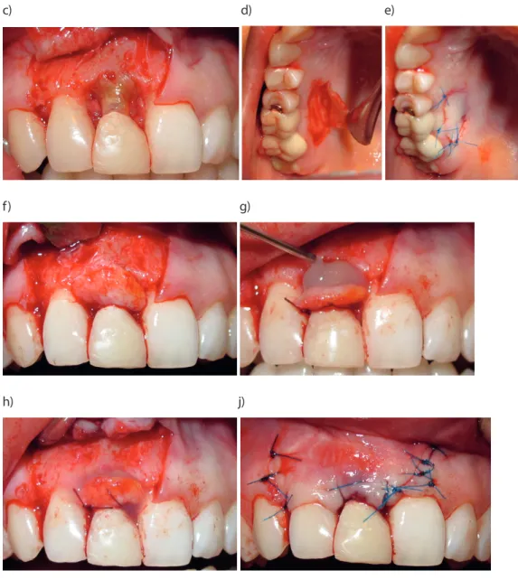
After the application of local anaesthesia, an intracrevicular incision was made at the buccal aspect of the tooth #11 (Figure 2b) and extended to the mesial line angle of tooth #21 and one tooth distally till the mesial line angle of tooth #13. Then, trapezoidal flap was prepared from the line angles and extended to the mucogingival junction reaching the alveolar mucosa in order to position the flap coronally without
59 JOURNAL OF HEALTH SCIENCES
A. J. Health Sci.
any tension. The flap was elevated in a split-full-split thickness approach (Figure 2c) (Pini Prato et al., 1992). The epithelium on the interdental papilla was de-epithelized for better vascularization. The exposed root surface was conditioned with EDTA 24%, pH 6.7 (PrefGel 0.6 ml gel, Straumann, Basel, Switzerland) for 2 min. Then, the root surface was rinsed with saline. The SCTG, obtained by trap door technique (Figures 2d, 2e) was adapted to cover the root surface to the level of cementoenamel junction (CEJ), and sutured by using sling suture technique with 5-0 resorbable sutures (Pegelak, Dogsan, Turkey) (Figure 2f). EMD was applied (Figures 2g, 2h). Finally, the flap was coronally advanced at least 1 mm coronal to CEJ and sutured to completely cover the SCTG using double sling suture technique with 5-0 non-resorbable polypropylene monofilament sutures (Propilen, Dogsan, Turkey). The releasing incisions were closed with interrupted sutures (Figure 2i).
For infection and pain control, systemic antibiotic (amoxicillin clavulanate, 2×625 mg, 5 days), oral analgesic (naproxen sodium, 550 mg, first day after surgery 2×1, afterwards as necessary), and oral rinse (chlorhexidine solution 0.12% rinse, 3×1 for 1 minute) were prescribed. After 2 weeks, sutures were removed. Four weeks after surgery, temporary crown was revised to achieve better soft tissue contour. At 6 months, permanent restoration was applied.
Post-operative healing was uneventful and oral hygiene remains stable. At 12 months, the recession was almost completely covered with thick keratinized tissue (Figure 3a). However there was still a slight gingival recession (0.5 mm) at tooth #11. An improvement in the thickness and height of the buccal keratinized tissue was observed when compared to baseline. CAL gain was 3.5 mm and this corresponds to a 1 mm reduction in PD and 2.5 mm recession reduction at 12 months. The buccal KTH was 6 mm with 3 mm of KTH increase. GT was increased to 2 mm. Patient satisfaction and esthetic evaluation was also performed by using patient satisfaction score (Mhajan et al., 2007) and root coverage esthetic score (Cairo et al., 2009). Patient satisfaction was assessed by using a three-point rating scale and patient was questioned with regard to root coverage attained, relief from dentinal hypersensitivity, colour, shape and contour of gums, surgical procedure, post-surgical phase and cost effectiveness. This score was detected as 19 out of 21. This result demonstrated that patient was highly satisfied. For the esthetic outcomes of the procedure; gingival margin level, marginal tissue contour, soft tissue texture, mucogingival junction alignment and gingival colour were evaluated. Zero, 3 and 6 points were used for the evaluation of the position of the gingival margin, whereas a score 0 or 1 point was used for each of the other variables. Patient’s root coverage esthetic score was found 7 which mean that the esthetic outcome was satisfying.
60
b) c)
Figure 2. (a) Pre-operative clinical view of recession defect (b) Insicion (c) Flap elevation (d) SCTG harvesting (e)
Suturing of the harvesting area (f) SCTG placement (g) EMD application (h) SCTG + EMD (i) Suturing
61 JOURNAL OF HEALTH SCIENCES
A. J. Health Sci.
c) d) e)
f) g)
h) j)
62
III recession at the esthetic zone caused by iatrogenic tooth preparation and improper crown restoration. When the biologic width is intruded by restorations, the periodontium reacts by recreating room between the alveolar bone and the restorative margin to allow space for tissue reattachment. This can result in gingival inflammation, increased probing depths and gingival recession as seen in this case (Bennani et al., 2017). The choice of surgical technique to treat such recessions will depend on different factors; area anatomy, the interdental attachment level and the amount and thickness of keratinized tissue apical and lateral to the recession. The use of EMD+SCTG+CAF has been deeply researched in the treatment of gingival recessions (Henriques et al., 2010, Rasperini et al., 2011, Shirakata et al., 2018). Some studies presented more favourable results with EMD+SCTG+CAF technique whereas other studies did not find any additive effects of EMD to evaluated clinical parameters. Since in this case there was interdental bone loss and wide naked root surface, EMD was also preferred in addition to CAF+SCTG application. With regard to Miller Class III recessions, data from the literature stated that the accomplishment of CRC was possible with SCTG and CAF application in defects presenting mild interdental attachment loss of 1 to 3 mm (Esteibar et al., 2011, Sculean et al., 2016). It has been suggested that additional grafting can stabilized marginal tissue and provide a scaffold to support wound healing with increasing the thickness of the wound area (Baldi et al., 1999). The clinical results from this case show that EMD used in conjunction with SCTG was successful in the treatment of Miller Class III recession. An incomplete, but at the same time substantial root coverage was achieved (83%). At this point we should keep in mind that, the tooth #11 is a restored tooth and in the literature, the demonstrated results of CRC with Miller Class III recessions are mostly at native tooth.
Considering the results of the present case report, application involving SCTG in combination with the use of EMD seems to be a predictable treatment procedure for root coverage and gain of clinical attachment in single Miller Class III recession defects. However, it is clear that further studies with a larger number of patients are warranted to reach a definitive clinical result.
Conflict of Interests
The authors declare that they have no conflicts of interest.
References
Baldi, C., Pini-Prato, G., Pagliaro, U., Nieri, M., Saletta, D., Muzzi, L., Cortellini, P. (1999). Coronally advanced flap procedure for root coverage. Is flap thickness a relevant predictor to achieve root coverage? A 19-case series. J Periodontol, 70(9), 1077–84.
63 JOURNAL OF HEALTH SCIENCES
A. J. Health Sci.
Bennani, V., Ibrahim, H., Al-Harthi, L., Lyons, K.M. (2017). The periodontal restorative interface: esthetic considerations. Periodontol 2000, 74(1), 74–101.
Cairo, F., Rotundo, R., Miller, P. D., Pini, Prato, G. P. (2009). Root coverage esthetic score: a system to evaluate th.e esthetic outcome of the treatment of gingival recession through evaluation of clinical cases. J Periodontol, 80(4), 705-710.
Cairo, F. (2017). Periodontal plastic surgery of gingival recessions at single and multiple teeth. Periodontol 2000, 75(1), 296–316.
De Waal, H., Castellucci, G. (1994). The importance of restorative margin placement to the biologic width and periodontal health. Part II. Int J Periodontics Restorative Dent, 14(1), 70–83.
Esteibar, J. R. V, Zorzano, L. A. A, Cundín, E. E, Blanco, J. D. M, Medina, J. R. O de G. (2011). Complete root coverage of Miller Class III recessions. Int J Periodontics Restorative Dent, 31(4), e1-7.
Gracis, S., Fradeani, M., Celletti, R., Bracchetti, G. (2001). Biological integration of aesthetic restorations: factors influencing appearance and long-term success. Periodontol 2000, 27, 29–44.
Henriques, P. S. G, Pelegrine, A. A, Nogueira, A. A, Borghi, M. M. (2010). Application of subepithelial connective tissue graft with or without enamel matrix derivative for root coverage: a split-mouth randomized study. J Oral Sci, 52(3), 463-471.
Mahajan, A., Dixit, J., Verma, U. P. (2007). A patient-centered clinical evaluation of acellular dermal matrix graft in the treatment of gingival recession defects. J Periodontol, 78(12), 2348-2355.
Miron, R. J., Sculean, A., Cochran, D. L., Froum, S., Zucchelli, G., Nemcovsky, C., Donos, N., Lyngstadaas, S. P., Deschner, J., Dard, M., Stavropoulos, A., Zhang, Y., Trombelli, L., Kasaj, A., Shirakata, Y., Cortellini, P., Tonetti, M., Rasperini, G., Jepsen, S., Bosshardt, D. D. (2016). Twenty years of enamel matrix derivative: the past, the present and the future. J Clin Periodontol, 43(8), 668-683.
Pini-Prato, G., Tinti, C., Vincenzi, G., Magnani, C., Coltellini, P., Clauser, C. (1992). Guided tissue regeneration versus mucogingival surgery in the treatment of human buccal gingival recession. J Periodontol, 63, 919-928. Rasperini, G., Roccuzzo, M., Francetti, L., Acunzo, R., Consonni, D., Silvestri, M. (2011). Subepithelial connective tissue graft for treatment of gingival recessions with and without enamel matrix derivative: a multicenter, randomized controlled clinical trial. Int J Periodontics Restorative Dent, 31(2), 133-139.
Sculean, A., Cosgarea, R., Stähli, A., Katsaros, C., Arweiler, N. B., Miron, R. J., Deppe, H. (2016). Treatment of multiple adjacent maxillary Miller Class I, II, and III gingival recessions with the modified coronally advanced tunnel, enamel matrix derivative, and subepithelial connective tissue graft: a report of 12 cases. Quintessence Int, 47(8), 653-659.
Shirakata, Y., Nakamura, T., Shinohara, Y., Hashiguchi, C., Takeuchi, N., Imafuji, T. (2018). Split-mouth evaluation of connective tissue graft with or without enamel matrix derivative for the treatment of isolated gingival recession defects in dogs. Clin Oral Investig, Dec 1. Doi: 10.1007/s00784-018-2750-1.