CLEO-PR 2015
Copyright @ IEEE Fig. 1: Laser system integrated with OCT. PC controls
scanner and elevator, and gets images from OCT
Fig. 2: 8-pulses repeated at 50kHz
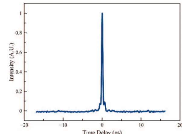
Fig. 3: Autocorrelation of output of laser system. 3μJ maximum pulse energies with 300 fs FWHM.
All-Fiber Burst Mode Laser System Integrated
With OCT for Cataract Surgery
D. K. Kesim
1, H. Kalaycıoğlu
2, C. Kerse
1and F. Omer Ilday
1, 21 Department of Electrical and Electronics Engineering, METU Ankara, 06800, Turkey 2 Department of Physics, Bilkent University, 06800 Ankara, Turkey
Abstract
We demonstrate a burst-mode Yb all-fiber femtosecond laser system integrated with OCT for cataract surgery and aim to enhance further the procedure with lower collateral tissue damage, cleaner, efficient cuts with compact and robust structure.
I. INTRODUCTION
Cataract condition is responsible for two thirds of preventable visual impairments afflicting nearly 190 million all over the world [1]. Every year, 19 million cataract surgeries are performed worldwide [2]. Femtosecond laser assisted cataract surgery has entered clinical use in recent years as an efficient and safe alternative to the traditional method. However, this laser-assisted surgery is in its infancy period and would benefit from lower pulse energies (to minimize collateral effects), shorter operation durations, and more compact laser systems. Fiber lasers can address these requirements with their robustness, compact size and minimal-alignment requiring structure. Further, burst mode operation [3], where high repetition rate pulses are delivered in packets repeated at a relatively low rate, have been shown to produce efficient ablation with minimal collateral thermal effects [4].
II. DETAILS OF SURGICAL SYSTEM
Here, we demonstrate the first fiber based burst-mode femtosecond laser device for cataract surgery. The laser is coupled to an optical coherence tomography (OCT) system with computerized controls (Fig. 1).
A home built all fiber Yb laser amplifier is seeded by a 91-MHz fiber oscillator, followed by a double-clad preamplifier, acusto optic modulator (AOM) pulse picker and a double-clad power amplifier. An FPGA based
electronic system triggered by the oscillator is used to impose the desired pulse train on the laser beam. The system is able to produce pulses up to the 5 μJ range compressible to sub-500 fs. The imaging and tissue processing functions are executed with OCT monitoring, using its galvo scanner with a common beam path. Samples are placed on an elevator controlled by RS232 for vertical scanning. Maximum imaging depth is 1.6 mm with 8 μm axial resolution. The galvo scanner can reach a scan speed of 1 m/s with any desired scanning pattern. In our
preliminary experiments, bursts comprising of 8 pulses with 3 μJ per pulse at 50 kHz burst repetition rate were applied to agar jells (Fig. 2), which have optical response similar to that of the cornea, as well as cornea obtained from cow eyes. The compressed pulses have ~300 fs FWHM when the grating compressor is optimized (Fig. 3).
CLEO-PR 2015
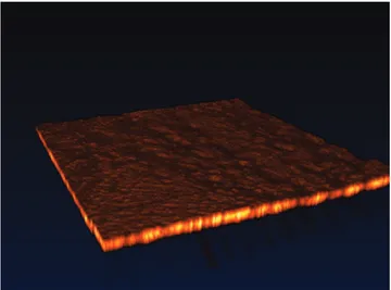
Copyright @ IEEE Fig. 5: (a)Agar surface treated with 8 pulse bursts of 1.4 μJ
per pulse energy at a burst repetition rate of 50 kHz (b) Crossection view
Fig. 4: Agar surface treated with 400 kHz repetition rate pulses of 1.4 μJ energy each
Fig. 6: Raster scanning with 8 pulse bursts containing 1 μJ pulses on cow cornea.
III. PRELIMINARY EXPERIMENTS
Effectiveness of burst mode is demonstrated on agar with bursts with 11.2 μJ (1.4 μJ per pulse energy) while the scan rate was adjusted to obtain 50% overlap between bursts. For the 400 kHz uniform repetition rate mode scan rate was adjusted to obtain equivalent exposure per spot. The OCT images of the results (Fig. 4(b)) show that while burst mode ablated a substantial volume, uniform repetition rate mode failed to ablate any visible volume (Fig. 5). The valley on figure is due to OCT imaging artifacts (Fig. 4(a)).
Additionally smooth cuts on ex vivo cow cornea by raster scan pattern with ~50% overlapping spots (Fig 6) were obtained. Agar and ex vivo cow eye trials show that we can create incisions below the surface, in addition to dotted patterns for corneal incision and smooth cuts for capsulorhexis.
IV. CONCLUSION
In summary, a femtosecond burst mode all-fiber Yb laser amplifier integrated with OCT for cataract surgery is demonstrated for the first time. We show promising results for burst mode with agar and ex vivo experiments on cornea. The system will ultimately be able to create corneal incisions, capsulorhexis and lens fragmentation using a single laser with a common optical path for laser processing and in-situ imaging.
ACKNOWLEDGMENT
This work was supported by the Scientific and Technological Research Council of Turkey TÜBİTAK under grant no. 112T980.
REFERENCES
[1] World Health Organization,”Universal eye health: a global action plan 2014-2019,” WHO Library Cataloguing-in-Publication Data (2013)
[2] K. E. Donaldson et al., “Femtosecond laser assisted cataract surgery,” J Caratact Refract Surg, 39, 1753-1763, (2013)
[3] M. Lapczyna, K.P. Chen, P.R. Herman, H.W. Tan, R.S. Marjoribanks, “Ultra high repetition rate (133MHz) laser ablation of aluminum with 1.2-ps pulses,” Appl. Phys. A 69, S883 (1999).
[4] C. Kerse, H. Kalaycioglu, F. O. Ilday, E. Atalar, ”Non-thermal Material and Tissue Processing with 100 MHz and 500 MHz Repetition Rate