PREGNANCY
Postpartum thyroiditis and hypothalamo-hypophysial insufficiency in
the same woman with successive pregnancies: a case report
SIBEL ERTEK & GURBUZ ERDOGAN
Department of Endocrinology and Metabolic Diseases, Ufuk University Medical Faculty, Dr. Ridvan Ege Hospital, Ankara, Turkey
(Received 13 April 2009; revised 24 May 2009; accepted 20 July 2009)
Abstract
Objective. Although the incidence of postpartum autoimmune disorders of endocrine glands are not rare, the presence of
two different entities in the same patient with two different pregnancies is uncommon.
Methods. We present a 35-year-old woman whose story starts with her first pregnancy when she was 29 years old, she had
the diagnosis of postpartum thyroiditis with hypothyroidism. We followed up the patient when she had her second pregnancy.
Results. When she was being followed up with levothyroxine replacement, 5 years later she had her second delivery after
which she had complaints of polydipsia, polyuria, weight loss and had the diagnosis of central diabetes insipitus and she has started desmopressin treatment and 17 months later the delivery she again applied with amenorrhea, continuation of lactation later she noticed oligomenorrhea, and her gonadotropin levels were found to be low as well as her TSH levels, although the L-thyroxine treatment dose was not changed. Dynamic tests of hypophysis revealed hypophyseal insufficiency and repeated hypophyseal MRI was in concordance with lymphocytic hypophysitis which explains the pattern of endocrinological abnormalities after the second delivery.
Conclusion. This case signals role of autoimmune mechanisms underlying the endocrinopathies seen after successive
pregnancies of the same patient.
Keywords: Postpartum, postpartum thyroiditis, hypophysitis, postpartum hypophysitis, lymphocytic hypophysitis
Introduction
Postpartum thyroiditis is an autoimmune disease with varying prevalence between 1.1% and 16.7% (mean prevalence rate 7.2%) and it commonly presents as hypothyroidism (43%) in previously euthyroid wo-man [1]. Lymphocytic infiltration of the thyroid is the most evident pathological feature and positive antith-yroid peroxidase antibodies are frequently observed [2].
Hypopituitarism arising postpartum may be caused frequently either by lymphocytic hypophysitis or by Sheehan syndrome [3]. Lymphocytic hypophysitis is pituicyte destruction with autoimmune basis, fre-quent in gestational and postpartum period [4]. Together with the granulomatous hypophysitis and xanthomatous hypophysitis, lymphocytic hypophysi-tis, (also called autoimmune hypophysihypophysi-tis, AH), constitute primary chronic inflammatory diseases of
hypophysis. If the inflammation is limited in adeno-hypophysis, it is called lymphocytic adenohypo-physitis (LAH). Other forms are lymphocytic infundibulohypophysitis and lymphocytic panhypo-physitis [5]. Most cases of LAH present in the last month of pregnancy or within the first 2 months after delivery [5]. In the first case of LAH of the medical literature, Hashimoto thyroiditis was also present and the authors emphasized the onset of autoimmune reaction to thyroid and pituitary antigens [6]. In the recent study of Manetti et al. [7] with 961 auto-immune thyroid disease patients, the frequency of antipituitary antibody positivity was significantly higher than patients without autoimmune thyroid disease. The link between these two disorders is not totally clarified [8].
Immune system changes and adaptations in pregnancy cause different effects on the course of autoimmune diseases. Generally, pregnancy
Correspondence: Sibel Ertek, Department of Endocrinology and Metabolic Diseases, Ufuk University, Faculty of Medicine, Dr. Ridvan Ege Hospital, Mevlana Bulvari 86-88 (Konya Yolu) 06520, Balgat, Ankara, Turkiye. Tel:þ90-312-204-40-00. Fax: þ90 312 204 40 55. E-mail: [email protected]
Gynecological Endocrinology, February 2010; 26(2): 105–108
ISSN 0951-3590 print/ISSN 1473-0766 online ª 2010 Informa UK Ltd. DOI: 10.3109/09513590903215532
Gynecol Endocrinol Downloaded from informahealthcare.com by JHU John Hopkins University on 05/19/10
improves the course of rheumatoid arthritis, Graves disease and Type1 diabetes mellitus, on the other side worsens Wegener syndrome [9–12]. Lymphocytic hypophysitis and postpartum thyroiditis are two unique examples of autoimmune diseases that the pregnancy is directly related with disease onset.
Lymphocytic hypophysitis can also be accompanied by other autoimmune diseases, like Addison disease, Type 1 diabetes mellitus, Graves disease, systemic lupus erythematosus, Sjogren syndrome, autoim-mune hepatitis, primary biliary cirrhosis and as in other autoimmune diseases there may be relapsing and remitting course, and spontaneous recovery has also been reported [13].
Searching the Embase and Pubmed both for the all case reports and reviews about the co-existence of these two postpartum diseases in same patient, with key words ‘postpartum hypophysitis’, ‘postpartum thyroiditis’, ‘lymphocytic hypophysitis’, ‘pregnancy’ and ‘hypophysitis’ and ‘thyroiditis’, we found that it was reported in the same pregnancy of a patient in the case report of Bevan et al. [14], another report of both diseases was presented by Iwaoka in 2001 [15].
In this article we present a case with a 9 years story of follow-up during two successive pregnancies.
Case presentation
In 1999, a 26-years-old female patient without any history of thyroid disease has applied to Endocrinol-ogy and Metabolic Diseases Department with com-plaints of fatigue and increase in weight at her fifth month of postpartum period, during lactation. Her story of menstruation was regular with age of menarche at 15 years, and she did not have any problem during her pregnancy and the labour. On physical examination she had diffuse goiter without palpable nodules. On laboratory tests, free T4: 9.59 ng/dl (Normal range:10–23 ng/dl), free T3:
4.38 ng/dl (Normal range: 2.8–7 ng/dl), TSH (thyr-oid-stimulating hormone): 18.6 mcIU/ml (Normal range:0.27–4.20 mcIU/ml), TPO antibody: 365 IU/ ml (Normal range: 0.01–34 IU/ml) and Thyroglo-bulin antibody: 1945 IU/ml (Normal range: 0.01– 115 mIU/ml). She had the diagnosis of postpartum thyroiditis and L-thyroxine treatment was started.
When she was being followed up with the diagnosis of permanent autoimmune thyroiditis with hypothyr-oidism after post-partum period, under L-thyroxine replacement, she had her second delivery at Septem-ber 2004. After the second delivery, she had the complaints of polydipsia, polyuria, prolonged lacta-tion, weight loss, tiredness and she came to endocrinologist in January 2005 when these com-plaints increased. With water deprivation test she had the diagnosis of central diabetes insipitus. In her hypophyseal magnetic resonance (MR) imaging, hyperintensity of neurohypophysis was disappeared. She had started desmopressin spray with the diag-nosis of central diabetes insipitus. Later, she had noticed that she had amenorrhea for 26 months after delivery, and 10 kg weight gain within last 6 months and her lactation continues although she was not giving breast feeding. She applied to her gynaecolo-gist and endocrinologynaecolo-gist with these complaints in December 2006. The tests revealed mild hyperpro-lactinemia and on February 2007 she had started bromocriptin (Table I).
At her control visit on December 2007, she had the complaint of oligomenorrhea and she was taking medical treatment for hypothyroidism and central diabetes insipitus. Her bromocriptin treatment was changed and cabergoline and lynesterol was started. She did not have complaint of galactorhea. Her tests for anterior hypophysis had revealed a suspicion for hypophyseal insufficiency and she was hospitalized for dynamic tests and evaluation for hypophyseal insufficiency (Table I).
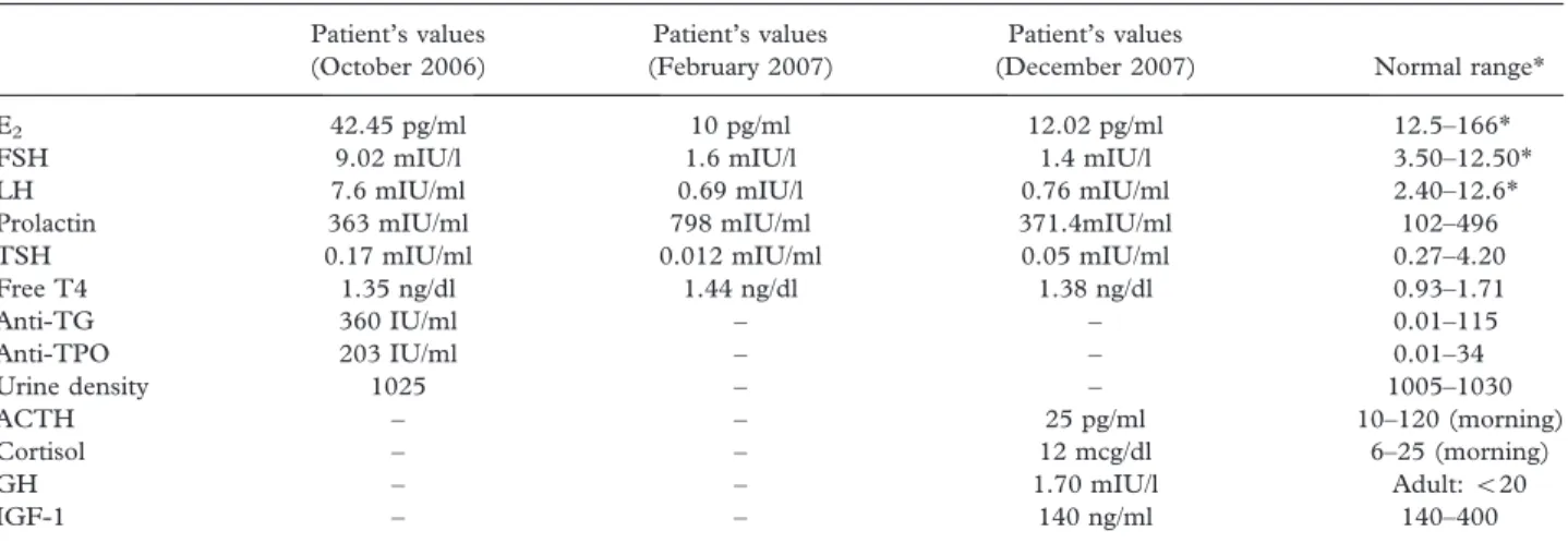
Table I. Laboratory tests of the patient on December 2006, February and December 2007.
Patient’s values (October 2006)
Patient’s values (February 2007)
Patient’s values
(December 2007) Normal range*
E2 42.45 pg/ml 10 pg/ml 12.02 pg/ml 12.5–166*
FSH 9.02 mIU/l 1.6 mIU/l 1.4 mIU/l 3.50–12.50*
LH 7.6 mIU/ml 0.69 mIU/l 0.76 mIU/ml 2.40–12.6* Prolactin 363 mIU/ml 798 mIU/ml 371.4mIU/ml 102–496 TSH 0.17 mIU/ml 0.012 mIU/ml 0.05 mIU/ml 0.27–4.20 Free T4 1.35 ng/dl 1.44 ng/dl 1.38 ng/dl 0.93–1.71 Anti-TG 360 IU/ml – – 0.01–115 Anti-TPO 203 IU/ml – – 0.01–34 Urine density 1025 – – 1005–1030 ACTH – – 25 pg/ml 10–120 (morning) Cortisol – – 12 mcg/dl 6–25 (morning) GH – – 1.70 mIU/l Adult:520 IGF-1 – – 140 ng/ml 140–400
*Normal range for follicular phase of the menstrual cycle. 106 S. Ertek & G. Erdogan
Gynecol Endocrinol Downloaded from informahealthcare.com by JHU John Hopkins University on 05/19/10
Insulin hypoglycaemia test, CRH (corticotrophin-releasing hormone), LHRH (luteinizing hormone-releasing hormone) and TRH (thyrotropin-hormone-releasing hormone) tests were performed. The results of the tests are shown on Table II. As it is seen from the dynamic tests of hypophysis, the cortisol and growth hormone responses to hypoglycaemia, and response of FSH (follicle-stimulating hormone) and LH (luteinizing hormone) to LHRH were not enough. It’s difficult to evaluate the TRH test, because the patient was under L-thyroxine treatment and she dropped the medicine only on the day of test.
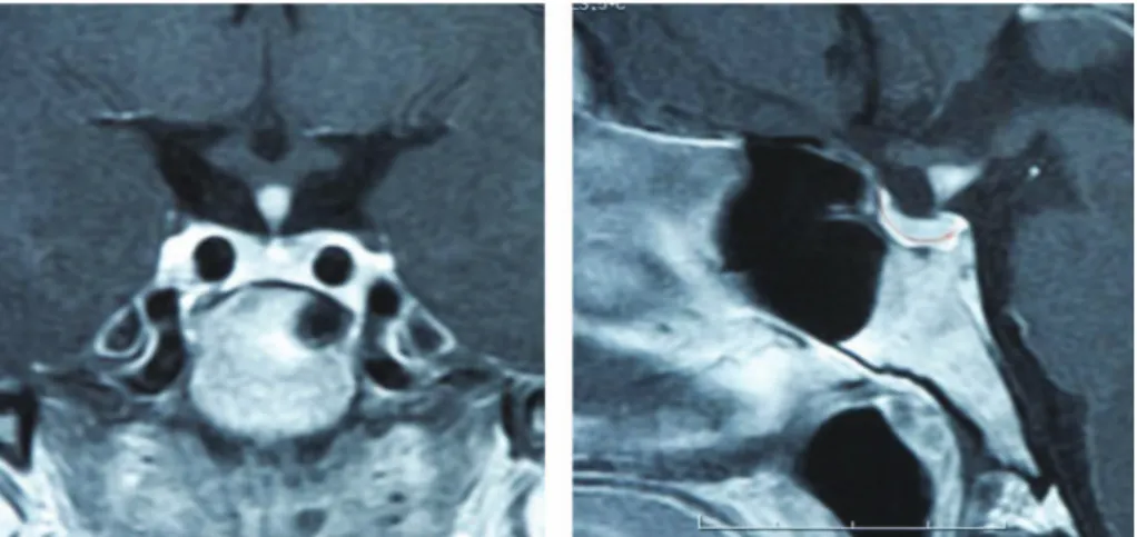
The hypophyseal MR was repeated and focal thickening and contrast enhancement in the middle of infundibulum was reported and this was relevant for lymphocytic hypophysitis (Figure 1).
Discussion
There are mainly three reasons of postpartum hypophyseal dysfunction; Sheehan syndrome, pitui-tary tumors and lymphocytic hypophysitis [5,14]. Absence of obstetrical problem and the presence of lactation after delivery decrease the suspicions of Sheehan syndrome, meanwhile the hormone levels and MR images were not relevant to this syndrome. In the presence of pituitary tumours the typical pattern of endocrinological abnormalities in the adenohypophyseal tests are known to be typical (i.e. theoretically GH and gonadotropin deficiencies occurs first, followed later by ACTH and TSH deficiency) and the MR rules out the presence of any hypophyseal tumour. Symmetrical enlargement and typical thickening of the infundibular stalk on MR series are typical features for lymphocytic hypophy-sitis. The mild hyperprolactinemia at the beginning of the story and presence of previous postpartum thyroiditis also support the diagnosis of this disease. Although the MR images resemble that of the ectopic bright spot of ectopic neurohypophysis, absence of musculoskeletal and midbrain anomalies and the
presence of neurohypophyseal dysfunction and clin-ical story in normal adult female in our case also gives a clue [16].
Table II. Dynamic endocrine tests of the patient.
Hormones
Basal values
With hypoglycaemia* Insulin hypoglycaemia test of the patient
Cortisol (mg/dl) 9.73 17.59
ACTH (pg/ml) 25 12
Growth_hormone (mIU/l) 2.18 2.48 IGF-1 (ng/ml) 140 180
Cortisol (mg/dl) ACTH (pg/ml) CRH test of the patient{
0 min 14.72 18.00
15 min 19.87 41.22
30 min 23.10 21.90
60 min 26.41 18.10
Time FSH (mIU/l) LH (mIU/l)
LHRH test{of the patient
Beginning 2.37 0.90
30 min 6.29 6.42
60 min 8.18 7.07
90 min 8.35 5.97
Time TSH (mcIU/ml) PRL (ng/ml)
TRH testxof the patient
Beginning 0.03 16.87
30 min 0.23 32.10
60 min 0.20 20.57
90 min 0.14 17.62
*Hypoglycemia was accepted as blood glucose value below 40 mg/ dl. Expected normal responses to hypoglycaemia were more than 20 mIU/l for GH, and increase to at least 18–20mg/dl for cortisol.
{
Normally peak cortisol of410 mg/dl should be observed within 30–60 min.
{
The normal response is five times increase in LH and two times increase in FSH within 20 min.
x
The patient stopped L-thyroxine intake only at the day of the test. Normal response should be46 mIU/ml increase in TSH within first 45 min and for this age of patient PRL should increase between 30 and 120 ng/ml in this test.
Figure 1. MRI imaging of the patient in 2008 showing the central homogenous symmetrical bright contrast enhanced area in hypophysis, and intact sellar floor, typical for lymphocytic hypophysitis.
Postpartum thyroiditis-hypophysitis 107
Gynecol Endocrinol Downloaded from informahealthcare.com by JHU John Hopkins University on 05/19/10
The diagnosis of lymphocytic hypophysitis is done by tissue biopsy, but because it is not feasible generally in the medical literature there are many patients who had this probable diagnosis by clinical presentation and MRI findings.
Anti-hypophyseal antibodies may be helpful for the diagnosis, but their significance is not clear. Some-times although biopsy proven hypophysitis is present, antibodies may be negative [17,18]. Because there are at least five types of cells in the pituitary gland, antibodies to different cell types, to different cell proteins or to different hormones may be the reason, and there are different techniques (like complement fixation, indirect immunoflorescence, immune blot-ting and enzyme-inked immunosorbent assay) to detect them. Autoantibodies to certain proteins may be positive in 70% of the patients [19] but they can be found in plasma of patients with other pituitary diseases [20].
Appropriate management of the lymphocytic hypophysitis remains contraversal. All patients should have treatment for deficient hormones, and long-term follow-up for new hormone deficiencies is necessary. The efficacy of corticosteroid therapy is unclear with known side effects [4]. For our patient, we decided to have close follow-up without steroid treatment.
Declaration of interest: The authors report no conflicts of interest. The authors alone are respon-sible for the content and writing of the paper.
References
1. Stagnaro-Green A. Postpartum thyroiditis. J Endocrinol Metab 2002;87:4042–4047.
2. Roti E, Uberti E. Post-partum thyroiditis a clinical update. Eur J Endocrinol 2002;146:275–279.
3. Molitch ME. Pituitary diseases in pregnancy. Semin Perinatol 1998;22:457–470.
4. Molitch ME, Gillam MP. Lymphocytic hypophysitis. Horm Res 2007;68(Suppl 5):145–150.
5. Caturegli P, Newschaffer C, Olivi A, Pomper MG, Burger PC, Rose NR. Autoimmune hypophysitis. Endocr Rev 2005;26: 599–614.
6. Goudie RB, Pinkerton PH. Anterior hypophysitis and hashimo-to’s disease in a woman. J Pathol Bacteriol 1962;83:584–585.
7. Manetti L, Lupi I, Morselli LL, Albertini S, Cosottini M, Grasso L, Genovesi M, Pinna G, Mariotti S, Bogazzi F, et al. Prevalance and functional significance of antipituitary anti-bodies in patients with autoimmune and non-autoimmune thyroid diseases. J Clin Endocrinol Metab 2007;92:2176– 2181.
8. Ozawa Y, Shishiba Y. Recovery from lymphocytic hypophy-sitis associated with painless thyroiditis: clinical implications of circulating antipituitary antibodies. Acta Endocrinol (Copenh) 1993;128:493–498.
9. Ostansen M, Villiger PM. Immunology of pregnancy-preg-nancy as a remission inducing agent in rheumatoid arthritis. Transpl Immunol 2002;9:155–160.
10. Amino N, Izumi Y, Hidaka Y, Takeoka K, Nakata Y, Tatsumi KI, Nagata A, Takano T. No increase of blocking type anti-thyrotropin receptor antibodies during pregnancy in patients with Grave’s disease. J Clin Endocrinol Metab 2003;88:5871– 5874.
11. Ilic S, Jovanovic L, Wollitzer AO. Is the paradoxical first trimester drop in insulin requirement due to an increase in C-peptide concentration in pregnant type 1 diabetic women? Diabetologia 2000;43:1329–1330.
12. Auzary C, Huong DT, Wechsler B, Vauthier-Brouzes D, Piette JC. Pregnency in patients with Wegener’s granuloma-tosis: report of five cases in three women. Ann Rheum Dis 2000;59:800–804.
13. Matta MP, Kany M, Delisle MB, Lagarrigue J, Caron PH. A relapsing-remitting lymphocytic hypophysitis. Pituitary 2002; 5:37–44.
14. Bevan JS, Othman S, Lazarus JH, Parkes AB, Hall R. Reversible adrenocorticotropin deficiency due to probable autoimmune hypophysitis in a woman with postpartum thyroiditis. J Clin Endocrinol Metab 1992;74:548–552. 15. Iwaoka T. A case of hypopituitarism associated with
Hashimaoto’s thyroiditis and candidiasis: lymphocytic hypophysitis or Sheehan’s syndrome? Endocr J 2001;48:585– 590.
16. Mitchell LA, Thomas PQ, Zacharin MR, Scheffer IE. Ectopic posterior pituitary lobe and periventricular heterotopia: cerebral malformations with the same underlying mechanism? Am Soc Neuroradiol 2002;23:1475–1481.
17. Guay AT, Agnello V, Tronic BC, Gresham DG, Freidberg SR. Lymphocytic hypophysitis in a man. J Endocrinol Metab 1987;64:631–634.
18. Jensen MD, Handwerger BS, Scheithauer BW, Carpenter PC, Mirakian R, Banks PM. Lymphocytic hypophysitis with isolated corticotropin deficiency. Ann Intern Med 1986;105: 200–203.
19. Stromberg S, Crock P, Lernmark A, Hulting AL. Pituitary autoantibodies in patients with hypopituitarism and their relatives. J Endocrinol 1998;157:475–480.
20. De Bellis A, Colao A, Pivonello R, Savoia A, Battaglia M, Ruocco G, Tirelli G, Lombardi G, Bellastella A, Bizzarro A. Antipituitary antibodies in idiopathic hyperprolactinemic patients. Ann N Y Acad Sci 2007;1107:129–135.
108 S. Ertek & G. Erdogan
Gynecol Endocrinol Downloaded from informahealthcare.com by JHU John Hopkins University on 05/19/10