ISSN: 2233-601X (Print) ISSN: 2093-6516 (Online)
− 281 −
1
Department of Cardiology, Erzurum Region Training and Research Hospital, 2Department of Cardiology, Arel University
†This case report was presented as an e-poster in 22th Annual Interventional Meeting of Turkish Society of Cardiology, in April 23-26, 2015. Received: May 13, 2015, Revised: June 18, 2015, Accepted: June 23, 2015, Published online: August 5, 2015
Corresponding author: Emrah Ipek, Department of Cardiology, Erzurum Region Training and Research Hospital, Cat yolu uzeri, Palandoken, 25050, Erzurum, Turkey
(Tel) 90-505-9140215 (Fax) 90-442-2325555 (E-mail) [email protected] C The Korean Society for Thoracic and Cardiovascular Surgery. 2015. All right reserved.
CC This is an open access article distributed under the terms of the Creative Commons Attribution Non-Commercial License (http://creative-commons.org/licenses/by-nc/4.0) which permits unrestricted non-commercial use, distribution, and reproduction in any medium, provided the original work is properly cited.
Traumatic Coronary Artery Dissection in a Young Woman after
a Kick to Her Back
Emrah Ipek, M.D.
1, Emrah Ermis, M.D.
1, Selami Demirelli, M.D.
1, Erkan Yıldırım, M.D.
1,
Mustafa Yolcu, M.D.
2, Bingül Dilekci Sahin, M.D.
1We present the case of a 38-year-old woman admitted to our outpatient clinic with accelerating back pain and fa-tigue following a kick to her back by her husband. Upon arrival, we detected ST segment elevation in the D1, aVL, and V2 leads and accelerated idioventricular rhythm. She had pallor and hypotension consistent with cardio-genic shock. We immediately performed coronary angiography and found a long dissection starting from the mid-left main coronary artery and progressing into the mid-left anterior descending (LAD) and circumflex arteries. She was then transferred to the operating room for surgery. A saphenous vein was grafted to the distal LAD. Since the pa-tient was hypotensive under noradrenaline and dopamine infusions, she was transferred to the cardiovascular sur-gery intensive care unit on an extracorporeal membrane oxygenator and intra-aortic balloon pump. During follow-up, her blood pressure remained low, at approximately 60/40 mmHg, despite aggressive inotropic and mechanical support. On the second postoperative day, asystole and cardiovascular arrest quickly developed, and despite ag-gressive cardiopulmonary resuscitation, she died.
Key words: 1. Coronary 2. Back 3. Dissection 4. Kick 5. Traumatic
CASE REPORT
A 38-year-old woman was admitted to our outpatient clinic with accelerating back pain and fatigue following a kick to her back by her husband two days previously. On her phys-ical examination, an ecchymotic area on her back between the scapulae was observed. She had pallor, her blood pressure was 80/60 mmHg in both arms, and was tachycardic on auscultation. ST segment elevations were observed in the D1,
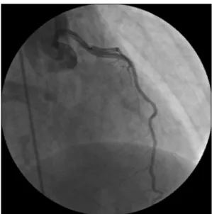
aVL, and V2 leads, along with accelerated idioventricular rhythm (Fig. 1). Transthoracic echocardiography demonstrated akinesia of the anterior septal, apical, basal-mid septal, and basal-mid anterior walls, and her ejection fraction was 20%. She was immediately transferred to the catheterization unit. We performed coronary angiography after introducing a 6-Fr sheath through the right femoral artery. We detected a dis-section of the left main artery, the left anterior descending ar-tery (LAD), and the circumflex arar-tery, originating from the
Korean J Thorac Cardiovasc Surg 2015;48:281-284 □ Case Report □
Emrah Ipek, et al
− 282 −
Fig. 1. Electrocardiogram of the patient on admission.
Fig. 3. Coronary angiography showing the dissection (right anterior
oblique view).
Fig. 2. Coronary angiography showing the dissection (anteroposterior
view with cranial angulation).
middle portion of the left main coronary artery (LMCA) (Figs. 2 and 3, Supplemental Videos 1, 2). After emergency cardiovascular surgery consultation, she was transferred to the operation room. A saphenous vein was grafted to the distal LAD. During the intraoperative evaluation of the epicardial vessels, our team of cardiac surgeons did not plan to place a bypass graft to the circumflex artery because it was thin and non-dominant. Since the patient was hypotensive under nora-drenaline and dopamine infusions, she was transferred to the cardiovascular surgery intensive care unit on an ex-tracorporeal membrane oxygenator (ECMO) and intra-aortic
balloon pump (IABP). During follow-up, her blood pressure remained low, at approximately 60/40 mmHg, despite ag-gressive inotropic and mechanical support. On the second postoperative day, although the patient’s LAD artery had been revascularized by a saphenous vein graft, her left ven-tricular ejection fraction remained as low as 10%–15%, lead-ing to ventricular failure. Asystole and cardiovascular arrest then quickly developed, and despite aggressive cardiopulmo-nary resuscitation, she died.
DISCUSSION
Coronary artery dissection after blunt chest trauma is an extremely rare condition that can be fatal, and some cases are detected in postmortem examinations [1]. Left main coronary artery dissection is even rarer [2]. Multiple mechanisms exist leading to coronary artery dissection, including intimal tears due to a deceleration injury, compression of the artery be-tween the heart and sternum, coronary spasm, and impairment of the coronary flow by a dissection flap or a superimposed thrombosis [3]. Coronary artery dissection is detected most commonly in the LAD (76%), the right coronary artery (12%), and the circumflex artery (6%) [4,5]. In necropsy ser-ies, the most common cause of acquired non-atherosclerotic coronary artery disease is spontaneous coronary artery
dis-A Kick to Back Leading to Death
− 283 − section, and the LAD is the artery in which this condition is most commonly detected [6]. The risk factors for spontaneous coronary artery dissection are exercise, arteriosclerosis, car-diovascular disease, oral contraceptive use, Marfan syndrome, systemic lupus erythematosis, and connective tissue disorders [7]. Our patient did not have any of these risk factors.
Patients with coronary artery dissection are usually admit-ted with myocardial infarction and sudden death. Early de-tection is crucial in order to provide adequate treatment. However, the time from injury to coronary artery occlusion may vary, ranging from immediately after the trauma to five weeks later [3]. Our patient was admitted to our clinic two days after the trauma. Bedside electrocardiography (ECG) provides important clues about coronary artery dissection after blunt anterior chest and back trauma. It has been previously reported that patients with baseline ECG changes [7] on ad-mission should be monitored for 24 hours. The ECG may be normal on presentation, but was found to demonstrate ST ab-normalities in 63% of patients who are admitted for blunt thoracic trauma within 24 hours of observation. Only 2% of such patients showed ST segment elevation [8]. Our patient presented with ST segment elevation the in D1, aVL, and V2 leads, along with accelerated idioventricular rhythm.
Some emergency therapeutic options exist for patients with spontaneous coronary artery dissection. In some previous case reports, the patients were managed by primary percutaneous coronary angioplasty, especially patients without LMCA le-sions [9]. Medical therapy with anticoagulants has also been utilized with successful outcomes [9]. However, surgical treat-ment remains most common treattreat-ment and is associated with the best outcomes. Unfortunately, surgical treatment was not successful in our patient. An internal mammary artery graft was not utilized in our patient, because she was in cardio-genic shock, and the surgical team tried to save time by us-ing a saphenous vein graft. In order to prevent death, all available measures were used by our surgical team, including intravenous inotropic agents, IABP, and ECMO. Nevertheless, using the internal mammary artery for revascularization may have been more effective for restoring left ventricular function. Additionally, complete revascularization, including the cir-cumflex artery, may have been helpful in our patient.
In conclusion, spontaneous coronary artery dissection is a
rare and potentially fatal complication of blunt chest trauma in younger patients, and early diagnosis and prompt treatment can be life-saving. Physicians should be aware of this possi-bility when evaluating patients in emergency conditions after blunt trauma of any kind. The 12-lead ECG, cardiac bio-markers, and transthoracic echocardiography, along with other imaging modalities, are important in the management of sus-pected coronary artery dissection in trauma patients.
CONFLICT OF INTEREST
No potential conflict of interest relevant to this article was reported.
SUPPLEMENTARY MATERIALS
Supplementary materials can be found via http://dx.doi. org/10.5090/kjtcs.2015.48.4.281. Video 1. Coronary angiog-raphy showing the dissection (left anterior oblique view). Video 2. Coronary angiography showing the dissection (right anterior oblique view).
REFERENCES
1. Kawakami Y, Inokuchi R, Tanji M, et al. Coronary artery
dissection after blunt trauma without abnormal electro-cardiogram findings. Am J Emerg Med 2014;32:1157.e5-6.
2. Li CH, Chiu TF, Chen JC. Extensive anterolateral
my-ocardial infarction caused by left main coronary artery dis-section after blunt chest trauma: a case report. Am J Emerg
Med 2007;25:858.e3-5.
3. Fanari Z, Hadid M, Hammami S, Qureshi WA. Traumatic
myocardial infarction in a young athletic patient after a football game. Del Med J 2014;86:213-5.
4. Hazeleger R, van der Wieken R, Slagboom T, Landsaat P.
Coronary dissection and occlusion due to sports injury.
Circulation 2001;103:1174-5.
5. Kim KI, Lee WY, Ko HH, Kim HS, Lee HS. Right
coro-nary artery fistula and occlusion causing myocardial in-farction after blunt chest trauma. Korean J Thorac
Cardiovasc Surg 2014;47:402-5.
6. Atay Y, Yagdi T, Turkoglu C, Altintig A, Buket S.
Spontaneous dissection of the left main coronary artery: a case report and review of the literature. J Card Surg
1996;11:371-5.
Emrah Ipek, et al
− 284 − C. Dissection of the left main coronary artery after blunt
thoracic trauma: case report and literature review. World J
Emerg Surg 2010;5:21.
8. Bjornstad JL, Pillgram-Larsen J, Tonnessen T. Coronary
ar-tery dissection and acute myocardial infarction following
blunt chest trauma. World J Emerg Surg 2009;4:14.
9. Sadr-Ameli MA, Amiri E, Pouraliakbar H, Heidarali M. Left
anterior descending coronary artery dissection after blunt chest trauma. Arch Iran Med 2014;17:86-90.